Abstract
AbstractBone regeneration requires a well-orchestrated cellular and molecular response including robust vascularization and recruitment of mesenchymal and osteogenic cells. In femoral fractures, angiogenesis and osteogenesis are closely coupled during the complex healing process. Here, we show with advanced longitudinal intravital multiphoton microscopy that early vascular sprouting is not directly coupled to osteoprogenitor invasion during calvarial bone regeneration. Early osteoprogenitors emerging from the periosteum give rise to bone-forming osteoblasts at the injured calvarial bone edge. Microvessels growing inside the lesions are not associated with osteoprogenitors. Subsequently, osteogenic cells collectively invade the vascularized and perfused lesion as a multicellular layer, thereby advancing regenerative ossification. Vascular sprouting and remodeling result in dynamic blood flow alterations to accommodate the growing bone. Single cell profiling of injured calvarial bones demonstrates mesenchymal stromal cell heterogeneity comparable to femoral fractures with increase in cell types promoting bone regeneration. Expression of angiogenesis and hypoxia-related genes are slightly elevated reflecting ossification of a vascularized lesion site. Endothelial Notch and VEGF signaling alter vascular growth in calvarial bone repair without affecting the ossification progress. Our findings may have clinical implications for bone regeneration and bioengineering approaches.
🔬 Techniques
💻 Software
✨ Fluorophores
🧪 Sample Preparation
🔬 Cell Lines
🏭 Microscope Brands
🧪 Reagent Suppliers
🔴 Lasers
📷 Detectors
💻 Software Details
🏛️ Research Organizations (ROR)
Affiliated research institutions:
📋 Methods
All animal experiments were performed according to the institutional guidelines and laws, approved by local animal ethical committee and were conducted at the University of Münster and the Max Planck Institute for Molecular Biomedicine with necessary permissions (84-02.04.2018.A171, 81-02.04.2019.A164) granted by the Landesamt für Natur, Umwelt und Verbraucherschutz (LANUV) of North Rhine-Westphalia, Germany.
Multiphoton imaging setup
We used a TriM Scope II multi photon system from LaVision BioTec (Bielefeld, Germany) to visualize immune labeling and SHG generated by collagen in the bone 41 . The setup is a single-beam instrument with an upright Olympus BX51 WI microscope stand that is equipped with highly sensitive NDD detectors close to the objective lens. The TriM Scope II is fitted with a Coherent Scientific Chameleon Ultra II Ti:Sapphire laser and a Coherent Chameleon Compact OPO. A 20x IR objective lens (Olympus XLUMPlanFL; NA 1.0) with a working distance of 2.0 mm was used. The microscope is equipped with a pair of x-y galvanometric mirrors used to scan at a scanning speed of up to 1200 lines/s. The maximal laser power (850 nm) on the object was 10–20 mW at superficial areas and 80–100 mW when imaging at large depths. Dichromatic mirrors and band pass filters spectrally separate the emitted light before the signal is detected using attached photomultiplier tubes (PMT; Hamamatsu H67080-01 (blue channel), H67080-20 (green and red channels). The following detection channels were used: red (560–680 nm), green (475–575 nm) and blue (370–470 nm). 3D-images were acquired and processed with LaVision BioTec ImSpector Pro.
Show full methods section
All animal experiments were performed according to the institutional guidelines and laws, approved by local animal ethical committee and were conducted at the University of Münster and the Max Planck Institute for Molecular Biomedicine with necessary permissions (84-02.04.2018.A171, 81-02.04.2019.A164) granted by the Landesamt für Natur, Umwelt und Verbraucherschutz (LANUV) of North Rhine-Westphalia, Germany.
Multiphoton imaging setup
We used a TriM Scope II multi photon system from LaVision BioTec (Bielefeld, Germany) to visualize immune labeling and SHG generated by collagen in the bone 41 . The setup is a single-beam instrument with an upright Olympus BX51 WI microscope stand that is equipped with highly sensitive NDD detectors close to the objective lens. The TriM Scope II is fitted with a Coherent Scientific Chameleon Ultra II Ti:Sapphire laser and a Coherent Chameleon Compact OPO. A 20x IR objective lens (Olympus XLUMPlanFL; NA 1.0) with a working distance of 2.0 mm was used. The microscope is equipped with a pair of x-y galvanometric mirrors used to scan at a scanning speed of up to 1200 lines/s. The maximal laser power (850 nm) on the object was 10–20 mW at superficial areas and 80–100 mW when imaging at large depths. Dichromatic mirrors and band pass filters spectrally separate the emitted light before the signal is detected using attached photomultiplier tubes (PMT; Hamamatsu H67080-01 (blue channel), H67080-20 (green and red channels). The following detection channels were used: red (560–680 nm), green (475–575 nm) and blue (370–470 nm). 3D-images were acquired and processed with LaVision BioTec ImSpector Pro.
Fluorescence microscopy
Bone immunostaining performed as described previously 76 , bone sections were washed in ice-cold PBS and permeabilized with ice-cold 0.3% Triton-X-100 in PBS for 10 mins at room temperature (RT). Samples were incubated in blocking solution (5% heat-inactivated donkey serum in 0.3% Triton-X-100) for 1 h at RT. Primary antibodies: rat monoclonal anti-Endomucin (1:200, Santa Cruz, Cat# sc-65495, clone V.7C7), goat polyclonal anti-CD31 (1:200, R&D, Cat# AF3628), goat polyclonal anti-Pdgfrb (1:200, R&D, Cat# AF1042), rabbit polyclonal anti-Osterix (1:200, Abcam, Cat# ab22552), rabbit polyclonal anti-Acan (1:200, Millipore, Cat#AB1031), rabbit polyclonal anti-vATPaseB1/B2 (1:200, Abcam, Cat# 200839), rabbit polyclonal anti-Runx2 (1:200, Abcam, Cat# 192256) were diluted in 5% donkey serum mixed PBS and incubated overnight at 4 °C. Next, slides were washed 3 to 5 times in PBS in 5-10 min intervals. Wholemount calvarial bones with drill hole lesions were fixed with PFA (2%) overnight at 4 °C and stained using primary antibody rabbit polyclonal anti-Osterix (1:200, Abcam, Cat# ab22552) for 3 days at 4 °C to allow for sufficient tissue penetration of the antibodies. Species-specific Alexa Fluor secondary antibodies (goat anti-rat IgG Alexa Fluor 594 (1:200, Thermo Fischer Scientific, Cat# A21209), donkey anti-rabbit IgG Alexa Fluor 647 (1:200, Thermo Fischer Scientific, Cat# A31573 ), donkey anti-goat IgG Alexa Fluor 647 (1:200, Thermo Fischer Scientific, Cat# A21447), donkey anti-rat IgG Alexa Fluor 488 (1:200, Thermo Fischer Scientific, Cat# A21208) were diluted in PBS were added and incubated for 3 h at RT or overnight at 4 °C.
Construction of fluorescent composite and tile scan images
Multiphoton images obtained with the microscope
ImspectorPro acquisition software were analyzed, processed and assembled using ImageJ (open source NIH software, http://imagej.nih.gov/ij ). For tile scans images a 20% overlap were recorded and stitched using the stitching plugin from ImageJ.
Animal models
All mice used were on a C57/Bl6J background. Female mice were used between 8–12 weeks old unless stated otherwise. Flk1-GFP + transgenic mice (STOCK Kdrtm2.1Jrt/J, RRID:IMSR_JAX:017006) 77 were used to label the bone vasculature. Sp7-mCherry + transgenic mice (STOCK Tg(Sp7/mCherry)2Pmay/J, RRID:IMSR_JAX:024850) 78 were used to visualize osteoprogenitor and osteoblasts. Vascular reporter Flk1-GFP mice were crossed to Fbxw7 lox/lox:Cdh5Cre-ERT2 (Fbxw7 iΔEC ) (B6;129-Fbxw7tm1Iaai/J, RRID:IMSR_JAX:017563: Tg(Cdh5-cre/ERT2)1Rha, MGI:3848982) 79 , 80 to study Notch GOF in endothelial cells. Notch LOF in endothelial cells was studied in Dll4 lox/lox:Cdh5Cre-ERT2 mice (Dll4 iΔEC ) (Dll4tm1Frad, MGI:3828266: Tg(Cdh5-cre/ERT2)1Rha, MGI:3848982) 62 . For inducible Cre-mediated recombination, mice received daily intraperitoneal (i.p.) injections of tamoxifen (4 mg/kg body weight) from day1 to day 5 (d1-d5) one week before the cranial window and calvarial drill hole lesion surgery. For fracture healing model, we used 10-week-old female mice for the fracture experiments. Mice were anesthetized by using a ketamine hydrochloride/xylazine mixture (80 /12 mg/kg body weight, i.p.) the left leg was fractured with three-point bending. It was stabilized with an intramedullary nail (hollow needle 23 G) as described previously 81 . Carprofen (4 mg/kg intra muscular) was given as an analgesic and further on at 24-hour intervals when required. Mice were sacrificed by cervical dislocation after post-fracture surgery day at 14 and 12 weeks wild-type female were used as control.
Bone sample preparation
Mice were sacrificed and femurs were harvested and fixed immediately in ice-cold 2% paraformaldehyde (PFA) for 6 to 8 h under gentle agitation. Bones were decalcified in 0.5 M EDTA for 16 to 24 h at 4 °C under gentle shaking agitation, which was followed by overnight incubation in cryopreservation solution (20% sucrose, 2% PVP) and embedded in in bone embedding medium (8% gelatine, 20% sucrose, 2% PVP). Samples were stored at −80 °C. For immunofluorescence staining 90 to 100 μm-thick cryosections were prepared.
Surgical preparation for longitudinal intravital imaging
We used a chronic cranial window to provide clear optical access to the calvarial bone and bone microvessels 41 , 82 – 84 . A schematic of the cranial window construction and fixation of the mouse using the head immobilization device is shown in Fig. 1a . Mice were anesthetized with a combination of ketamine/xylazine. A circular incision was made in the scalp to expose the underlying dorsal skull surface. A dental drill (diameter: 500 µm) was used to insert a drill hole lesion into the parietal bone (Fig. 1b ). A round glass cover slip with a hanging drop of sterile PBS was placed over the bone lesion and fixed with dental acrylic. A custom-designed titanium ring was glued on top to allow head immobilization and the elimination of motion artifacts. Dexamethason was used to prevent inflammatory responses. Ketamine/xylazine anesthesia was used for intravital imaging and mice were kept on a 37 °C heat pad to keep the body temperature constant 41 , 82 . The physical condition of the mice was frequently controlled by observing their breathing frequency and their overall condition. Bone stromal cell preparation for single cell RNA sequencing (scRNA-seq) Skulls with calvarial lesions (diameter: 1 mm) at PLD14 with their corresponding age-matched controls were harvested, cleaned from attached surrounding tissue and collected in digestion enzyme solution (Collagenase type I and IV, 2 mg/ml). Next, bones were cut into small pieces. Samples were digested for 30 min at 37 °C under gentle agitation. Digested samples were transferred to 70 µm strainers in 50 ml tubes to obtain a single–cell suspension, which was resuspended in blocking solution (1% BSA, 1 mM EDTA in PBS without Ca 2+ /Mg 2+ ), centrifuged at 300 × g for 5 mins, washed 2–3 times with ice-cold blocking solution, and filtered through 50 µm strainers. Pellets were resuspended in respective volume of blocking solution. Single cell suspensions were subjected to lineage depletion using lineage cell depletion kit (MACS, cat#130-090-858) following the manufacturer’s instructions. Next, lineage negative cells were depleted by CD45 and CD117 using microbeads (MACS, cat#130-052-301 and cat#130-091-224) from lineage negative (Lin - ) bone cells to enrich bone stromal cells. Single cell suspensions were processed with BD Rhapsody and scRNA-seq libraries were evaluated and quantified by Agilent Bioanalyzer using High sensitivity DNA kit (cat#5067-4626) and Qubit (ThermoFisher Scientific, Cat# Q32851 ). Individual libraries were diluted to 4 nM and pooled for sequencing. Pooled libraries were sequenced by using High Output kit (150 cycle) (Illumina cat#TG-160-2002) with a NextSeq500 sequencer (Illumina).
Single cell RNA-seq data analysis
Sequencing data of FASTQ format were processed with BD Rhapsody WTA Analysis pipeline (version 1.0) on SevenBridges Genomics online platform (SevenBridges) and expression matrix were used for further data analysis. Data normalization, dimensionality reduction and visualization were performed using Seurat (version 4.3.0) if not specified otherwise. For initial quality control of the extracted gene-cell matrices, we filtered cells with parameters nFeature_RNA > 500 & nFeature_RNA < 6000 for number of genes per cell and percent.mito
📊 Figures
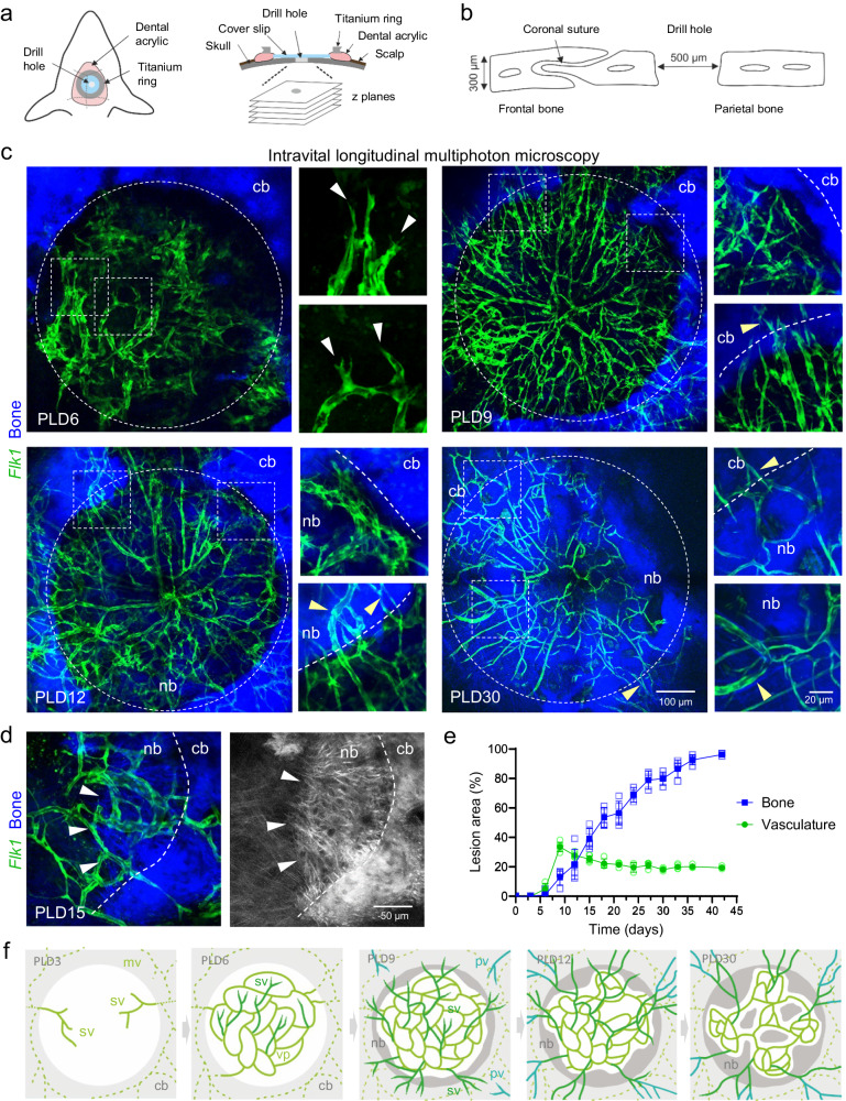
Fig. 1
Longitudinal intravital multiphoton imaging of calvarial bone healing after lesion injury.
a Schematic of a chronic cranial window for intravital imaging of calvarial bone repair. A drill hole lesion is inserted in the parietal calvarial bone. Coverslip and titanium fixation ring are mounte...
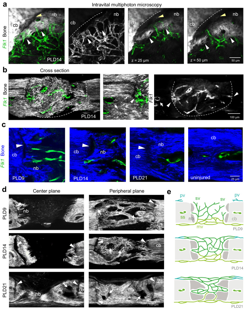
Fig. 2
Vascular and bone connectivity during calvarial bone healing.
a Intravital multiphoton microscopy showing newly formed calvarial bone matrix and regenerating microvasculature at PLD14. Maximum intensity projections (left) of SHG + (white) injured calvarial bone ...
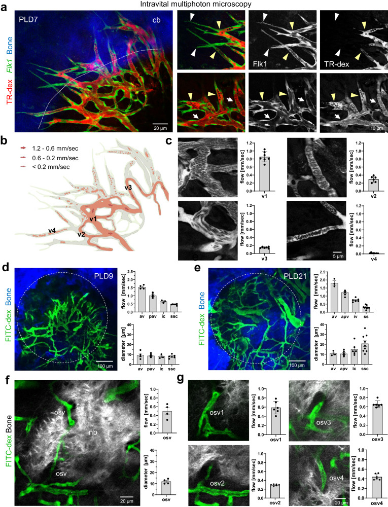
Fig. 3
Blood flow in sprouting and regenerating bone microvessels.
a Intravital multiphoton imaging of sprouting Flk1-GFP + (green) microvasculature near the SHG + (blue) calvarial bone edge in Flk1-GFP + reporter mice at PLD7. Intravenously injected TexasRed-dextran...
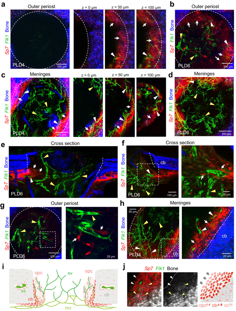
Fig. 4
Osteoprogenitors emerge from the periosteum and collectively colonize the calvarial bone lesion, while blood vessel vascularizes the entire lesion.
a, b Multiphoton imaging of wholemount calvarial bone at PLD4 ( a ) and PLD6 ( b ) from the outer periosteal side. a Sp7-mCherry + (red) osteoblastic cells colonize the injured SHG + (blue) calvarial ...
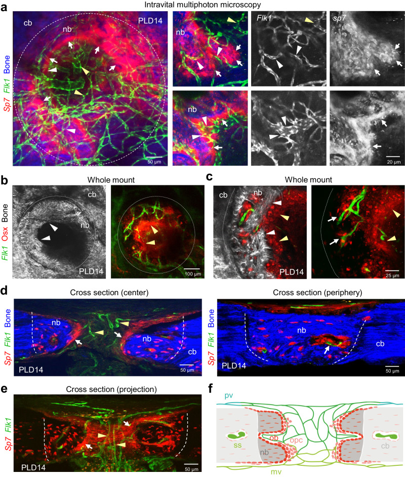
Fig. 5
Osteoblasts lining the growing bone collectively invade the vascularized calvarial bone lesion.
a Intravital multiphoton microscopy showing the expanding SHG + (blue) calvarial bone lined by a multicellular layer of Sp7-mCherry + (red) osteoblastic cells and Flk1-GFP + (green) microvessels vascu...
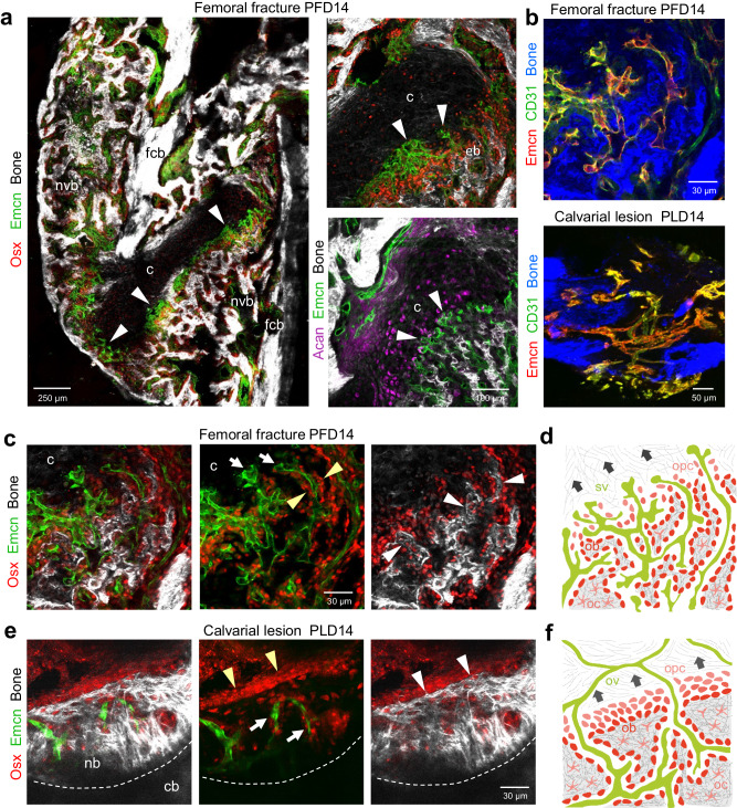
Fig. 6
Vascular and bone regeneration in femoral fractures and calvarial bone lesions.
a Tile-scan multiphoton microscopy of a PFD14 femoral fracture. Overview image shows maximum intensity projections of Emcn + (green) microvasculature, Osx + (red) osteoblasts and progenitors and SHG +...
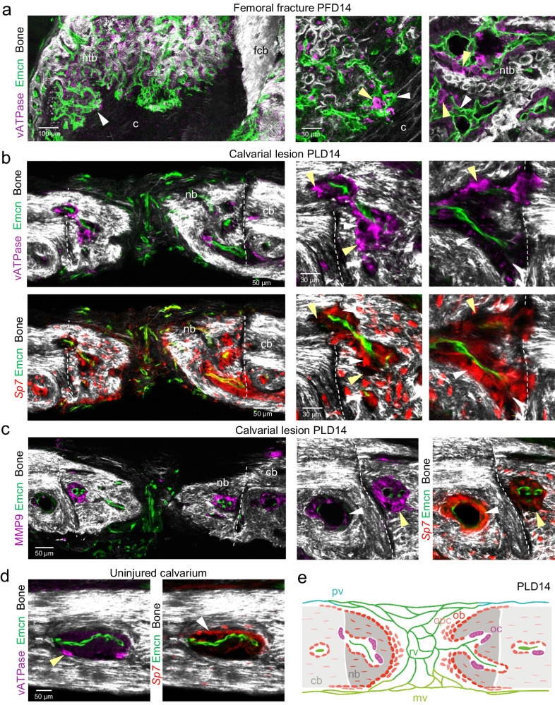
Fig. 7
Bone remodeling by osteoclasts in femoral fractures and calvarial bone lesions.
a Tile-scan multiphoton microscopy of PFD14 femoral fracture. Overview image shows maximum intensity projections of Emcn + (green) microvasculature, vATPase + (purple) osteoclasts and SHG + (white) ne...
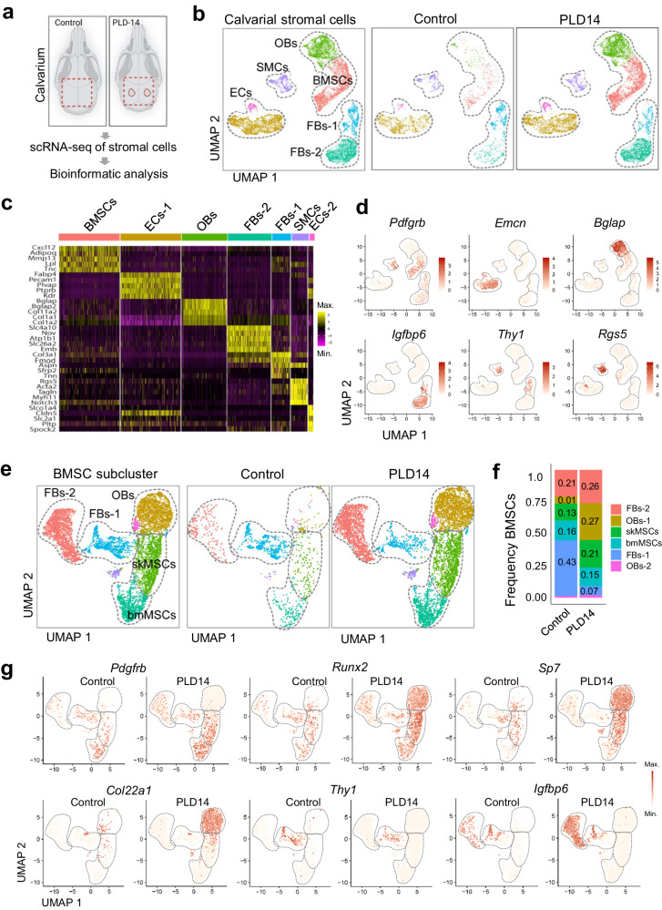
Fig. 8
Single cell RNA-sequencing analysis of bone non-hematopoietic cells after calvarial bone injury.
a Preparation of calvarial bones at PLD14 for scRNA-seq data analysis. b UMAP plots showing color-coded merged cell clusters from calvarial bones at PLD14 (left), group-selected color-coded cell clust...
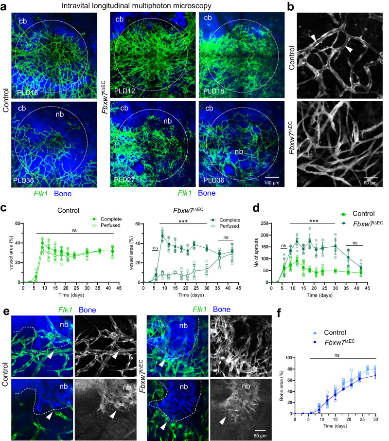
Fig. 9
Notch activation enhances endothelial sprouting after calvarial bone injury.
a Longitudinal intravital multiphoton microscopy showing hypersprouting of Flk1-GFP + (green) microvessels in Fbxw7 iu0394EC Flk1-GFP transgenic mice after calvarial bone lesions at PLD12, PLD18, PLD2...
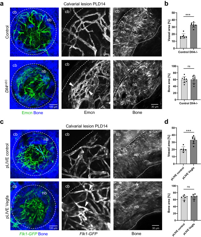
Fig. 10
Vascular changes via Notch or VEGF-A signaling do not affect with calvarial bone regeneration.
a Whole mount multiphoton microscopy showing Emcn + (green) microvessels in Dll4 iu0394EC and control mice after calvarial bone lesions at PLD14. SHG + calvarial bone (cb) and new bone (nb) are shown ...
Figure images are served from the NIH/NLM PubMed Central Open Access Subset or Europe PMC; copyright remains with the publishers and authors.
💬 Discussion
0 commentsNo comments yet. Be the first to start a discussion!
Leave a Comment