Abstract
AIMS: CLARITY is a novel technique which enables three-dimensional visualization of immunostained tissue for the study of circuitry and spatial interactions between cells and molecules in the brain. In this study, we aimed to compare methodological differences in the application of CLARITY between rodent and large human post mortem brain samples. In addition, we aimed to investigate if this technique could be used to visualize Lewy pathology in a post mortem Parkinson's brain. METHODS: Rodent and human brain samples were clarified and immunostained using the passive version of the CLARITY technique. Samples were then immersed in different refractive index matching media before mounting and visualizing under a confocal microscope. RESULTS: We found that tissue clearing speed using passive CLARITY differs according to species (human vs. rodents), brain region and degree of fixation (fresh vs. formalin-fixed tissues). Furthermore, there were advantages to using specific refractive index matching media. We have applied this technique and have successfully visualized Lewy body inclusions in three dimensions within the nucleus basalis of Meynert, and the spatial relationship between monoaminergic fibres and Lewy pathologies among nigrostriatal fibres in the midbrain without the need for physical serial sectioning of brain tissue. CONCLUSIONS: The effective use of CLARITY on large samples of human tissue opens up many potential avenues for detailed pathological and morphological studies.
🔬 Techniques
🔭 Microscopes
💻 Software
✨ Fluorophores
🧪 Sample Preparation
🔬 Cell Lines
🏭 Microscope Brands
🧪 Reagent Suppliers
💻 Software Details
🏛️ Research Organizations (ROR)
Affiliated research institutions:
📋 Methods
Rodent and human brain samples were clarified and immunostained using the passive version of the CLARITY technique. Samples were then immersed in different refractive index matching media before mounting and visualizing under a confocal microscope.
Methods
CLARITY hydrogel embedding for rodent brain tissue 12‐week‐old C57BL/6 mice and Sprague–Dawley rats weighing between 250–300 g were terminally anaesthetized with sodium pentobarbital (intraperitoneal injection; 120 mg/kg body weight) and transcardially perfused with ice‐cold phosphate‐buffered saline (PBS, pH7.4) and then with hydrogel monomer solution consisting of 4% paraformaldehyde (PFA), 2% or 4% (vol/vol) acrylamide (Bio‐Rad, UK), 0.05% (vol/vol) bis‐acrylamide (Bio‐Rad, UK), 0.25% (wt/vol) VA‐044 photoinitiator (Wako, Alpha‐Labs, Eastleigh, UK) and PBS. Brains were then extracted and immersed in the same solution for 2 days at 4°C. Next, the brains with hydrogel solution were transferred into a 50 ml container and a layer of olive oil was poured on top of the solution with the lid tightly screwed to prevent oxygen inhibition of the subsequent polymerization of the hydrogel by incubating at 37°C for 3 h (or 4 h for 2% acrylamide solution). After removal of excess hydrogel from the hydrogel‐tissue hybrid, brain tissues were left either undissected, bisected into two hemispheres in the case for mouse brains or cut into 3‐mm‐thick coronal sections for rat brains using a rat brain matrix before proceeding to the subsequent clarification step. CLARITY hydrogel embedding for human brain tissue Human brain tissues from the Parkinson's UK Tissue Bank at Imperial College London were used in this study. Both fresh and formalin‐fixed (>4 weeks fixation) tissues from various brain regions were obtained. Tissue blocks were cut into 0.5–1‐cm‐thick and incubated in hydrogel monomer solution as described above. Fresh brain tissues were incubated for 5–7 days and formalin‐fixed tissues were incubated for 7–10 days (depending on the block thickness) at 4°C until a rubbery‐firm consistency was reached. The tissue blocks with hydrogel solution were then transferred into 50 ml containers and a layer of olive oil was poured on top of the solution, with the lid tightly screwed on to prevent oxygen inhibition of the subsequent polymerization of the hydrogel by incubating at 37°C for 3 h (or 4 h for 2% acrylamide solution). After removal of excess hydrogel from the hydrogel‐tissue hybrid, tissues were then sectioned into 3 mm blocks using a scalpel prior to clearing.
Show full methods section
Rodent and human brain samples were clarified and immunostained using the passive version of the CLARITY technique. Samples were then immersed in different refractive index matching media before mounting and visualizing under a confocal microscope.
Methods
CLARITY hydrogel embedding for rodent brain tissue 12‐week‐old C57BL/6 mice and Sprague–Dawley rats weighing between 250–300 g were terminally anaesthetized with sodium pentobarbital (intraperitoneal injection; 120 mg/kg body weight) and transcardially perfused with ice‐cold phosphate‐buffered saline (PBS, pH7.4) and then with hydrogel monomer solution consisting of 4% paraformaldehyde (PFA), 2% or 4% (vol/vol) acrylamide (Bio‐Rad, UK), 0.05% (vol/vol) bis‐acrylamide (Bio‐Rad, UK), 0.25% (wt/vol) VA‐044 photoinitiator (Wako, Alpha‐Labs, Eastleigh, UK) and PBS. Brains were then extracted and immersed in the same solution for 2 days at 4°C. Next, the brains with hydrogel solution were transferred into a 50 ml container and a layer of olive oil was poured on top of the solution with the lid tightly screwed to prevent oxygen inhibition of the subsequent polymerization of the hydrogel by incubating at 37°C for 3 h (or 4 h for 2% acrylamide solution). After removal of excess hydrogel from the hydrogel‐tissue hybrid, brain tissues were left either undissected, bisected into two hemispheres in the case for mouse brains or cut into 3‐mm‐thick coronal sections for rat brains using a rat brain matrix before proceeding to the subsequent clarification step. CLARITY hydrogel embedding for human brain tissue Human brain tissues from the Parkinson's UK Tissue Bank at Imperial College London were used in this study. Both fresh and formalin‐fixed (>4 weeks fixation) tissues from various brain regions were obtained. Tissue blocks were cut into 0.5–1‐cm‐thick and incubated in hydrogel monomer solution as described above. Fresh brain tissues were incubated for 5–7 days and formalin‐fixed tissues were incubated for 7–10 days (depending on the block thickness) at 4°C until a rubbery‐firm consistency was reached. The tissue blocks with hydrogel solution were then transferred into 50 ml containers and a layer of olive oil was poured on top of the solution, with the lid tightly screwed on to prevent oxygen inhibition of the subsequent polymerization of the hydrogel by incubating at 37°C for 3 h (or 4 h for 2% acrylamide solution). After removal of excess hydrogel from the hydrogel‐tissue hybrid, tissues were then sectioned into 3 mm blocks using a scalpel prior to clearing.
Passive tissue clearing
A passive clearing protocol, outlined by Tomer and colleagues 17 , was adopted in preference to active electrophoretic clearing as described in the original protocol 13 . Hydrogel‐embedded samples were washed with SDS clearing solution (4% wt/vol SDS in 1 M boric acid solution, pH8.5; 2 × 24 h at room temperature, RT) to remove remaining hydrogel monomers. Samples were then incubated at 50°C in SDS clearing solution to begin the passive clearing process. Clearing solution was replaced every 2–3 days. Tissue remained in the clearing solution until transparency was achieved.
Immunostaining on clarified rodent and human brain tissues
After clearing, samples were washed thoroughly with PBS with Triton‐X (PBST; 0.1% vol/vol triton‐X and 0.01% wt/vol sodium azide in PBS) (2 × 24 h at 37°C) to remove residual SDS micelles. Samples were incubated with primary antibody in 15 ml containers with PBST as diluent. Working concentrations for various antibodies used are listed in Table 2 . Typically, 1 ml of antibody solution is sufficient to cover a 3‐mm‐thick sample in the container. Tissue samples were incubated at 37°C for 5 days. Primary antibody was washed off with PBST (2 × 24 h at 37°C). Secondary antibody was applied at a concentration of 1:50 and incubated at 37°C for 5 days. A nuclear counterstain, 4′,6‐diamidino‐2‐phenylindole (DAPI; 1:100 from a stock of 1 μ g/ml diluted with 1:1 water: dimethyl sulphoxide), can be added at this stage. Tissue was then washed with PBST (2 × 24 h at 37°C) to remove excess secondary antibody. Table 2 Antibodies used in the study Primary antibodies Catalogue no. Company Host Clonality Dilution Antigen Neurofilament (NF, 2F11) M0762 Dako Mouse Monoclonal 1:50–1:100 Ionized calcium‐binding adapter molecule 1 (IBA1) 019‐19741 Wako Rabbit Polyclonal 1:50 α‐ synuclein ( α ‐syn, 42) 610787 Becton Dickson (BD) Mouse Monoclonal 1:50 Tyrosine hydroxylase (TH) AB152 Millipore Rabbit Polyclonal 1:50–1:100 Secondary antibodies Catalogue no. Company Host Fluorophore conjugates Dilution Name AlexaFluor anti‐goat A‐11055 Invitrogen Donkey 488 IgG (H+L) 1:50 AlexaFluor anti‐goat A‐11057 Invitrogen Donkey 568 IgG (H+L) 1:50 AlexaFluor anti‐rabbit A10042 Invitrogen Donkey 568 IgG (H+L) 1:50 AlexaFluor anti‐mouse A‐21202 Invitrogen Donkey 488 IgG (H+L) 1:50 AlexaFluor anti‐rabbit A‐11011 Invitrogen Goat 568 IgG (H+L) 1:50 AlexaFluor anti‐mouse A‐11001 Invitrogen Goat 488 IgG (H+L) 1:50 John Wiley & Sons, Ltd Comparison of various refractive index matching media with human brain tissues For optimizing tissue refractive index (RI) matching, roughly 1 × 1 × 1 cm 3 of clarified human cerebellum was each stained in 1:100 dilution of antibodies against neurofilament in 1 ml of PBST at 37°C for 2 days, followed by washing in excess PBST at 37°C for 6 h, and stained with secondary antibody at 1:100 dilution in 1 ml of PBST at 37°C for 2 days. After several washings, the samples were immersed in 5 ml of each compared RI matching formula or cleared as described by various techniques (Table 3 ). Table 3 Comparison of different refractive index matching solutions Reagent Fluorescent signal preservation Transparency Time to maximal clearance (3 × 3 × 3 mm 3 tissue block) Preparation Storage Other remarks PBST Good + Never Simple Indefinitely N/A FocusClear Good +++ 2 h (Bought) Cannot be stored Occasional yellow precipitates or discoloration observed 87% Glycerol Good ++ Overnight Simple Not recommended, slight loss of signal after 2 days Cheapest and simplest BABB Poor ++ 3 h Simple but hazardous Cannot be stored Over‐shrinkage Histodenz‐RIMS Moderate in humans, good in mice +++ Overnight Complex Up to 6 months for mice, up to 1.5 months for humans Works best for mice brains Sorbitol (sRIMS) Good +++ Overnight Complex Up to 6 months for mice, up to 1.5 months for humans Take extra care to avoid fungal growth SeeDB Moderate ++ 6 h Complex Up to 1.5 months for humans Take extra care to avoid fungal growth Sca l eA2 Good ++++ 3 h Simple Up to 1.5 months for humans Tissue remains swollen/swells even more; most transparent THF/DBE Moderate ++ 18 h Simple but hazardous Up to 1.5 months for humans Over‐shrinkage 47% TDE in PBS Good ++++ 3 h Simple Up to 1.5 months for humans Remains slightly swollen 63% TDE in PBS Good +++ 3 h Simple Up to 1.5 months for humans Shrinks back to original size BABB, benzyl alcohol–benzyl benzoate; DBE, dibenzyl ether; PBS, phosphate‐buffered saline; PBST, phosphate‐buffered saline with Triton‐X; RIMS, refractive index matching solution; TDE, 2,2′‐Thiodiethanol; THF, tetrahydrofuran. John Wiley & Sons, Ltd Imaging For imaging of immunostained whole or hemisected mouse brain samples, the CLARITY‐treated brain was incubated in 87% glycerol overnight at room temperature. For immunostained 3‐mm‐thick coronal rat brain or human brain samples, the clarified tissues were incubated in 87% glycerol or Sca l eA2 solution for 3 h prior to imaging. Samples were mounted in a 50‐mm diameter standard bottom imaging dish (Ibidi, Germany) with a surrounding ring of Blu‐Tack to prevent sample movement. Samples were placed carefully at the bottom of the dish. RI matching solution was slowly pipetted to the dish, so as not to agitate the sample. It was paramount that the sample remained in contact with the bottom of the dish to ensure the sample was in focus during imaging, due to the short working distance of the objective. Imaging on CLARITY‐treated samples was performed using a Zeiss LSM‐780 inverted confocal and Zeiss LSM‐710 upright confocal laser scanning microscopes (Carl Zeiss, Oberkochen, Germany) either at the Facility for Imaging by Light Microscopy (FILM) facility in Hammersmith Hospital or at the University of Hong Kong Li Ka Shing Faculty of Medicine Faculty Core Facility. A ×20 objective (W Plan Apochromat DIC M27, numerical aperture 1.0; working distance, 1.7 mm) with laser excitation at 800 nm was used to image the whole mouse brain tissue. To image various human tissues, a ×10 objective (EC Plan‐Neofluar, numerical aperture, 0.3; working distance, 5.2 mm) and ×20 objective (Plan Apochromat DIC, numerical aperture, 0.8; working distance, 0.55 mm) with laser excitation at 405 nm, 488 nm, 543 nm and 594 nm were used. Image capture and processing were performed using the Zen Black (Carl Zeiss, Germany) software. Three‐dimensional rendering and video production were performed on Zen Black (Carl Zeiss, Germany), Volocity (PerkinElmer, MA, USA) and Fiji (Image J, NIH) software. For the comparison of fluorescent signal intensity after immersion in various RI matching solution, a Leica SP5 inverted confocal microscope with a ×10 objective (HC PL APO CS, numerical aperture, 0.40; working distance 2.2 mm) and laser excitation at 514 nm and 561 nm were used. Image capture was performed using the Leica Application Suite Advanced Fluorescence (LAS AF) software. All imaging settings were the same for each RI‐matched sample. Intensity analysis was done using Fiji macros made by Steve Rothery which is downloadable in http://www3.imperial.ac.uk/imagingfacility/resources/macros-n-scripts , using which intensity profile across the diagonals through the whole image Z‐stacks was analysed, and the overall maximum intensity was found and compared. We did not construct a scoring scheme to quantitatively evaluate each RI matching formula, otherwise many samples would be required yet inaccurate due to intervening variables. Overall, the description of fluorescent signal intensity in Table 3 is based on the intensity evaluation, our overall impression of the samples under the microscope, and our experience with different RI matching formulas, which is sufficient as most histological evaluation focus on morphology rather than quantitative analysis. Ethical considerations The work conducted on human tissue was under ethical approval held by the Parkinson's UK Brain Bank at Imperial College London (Registered charity in England and Wales (258197) and in Scotland (SC037554); Multicentre Research Ethics Committee approval reference number: 07/MRE09/72). Parkinson's UK Brain Bank is an approved Research Tissue Bank by the Wales Research Ethics Committee (Ref. No. 08/MRE09/31+5). Informed consent was obtained prospectively for the use of post mortem brain tissues and brain samples were obtained and prepared in accordance to the Wales Research Ethics Committee approved protocols. All animal work in this study was performed at the University of Hong Kong with approval from the Committee on the Use of Live Animals in Teaching and Research (CULATR) in the Laboratory Animal Unit (CULATR reference numbers: 3161‐13 and 3494‐14), a fully accredited unit awarded by the Association for Assessment and Accreditation of Laboratory Animal Care International (AAALAC).
Supporting information Figure S1. Comparison of clearing speed between different tissues. a – c : Clearing of a whole mouse brain to transparency in 21 days. d – f : Clearing of a 3‐mm block of rat brain to transparency in 10 days. g , h : Clearing of a 3‐mm block of human cortical tissue to transparency in 39 days. i : human brain tissue cross‐linked with 2% instead of 4% acrylamide does not improve clearing speed. Tissue integrity is also considerably worse than that with 4% acrylamide. Click here for additional data file. Figure S2. 1 × 1 × 1 cm 3 of human cerebellum stained with anti‐neurofilament and refractive index‐matched for 28 h at room temperature in different formula/techniques, upper row from left to right: FocusClear, 87% Glycerol, Histodenz‐RIMS, Sorbitol (sRIMS), ScaleA2, SeeDB, PBST (control), 47% TDE in water, 63% TDE in water, 47% TDE in PBS, 63% TDE in PBS; lower left: clearing by dehydration (methanol series)‐BABB method; lower right: clearing by dehydration (THF series)‐DBE method. Click here for additional data file. Figure S3. 1 × 1 × 1 cm 3 of human cerebellum stained with anti‐neurofilament and RI‐matched for 28 h at room temperature in 47% TDE with different diluents, from left to right: in PBS, in 1.37 M NaCl, in 10 mM phosphate buffer, in water. Click here for additional data file. Figure S4. Z‐projections of samples in Figure S3 RI‐matched in 47% TDE diluted in different medium. From left to right: in PBS, in 1.37 M NaCl, in 10 mM phosphate buffer, in water. Click here for additional data file. Figure S5. A 3 × 3 tiled Z‐projection (z‐stack depth = 419.95 μ m, step size = 2.295 μ m) of a basal ganglia section stained using anti‐TH antibody with a nuclear counterstain 4′,6‐diamidino‐2‐phenylindole (DAPI), revealing the sparse monoaminergic fibres in a Parkinson's case. Scale bar = 80 μ m. Click here for additional data file. Video Clip S1. Three‐dimensional visualization of neurofilament immunohistochemical staining in post mortem human cortical tissue. A 3‐mm block of human cortex was immunostained using anti‐neurofilament antibody (green). Stained tissue was visualized using a 10× objective (EC Plan‐Neofluar, numerical aperture, 0.3; working distance, 5.2 mm) with an imaging depth to 771.67 μ m (z‐stack step size 5.3 μ m). Click here for additional data file. Video Clip S2. Three‐dimensional visualization of a Lewy body in the human nucleus basalis of Meynert. A 3‐mm block of human nucleus basalis of Meynert was immunostained using anti‐alpha‐synuclein antibody (green). Stained tissue was visualized using a 20× objective (Plan Apochromat DIC, numerical aperture, 0.8; working distance, 0.55 mm; z‐stack step size 1.1 μ m). Click here for additional data file. Video Clip S3. Three‐dimensional visualization of monoaminergic fibres and Lewy pathologies in the post mortem human midbrain tissue. A 3‐mm block of human midbrain tissue was immunostained using anti‐alpha‐synuclein antibody (green) and tyrosine hydroxylase (red). Stained tissue was visualized using a 20× objective (Plan Apochromat DIC, numerical aperture, 0.8; working distance, 0.55 mm) with an imaging depth to 312.62 μ m (z‐stack step size 1.5 μ m). Click here for additional data file.
📊 Figures
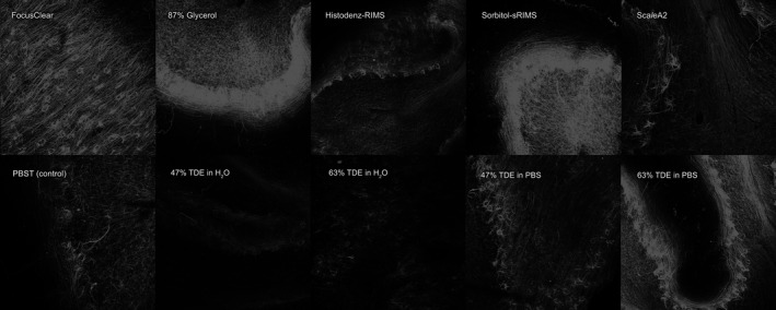
Figure 1
Human cerebellar cortex stained with antiu2010neurofilament primary antibody and subsequently AlexaFluoru2010568u2010conjugated donkey antiu2010mouse secondary antibody and imaged using Leica SP 5 con...
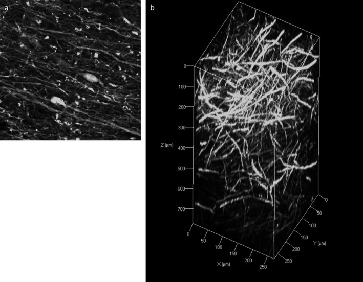
Figure 2
Immunofluorescence with neurofilament ( NF ) staining on human cortical tissue. a : A twou2010dimensional image of NF staining showing fine axonal processes and neuronal somas. Scale bar = 50 u03bc m....
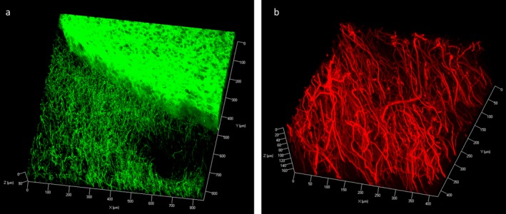
Figure 3
Zu2010stack image of Immunofluorescence with tyrosine hydroxylase ( TH ) staining on rat coronal block showing TH u2010positive neuronal processes at the cortex and dense, homogenous staining within t...
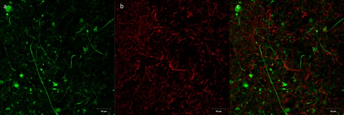
Figure 4
Double immunofluorescence with antiu2010neurofilament ( a , green) and antiu2010Ibau20101 ( b , red) antibodies on human cortical block. c : Combined figure showing surveillance network of Ibau20101u2...
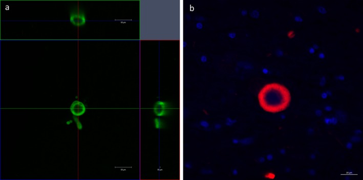
Figure 5
An orthogonal projection of a Lewy bodyu2010like inclusion in a block of cleared tissue containing the nucleus basalis of Meynert in human stained with antiu2010 u03b1 SN antibody (green), showing a n...
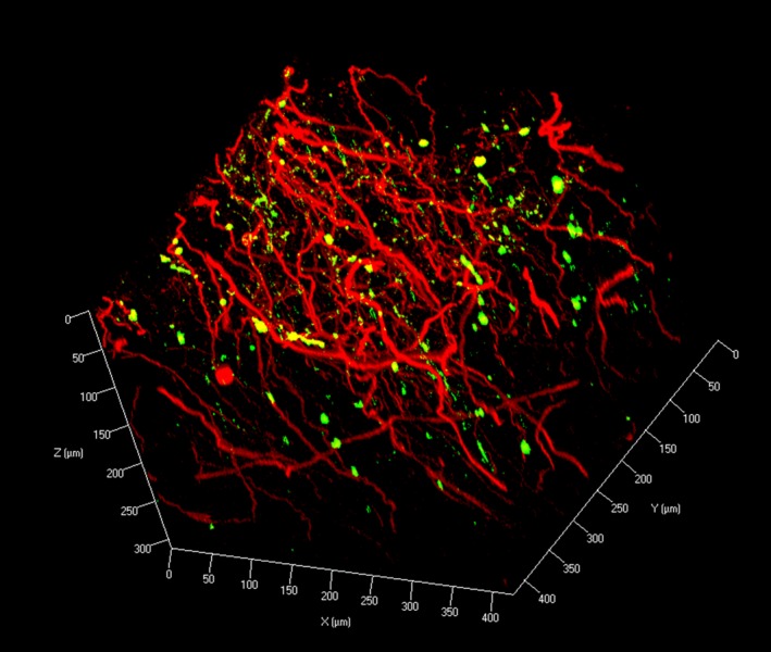
Figure 6
Zu2010stack image of double immunofluorescence with antiu2010 u03b1 SN (green) and antiu2010 TH (red) antibodies on human midbrain block (zu2010stack step size 1.5 u03bc m).
Figure images are served from the NIH/NLM PubMed Central Open Access Subset or Europe PMC; copyright remains with the publishers and authors.
💬 Discussion
0 commentsNo comments yet. Be the first to start a discussion!
Leave a Comment