Abstract
Although a critical role for caveolae-mediated albumin transcytosis in pulmonary endothelium is well established, considerably less is known about caveolae-independent pathways. In this current study, we confirmed that cultured rat pulmonary microvascular (RPMEC) and pulmonary artery (RPAEC) endothelium endocytosed Alexa488-labeled albumin in a saturable, temperature-sensitive mode and internalization resulted in co-localization by fluorescence microscopy with cholera B toxin and caveolin-1. Although siRNA to caveolin-1 (cav-1) in RPAEC significantly inhibited albumin uptake, a remnant portion of albumin uptake was cav-1-independent, suggesting alternative pathways for albumin uptake. Thus, we isolated and cultured mouse lung endothelial cells (MLEC) from wild type and cav-1(-/-) mice and noted that ~ 65% of albumin uptake, as determined by confocal imaging or live cell total internal reflectance fluorescence microscopy (TIRF), persisted in total absence of cav-1. Uptake of colloidal gold labeled albumin was evaluated by electron microscopy and demonstrated that albumin uptake in MLEC from cav-1(-/-) mice was through caveolae-independent pathway(s) including clathrin-coated pits that resulted in endosomal accumulation of albumin. Finally, we noted that albumin uptake in RPMEC was in part sensitive to pharmacological agents (amiloride [sodium transport inhibitor], Gö6976 [protein kinase C inhibitor], and cytochalasin D [inhibitor of actin polymerization]) consistent with a macropinocytosis-like process. The amiloride sensitivity accounting for macropinocytosis also exists in albumin uptake by both wild type and cav-1(-/-) MLEC. We conclude from these studies that in addition to the well described caveolar-dependent pulmonary endothelial cell endocytosis of albumin, a portion of overall uptake in pulmonary endothelial cells is cav-1 insensitive and appears to involve clathrin-mediated endocytosis and macropinocytosis-like process.
🔬 Techniques
💻 Software
✨ Fluorophores
🧪 Sample Preparation
🔬 Cell Lines
🏭 Microscope Brands
🧪 Reagent Suppliers
📷 Detectors
💻 Software Details
🏛️ Research Organizations (ROR)
Affiliated research institutions:
📋 Methods
Cell Culture Rat pulmonary microvascular endothelial cells
(RPMEC) were purchased from VEC Technologies (VEC Technologies, Rensselaer, NY, USA) and cultured in MCDB-131 complete medium (VEC Technologies).
Rat pulmonary artery endothelial cells
(RPAEC), isolated [ 27 ] and donated by Dr. Troy Stevens (University of South Alabama, Tuscaloosa, AL), were cultured in Dulbecco’s Modified Eagle Medium (DMEM, Life Technologies, Carlsbad, CA, USA) supplemented with 10% fetal bovine serum (FBS, Life Technologies, Carlsbad, CA, USA), 100 U/ml penicillin and 100 µg/ml streptomycin (Life Technologies, Carlsbad, CA, USA). RPMEC and RPAEC were incubated at 37°C in humidified atmosphere with 21% O 2 and 5% CO 2 .
Mouse lung endothelial cells
(MLEC) were isolated from wild type or caveolin-1 (cav-1) null mice [ 28 ] by immunobeads coated with anti-mouse PECAM (CD31) antibody (BD Pharmingen, San Diego, CA, USA) followed by cell sorting with fluorescently-labeled acetylated-Low Density Lipoprotein (Dil-AcLDL, Biomedical Technologies Inc., Stoughton, MA, USA) uptake. MLEC were subcultured at 37°C in humidified atmosphere with 2% O 2 and 5% CO 2 in OPTI-MEM (Life Technologies, Carlsbad, CA, USA) supplemented with 10% FBS, 100 U/ml penicillin, 100 µg/ml streptomycin, and EC growth supplement (ENDOGRO, VEC Technologies) as previously described [ 29 ]. MLEC were studied before passage 7 and were virtually (98-100%) homogeneously positive for PECAM and uptake of Dil-AcLDL. Animal care and use were carried out in strict accordance with the recommendations in the Guide for the Care and Use of Laboratory Animals of the National Institutes of Health. The protocol was approved by the Committee on the Ethics of Animal Experiments of the University of Pittsburgh (Protocol Number: 1111998). Mouse sacrifice was performed under anesthesia and all efforts were made to minimize suffering. Alexa488-BSA Uptake Cells were plated into Lab-Tek II 4-well chamber slides (Nalge Nunc, Naperville, IL, USA) and grown until 90% confluent. After serum starvation for 4 h, cells were incubated with 50 μg/ml of Alexa488-BSA (Life Technologies, Carlsbad, CA, USA) in HBSS for indicated time at 37°C. Cells were fixed with 2% paraformaldehyde and stained with DAPI (Life Technologies, Carlsbad, CA, USA) to reveal cell nuclei and then slides were peeled off and sealed with Fluoromount-G (Southern Biotechnology Associates, Birmingham, AL, USA) under coverslips. Images were taken with Nikon Eclipse 80i fluorescence microscope (Nikon, Melville, NY, USA) and a Fluoview 500 confocal Microscope (Olympus, Center Valley, PA, USA).
Show full methods section
Cell Culture Rat pulmonary microvascular endothelial cells
(RPMEC) were purchased from VEC Technologies (VEC Technologies, Rensselaer, NY, USA) and cultured in MCDB-131 complete medium (VEC Technologies).
Rat pulmonary artery endothelial cells
(RPAEC), isolated [ 27 ] and donated by Dr. Troy Stevens (University of South Alabama, Tuscaloosa, AL), were cultured in Dulbecco’s Modified Eagle Medium (DMEM, Life Technologies, Carlsbad, CA, USA) supplemented with 10% fetal bovine serum (FBS, Life Technologies, Carlsbad, CA, USA), 100 U/ml penicillin and 100 µg/ml streptomycin (Life Technologies, Carlsbad, CA, USA). RPMEC and RPAEC were incubated at 37°C in humidified atmosphere with 21% O 2 and 5% CO 2 .
Mouse lung endothelial cells
(MLEC) were isolated from wild type or caveolin-1 (cav-1) null mice [ 28 ] by immunobeads coated with anti-mouse PECAM (CD31) antibody (BD Pharmingen, San Diego, CA, USA) followed by cell sorting with fluorescently-labeled acetylated-Low Density Lipoprotein (Dil-AcLDL, Biomedical Technologies Inc., Stoughton, MA, USA) uptake. MLEC were subcultured at 37°C in humidified atmosphere with 2% O 2 and 5% CO 2 in OPTI-MEM (Life Technologies, Carlsbad, CA, USA) supplemented with 10% FBS, 100 U/ml penicillin, 100 µg/ml streptomycin, and EC growth supplement (ENDOGRO, VEC Technologies) as previously described [ 29 ]. MLEC were studied before passage 7 and were virtually (98-100%) homogeneously positive for PECAM and uptake of Dil-AcLDL. Animal care and use were carried out in strict accordance with the recommendations in the Guide for the Care and Use of Laboratory Animals of the National Institutes of Health. The protocol was approved by the Committee on the Ethics of Animal Experiments of the University of Pittsburgh (Protocol Number: 1111998). Mouse sacrifice was performed under anesthesia and all efforts were made to minimize suffering. Alexa488-BSA Uptake Cells were plated into Lab-Tek II 4-well chamber slides (Nalge Nunc, Naperville, IL, USA) and grown until 90% confluent. After serum starvation for 4 h, cells were incubated with 50 μg/ml of Alexa488-BSA (Life Technologies, Carlsbad, CA, USA) in HBSS for indicated time at 37°C. Cells were fixed with 2% paraformaldehyde and stained with DAPI (Life Technologies, Carlsbad, CA, USA) to reveal cell nuclei and then slides were peeled off and sealed with Fluoromount-G (Southern Biotechnology Associates, Birmingham, AL, USA) under coverslips. Images were taken with Nikon Eclipse 80i fluorescence microscope (Nikon, Melville, NY, USA) and a Fluoview 500 confocal Microscope (Olympus, Center Valley, PA, USA).
Immunofluorescent staining of cav-1
After incubating with Alexa488-BSA and washing with HBSS, cells were fixed with 2% paraformaldehyde for 15 min at room temperature and then permeabilized with 0.1% Triton X-100 (Sigma-Aldrich, St Louis, MO, USA) for 15 min. Blocking was performed with 20% goat serum (Sigma-Aldrich, St Louis, MO, USA) in PBS containing 0.5% BSA (Fraction V, Roche, Mannheim, Germany) for 1 h, then cells were incubated with anti-cav-1 primary polyclonal antibody (Santa Cruz Biotechnology, Santa Cruz, CA, USA) for 1 h followed by 1 h incubation with Cy3-conjugated secondary antibody (Jackson ImmunoResearch, West Grove, PA, USA). Cell nuclei were stained with DAPI (Life Technologies, Carlsbad, CA, USA) and coverslips were sealed on slides. Images were taken with Nikon Eclipse 80i fluorescence microscope and an Olympus Fluoview 500 confocal Microscope. Cav-1 knockdown with siRNA Small interfering RNA (siRNA) was designed against the coding sequence of cav-1 cDNA ( AAGAGCTTCCTGATTGAGATT ) and both siRNA and sham siRNA (with scrambled sequence) were purchased from Dharmacon (Chicago, IL, USA). Transfection of siRNA (100 nM) into RPAEC was carried out by using Lipofectamine 2000 (Life Technologies, Carlsbad, CA, USA) and 72 h later Alexa488-BSA endocytosis and immunofluorescent staining of cav-1 were performed.
Immunoblot of cav-1
Cells were washed three times with ice-cold PBS, and then lysed on ice in SDS sample buffer (62.5 mM Tris-HCl, pH 6.8, 2% w/v SDS, 10% glycerol, 50 mM DTT, 0.01% w/v bromophenol blue). The extracts were sonicated briefly, boiled for 5 min, then centrifuged at 14,000 rpm for 5 min. Equivalent amounts of protein were separated by electrophoresis using 10% SDS-PAGE gels. The proteins were then transferred to PVDF membrane (Life Technologies, Carlsbad, CA, USA) and blocked with PBS containing 5% nonfat milk for 1 h at RT. Membranes were incubated with primary antibody against cav-1 (BD Pharmingen, San Diego, CA, USA) overnight at 4°C and HRP-conjugated secondary antibody (Santa Cruz Biotechnology, Santa Cruz, CA, USA) was used for visualization by chemiluminescence using ECL reagent (Perkin-Elmer Life Science, Boston, MA, USA). Blots were stripped with stripping buffer (Pierce, Rockford, IL, USA) and reprobed with antibody against β-actin (Sigma-Aldrich, St Louis, MO, USA).
Transmission electron microscopy
Bovine serum albumin (BSA, Roche, Mannheim, Germany) was conjugated to colloidal gold particles (5 nm) as described by Baschong and Wrigley [ 30 ]. Unconjugated colloidal gold were removed by centrifugation (45,000 × g for 15 min at 4°C) of protein-gold complex, and the soft pellet was resuspended in low-salt buffer (10 mM HEPES, 1 mM KCl, 0.5 mM MgCl 2 , pH 7.5). Cells were plated into 6-well plate and grown until confluent. After serum starvation for 4 h, cells were moved to 4°C for 20 min, then incubated with Gold-labeled BSA in cold HBSS for 40 min. Cells were fixed and embedded by inverting Polybed 812-filled Better Equipment for Electron Microscopy (BEEM) capsules on top of the cells. Blocks were cured overnight at 37°C, and then cured for two days at 65°C. Monolayers were pulled off the plastic and re-embedded for cross-sectioning. Ultrathin cross-sections (60 nm) of the cells were obtained on a Riechert Ultracut E microtome, post-stained in 4% uranyl acetate for 10 min and 1% lead citrate for 7 min. Sections were viewed on a JEM 1011 transmission electron microscope (JEOL, Peabody, MA, USA) at 80 KV. Images were acquired with a side-mount AMT 2K digital camera (Advanced Microscopy Techniques, Danvers, MA, USA). Total Internal Reflection Fluorescence (TIRF) Microscopy TIRF imaging was performed as described [ 31 ]. Cells were imaged on a Nikon (Melville, NY, USA) TIRF system using a 60x (1.45 NA) optic and a Prairie (Madison, WI, USA) acousto-optic tunable filter controlled laser bench. All experiments used the 488 line on a 150-milliwatt argon source. Images were collected using MetaMorph software (Molecular Devices, Sunnyvale, CA) and a Orca II ER camera (Hamamatsu, Tokyo, Japan). Time-based series were collected and processed using MeatMorph. Protocol for macropinocytosis study Inhibitors of macropinocytosis (Amiloride, Cytochalasin D and Gö6976) were purchased from Sigma-Aldrich (St Louis, MO, USA). RPMEC were serum starved for 4 h followed by pretreatment with amiloride (Na/H exchange inhibitor, 3 mM), Cytochalasin D (actin polymerization inhibitor, 10 µM), and Gö6976 (PKC inhibitor, 10 µM) for 30 min, then cells were incubated with Alexa488-BSA (50 µg/ml) for 15 min and fixed for imaging.
Image quantification and statistical analysis
Image quantification was performed with Imaris software (Bitplane Inc, South Windsor, CT). Images were thresholded and the spots function was used to define and quantify the regions of albumin uptake within the cells. Data are present as mean ± SEM. Statistical significance differences (* P < 0.05, ** P < 0.01, *** P < 0.001) were determined by t-test or one-way analysis of variance (ANOVA) followed by Tukey's multiple comparisons using Graphpad Prism ver. 5.0 (GraphPad Software, San Diego, CA, USA).
Protocol for macropinocytosis study Inhibitors of macropinocytosis (Amiloride, Cytochalasin D and Gö6976) were purchased from Sigma-Aldrich (St Louis, MO, USA). RPMEC were serum starved for 4 h followed by pretreatment with amiloride (Na/H exchange inhibitor, 3 mM), Cytochalasin D (actin polymerization inhibitor, 10 µM), and Gö6976 (PKC inhibitor, 10 µM) for 30 min, then cells were incubated with Alexa488-BSA (50 µg/ml) for 15 min and fixed for imaging.
📊 Figures
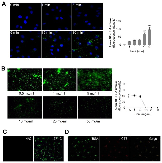
Figure 1
Albumin endocytosis is a competitive, temperature sensitive, and caveolae-associated process in pulmonary endothelial cells.
( A ) Time course of Alexa488-BSA endocytosis in rat pulmonary microvascular endothelial cells (RPMEC). Cells were serum starved and then incubated with Alexa488-BSA (50 u00b5g/ml) for 0 to 30 min at ...
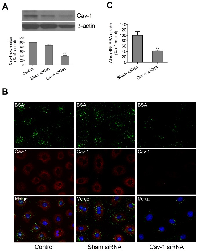
Figure 2
Caveolin-1 RNAi reduced Alexa488-BSA endocytosis in pulmonary artery endothelial cells.
( A ) Representative immunoblot of cav-1 expression in RPAEC treated with transfection reagent only, sham siRNA, and cav-1 siRNA is shown with u03b2-actin as loading control. Densitometric measurement...
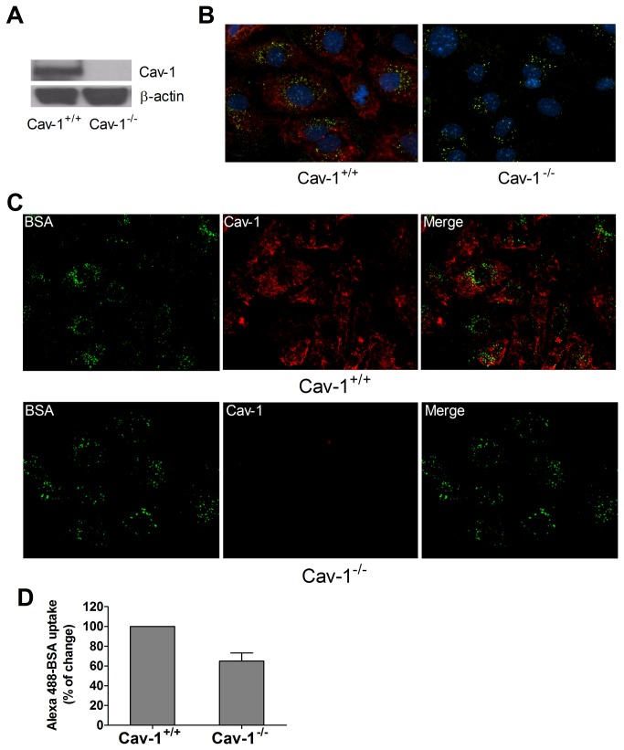
Figure 3
Endocytosis of Alexa488-BSA occurs in cav-1 -/- MLEC through a caveolae-independent pathway.
( A ) Representative immunoblot of cav-1 expression in cav-1 +/+ and cav-1 -/- MLEC is shown with u03b2-actin as loading control. ( B ) Fluorescent images and ( C ) confocal images of Alexa488-BSA (gr...
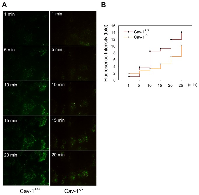
Figure 4
Dynamic process of Alexa488-BSA endocytosis in cav-1 +/+ and cav-1 -/- MLEC.
( A ) Cav-1 +/+ and cav-1 -/- MLEC were serum starved for 4 h then incubated with Alexa488-BSA (50 u00b5g/ml) in HBSS. Fluorescence signals were recorded from 0 min to 30 min under total internal refl...
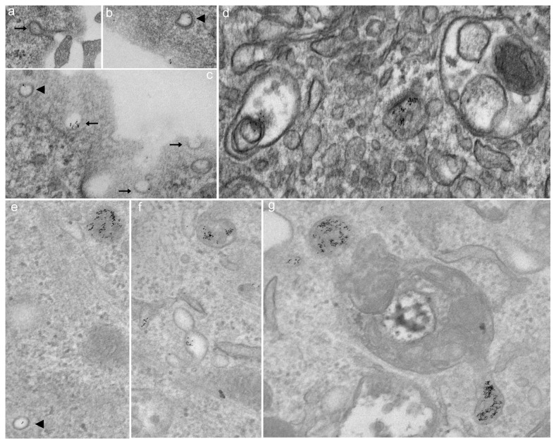
Figure 5
Endocytosis of gold-labeled albumin occurs in cav-1 +/+ MLEC (a-d) through caveolae-dependent and -independent pathways but only through caveolae-independent pathway in cav-1 -/- MLEC (e-g).
Both caveolae vesicles (black arrow) and clathrin-coated pits (black arrowhead) were positive for gold-labeled albumin ( a - c ) suggestive of internalization via a non-caveolin-1 dependent endocytic ...
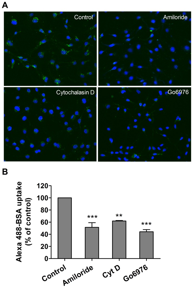
Figure 6
Inhibition of macropinocytosis prevents Alexa488-BSA uptake by rat pulmonary endothelial cells.
( A ) RPMEC were serum starved for 4 h followed by pretreatment with various inhibitors of macropinocytosis for 30 min including amiloride (Na/H exchange inhibitor, 3 mM), Cytochalasin D (actin polyme...
Figure images are served from the NIH/NLM PubMed Central Open Access Subset or Europe PMC; copyright remains with the publishers and authors.
💬 Discussion
0 commentsNo comments yet. Be the first to start a discussion!
Leave a Comment