Abstract
Background: The liver-derived plasma protein fetuin-A is a systemic inhibitor of ectopic calcification. Fetuin-A stabilizes saturated mineral solutions by forming colloidal protein-mineral complexes called calciprotein particles (CPP). CPP are initially spherical, amorphous and soft, and are referred to as primary CPP. These particles spontaneously convert into secondary CPP, which are larger, oblongate, more crystalline, and less soluble. CPP mediate excess mineral transport and clearance from circulation. Methods: We studied by intravital two-photon microscopy the clearance of primary vs. secondary CPP by injecting i.v. synthetic fluorescent CPP in mice. We analyzed CPP organ distribution and identified CPP endocytosing cells by immunofluorescence. Cellular clearance was studied using bone marrow-derived mouse wildtype and scavenger receptor A (SRA)-deficient macrophages, as well as human umbilical cord endothelial cells (HUVEC), monocyte-derived macrophages (hMDM), and human aortic endothelial cells (haEC). We employed mouse wildtype and mutant immortalized macrophages to analyze CPP-induced inflammasome activation and cytokine secretion. Results: In live mice, only primary CPP were rapidly cleared by liver sinusoidal endothelial cells (LSEC), whereas primary and secondary CPP were cleared by Kupffer cells. Scavenger receptor A (SRA)-deficient bone marrow macrophages endocytosed secondary CPP less well than did wildtype macrophages. In contrast, primary CPP endocytosis did not depend on the presence of SRA, suggesting involvement of an alternative clearance pathway. CPP triggered TLR4 dependent TNFα and IL-1β secretion in cultured macrophages. Calcium content-matched primary CPP caused twice more IL-1β secretion than did secondary CPP, which was associated with increased calcium-dependent inflammasome activation, suggesting that intracellular CPP dissolution and calcium overload may cause this inflammation. Conclusions: Secondary CPP are endocytosed by macrophages in liver and spleen via SRA. In contrast, our results suggest that primary CPP are cleared by LSEC via an alternative pathway. CPP induced TLR4-dependent TNFα and inflammasome-dependent IL-1β secretion in macrophages suggesting that inflammation and calcification may be considered consequences of prolonged CPP presence and clearance.
🔬 Techniques
✨ Fluorophores
🧪 Sample Preparation
🔬 Cell Lines
🏭 Microscope Brands
🧪 Reagent Suppliers
🔎 Objectives
💻 Software Details
🏛️ Research Organizations (ROR)
Affiliated research institutions:
📋 Methods
Animal experimentation All animal experiments were conducted in agreement with the recommendation of the Federation for Laboratory Animal Science Associations (FELASA), and were approved by the animal welfare committee of the Landesamt für Natur-, Umwelt- und Verbraucherschutz (LANUV, 84-02.04.2013.A113 and 84.02.04.2015.A294). At least three mice each were injected in the clearance experiments and three mice of the genotypes were used to isolate bone marrow-derived macrophages. All mice were maintained in a temperature-controlled room on a 12-h day/night cycle. Food and water were given ad libitum .
Protein purification and CPP preparation
Bovine fetuin-A (Sigma F2379) was purified by gel-filtration and labeled with Alexa-488 or Alexa-546 NHS ester (Thermo Scientific). Purified fetuin-A was routinely analyzed for LPS activity using the Endosafe ultrasensitive cartridge assay (Charles River). LPS content was
📊 Figures
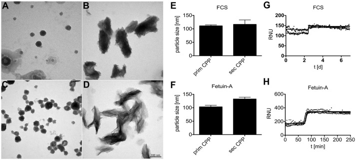
Figure 1
Calciprotein particle CPP formation in protein solutions supersaturated with calcium and phosphate. (A,B,E,G) CPP were prepared in DMEM supplemented with 1.0 mM calcium (total Ca 3.8 mM), 3.5 mM phosp...
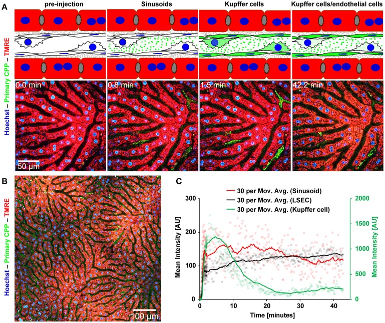
Figure 2
Functional imaging of primary CPP clearance using 2-photon intravital microscopy. Mice received intravenous injections of primary CPP prepared with fluorescent fetuin-A, and the major clearance organ ...
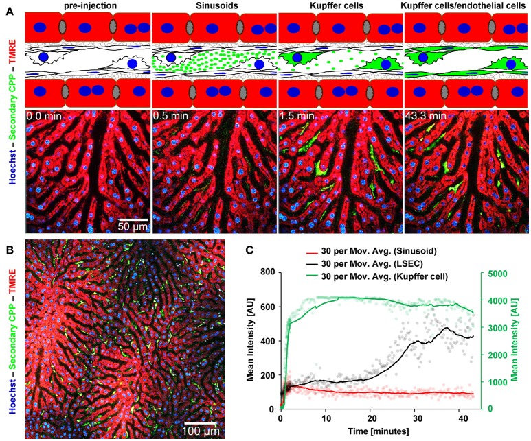
Figure 3
Functional imaging of secondary CPP clearance using 2-photon intravital microscopy. Mice received intravenous injections of secondary CPP prepared with fluorescent fetuin-A, and the major clearance or...
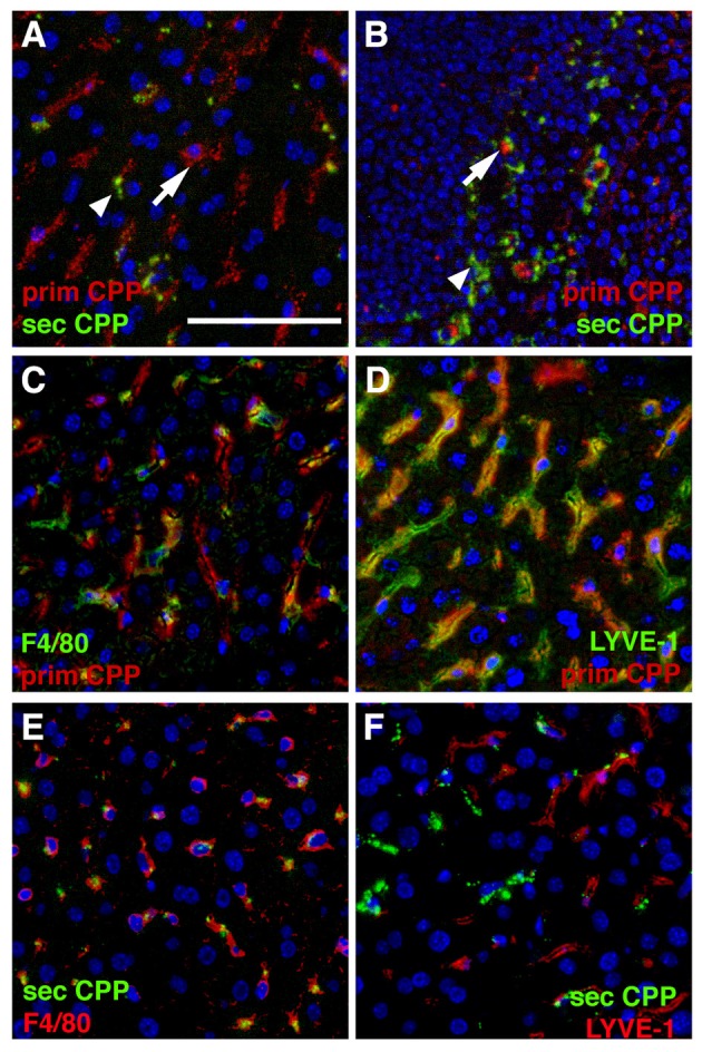
Figure 4
Differential clearance of primary and secondary CPP. Mice were injected with a mixture of fluorescence labeled primary (red) and secondary CPP (green) and the major clearance organs liver (A,Cu2013F) ...
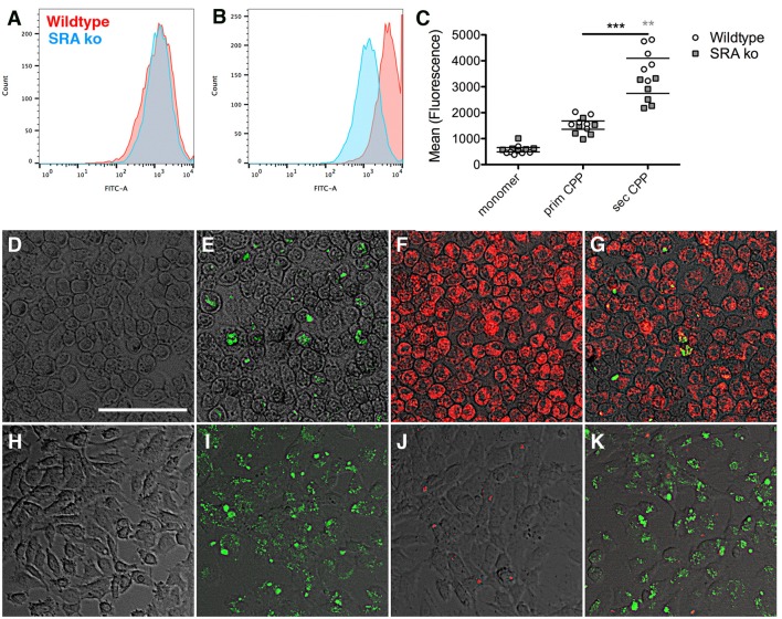
Figure 5
Differential endocytosis of primary and secondary CPP in mouse macrophages and endothelial cells. (Au2013C) Primary bone marrow macrophages derived from wildtype and scavenger receptor A-deficient mic...
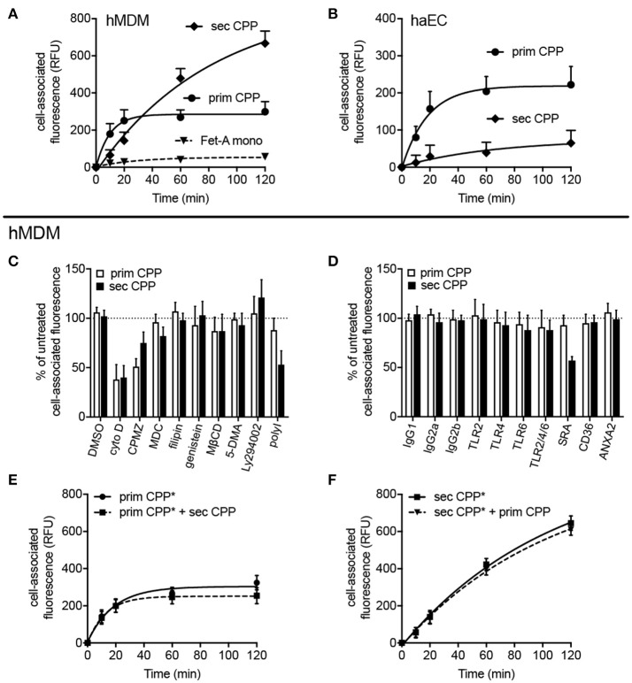
Figure 6
Differential endocytosis of primary and secondary CPP in human monocyte-derived macrophages and aortic endothelial cells. Human monocyte-derived macrophages (hMDM) or human aortic endothelial cells (h...
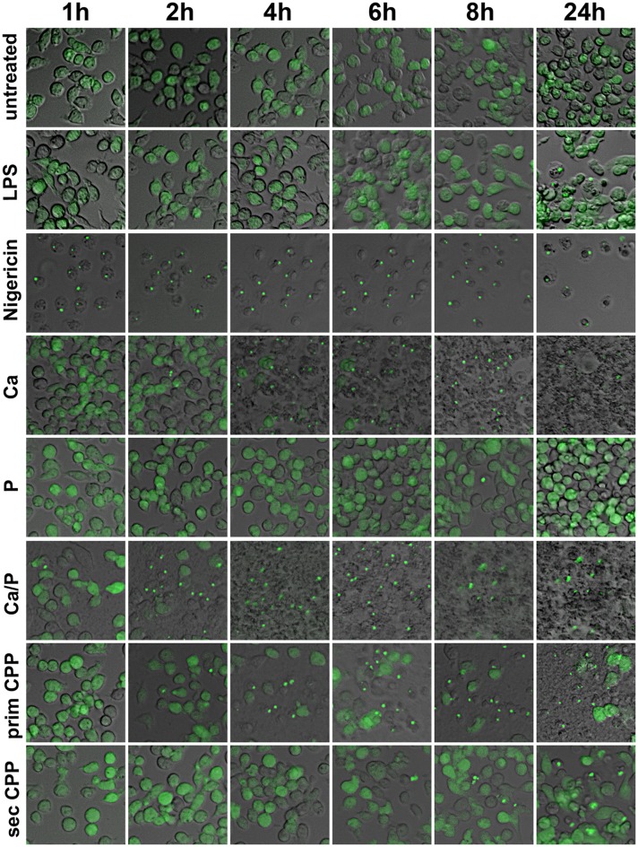
Figure 7
Calcification media induce differential cellular calcium overload and inflammasome activation. Immortalized macrophages expressing the inflammasome adaptor protein labeled apoptosis-associated speck l...
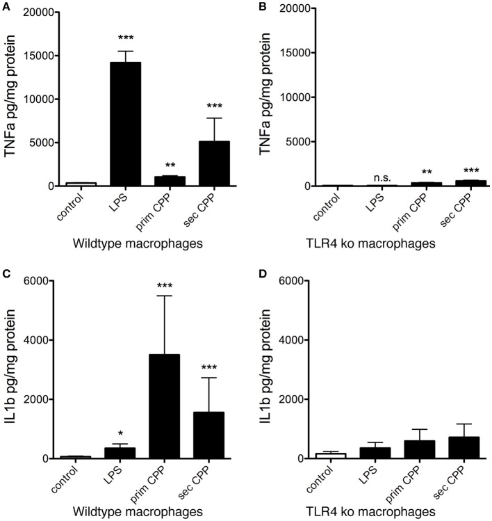
Figure 8
CPP-induced inflammatory cytokine secretion by macrophages is TLR4 dependent. Serum-starved wildtype and TLR4-deficient macrophages (TLR ko) where treated with LPS, primary or secondary CPP. (A,B) Aft...
Figure images are served from the NIH/NLM PubMed Central Open Access Subset or Europe PMC; copyright remains with the publishers and authors.
💬 Discussion
0 commentsNo comments yet. Be the first to start a discussion!
Leave a Comment