Abstract
Successful cancer therapy requires drugs being precisely delivered to tumors. Nanosized drugs have attracted considerable recent attention, but their toxicity and high immunogenicity are important obstacles hampering their clinical translation. Here we report a novel "cocktail therapy" strategy based on excess natural killer cell-derived exosomes (NKEXOs) in combination with their biomimetic core-shell nanoparticles (NNs) for tumor-targeted therapy. The NNs were self- assembled with a dendrimer core loading therapeutic miRNA and a hydrophilic NKEXOs shell. Their successful fabrication was confirmed by transmission electron microscopy (TEM) and confocal laser scanning microscopy (CLSM). The resulting NN/NKEXO cocktail showed highly efficient targeting and therapeutic miRNA delivery to neuroblastoma cells in vivo, as demonstrated by two-photon excited scanning fluorescence imaging (TPEFI) and with an IVIS Spectrum in vivo imaging system (IVIS), leading to dual inhibition of tumor growth. With unique biocompatibility, we propose this NN/NKEXO cocktail as a new avenue for tumor therapy, with potential prospects for clinical applications.
🔬 Techniques
🔭 Microscopes
💻 Software
✨ Fluorophores
🧪 Sample Preparation
🔬 Cell Lines
🏭 Microscope Brands
🧪 Reagent Suppliers
💻 Software Details
🏛️ Research Organizations (ROR)
Affiliated research institutions:
📋 Methods
4.1.
Cell Culture
The human breast cancer cell lines MDA-MB-231 and human embryonic kidney cell lines 293T were propagated in DMEM (Gibco, Grand Island, NY, USA) supplemented with 10% FBS (Gibco) and 1% penicillin-streptomycin (Hyclone, Logan, UT, USA).
Human neuroblastoma cells
CHLA-255 were incubated in IMDM (Gibco) with 20% FBS and 1% penicillin-streptomycin. All cell lines were purchased from the American Type Culture Collection (ATCC). Both MDA-MB-231 and CHLA-255 cells were transfected by Nucleofection™ (Lonza, Cologne, Germany) with a firefly luciferase expression plasmid. All cells were maintained in a humidified 5% CO 2 atmosphere at 37 °C. 4.2. Preparation and Function of NK Cell Derived Exosomes 4.2.1.
Isolation of Exosomes
We isolated NK cell-derived exosomes using differential ultracentrifugation according to the literature. On day 21, NK cell culture supernatants were harvested after a preliminary centrifugation at 400× g for 10 min to eliminate cells. The supernatants were centrifuged first at 2000× g for 10 min and then at 10,000× g for 70 min to remove dead cells and cell debris, followed by an additional centrifugation step at 100,000× g for 70 min in a 70Ti ultracentrifuge rotor (Optima™XPN-100, Beckman, Brea, CA, USA) to obtain pellets containing raw exosomes. The pellets were then purified by washing them with PBS and centrifuged at 100,000× g for another 70 min. The isolated exosomes were resuspended in PBS (100:1 enrichment) and stored at −80 °C before use. All procedures were carried out at 4 °C. We isolated exosomes from the 293T culture medium in the same way. 4.2.2.
Show full methods section
4.1.
Cell Culture
The human breast cancer cell lines MDA-MB-231 and human embryonic kidney cell lines 293T were propagated in DMEM (Gibco, Grand Island, NY, USA) supplemented with 10% FBS (Gibco) and 1% penicillin-streptomycin (Hyclone, Logan, UT, USA).
Human neuroblastoma cells
CHLA-255 were incubated in IMDM (Gibco) with 20% FBS and 1% penicillin-streptomycin. All cell lines were purchased from the American Type Culture Collection (ATCC). Both MDA-MB-231 and CHLA-255 cells were transfected by Nucleofection™ (Lonza, Cologne, Germany) with a firefly luciferase expression plasmid. All cells were maintained in a humidified 5% CO 2 atmosphere at 37 °C. 4.2. Preparation and Function of NK Cell Derived Exosomes 4.2.1.
Isolation of Exosomes
We isolated NK cell-derived exosomes using differential ultracentrifugation according to the literature. On day 21, NK cell culture supernatants were harvested after a preliminary centrifugation at 400× g for 10 min to eliminate cells. The supernatants were centrifuged first at 2000× g for 10 min and then at 10,000× g for 70 min to remove dead cells and cell debris, followed by an additional centrifugation step at 100,000× g for 70 min in a 70Ti ultracentrifuge rotor (Optima™XPN-100, Beckman, Brea, CA, USA) to obtain pellets containing raw exosomes. The pellets were then purified by washing them with PBS and centrifuged at 100,000× g for another 70 min. The isolated exosomes were resuspended in PBS (100:1 enrichment) and stored at −80 °C before use. All procedures were carried out at 4 °C. We isolated exosomes from the 293T culture medium in the same way. 4.2.2.
Measurement of Particle Size
A dynamic light scattering (DLS) system equipped with a 532-nm laser (Malvern Instruments, Malvern, UK) was used for exosome particle size analysis. Samples were diluted before analysis (10 μL, 1:100 diluted in PBS) and each sample was measured three times. Data was collected and analyzed by Dispersion Technology Software (Malvern Instruments). The size distribution was measured by signal intensity and the Z-average diameter obtained from the autocorrelation function using the general-purpose mode. 4.2.3.
Determination of Protein Concentration
Protein concentration in isolated exosomes was calculated by the bicinchoninic acid protein assay. Briefly, 25 µL of an aqueous suspension of isolated exosomes after lysis were added to 200 µL of standard working reagent in wells of a 96-well plate. Absorbance of the mixture was determined at 562 nm with a microplate reader and the protein concentration estimated. A calibration curve was constructed using bovine serum albumin as protein standard. 4.2.4. Western Blotting (WB) Analysis Presence of marker proteins in exosomes was analyzed and confirmed by WB. Proteins from lysed cells or isolated exosomes were denatured and loaded onto sodium dodecyl sulfate polyacrylamide gels, transferred to polyvinylidene difluoride membranes (Millipore, Billerica, MA, USA), and subsequently stained with the corresponding primary and secondary antibodies. The following antibodies were used for WB analysis according to manufacturer’s instructions: Anti-ALIX polyclonal antibody (12422-1-AP, Proteintech, Rosemont, IL, USA), anti-CD47 polyclonal antibody (ab108415, Abcam, Cambridge, MA, USA), anti-CXCR4 polyclonal antibody (35-8800, Invitrogen, Waltham, MA, USA ), anti-TSG101 monoclonal antibody (4A10, Abcam), anti-human CD63 polyclonal antibody (556019, BD Biosciences) and anti-cytochrome C antibody (10993-1-AP, Proteintech). The BioRad ChemiDoc Touch imaging system (BioRad, Hercules, CA, USA) was used to analyze the bands after incubation with the corresponding goat anti-mouse- or goat anti-rabbit-HRP secondary antibody conjugates (1:5000 dilutions). The densitometry readings of each band were calculated by ImagJ (NIH, Bethesda, MD, USA). 4.2.5. Transmission Electron Microscopy (TEM) Morphology of the isolated NKEXOs was studied by TEM. A small drop (about 10 μL) of exosomes was dropped onto the Formvar/carbon-coated TEM grid. After settling for approximately 1 min, excess water was removed by touching the grid with a piece of filter paper. Next, the grid was covered with a small drop of 2% uranyl acetate. After drying for ten seconds, the copper nets were examined with a Tecnai T10 TEM (FEI, Oregon, OH, USA) operated at 120 kV. The morphology of the PAMAM dendrimers and NNs was also imaged by TEM following similar steps. 4.2.6.
Samples Labeling
To label isolated exosomes and let-7a loaded PAMAM, different labeling assays were performed. We added 2 μL of red lipophilic fluorescent dye PKH26 (Sigma-Aldrich, St. Louis, MO, USA) to 500 μL of Diluent C (Sigma-Aldrich). The mixture was then added to the isolated NK derived exosomes and mixed gently for 5 min at room temperature followed by addition of 1% BSA (1 mL) to facilitate the binding of the excess dye. Next, samples were washed with PBS and ultracentrifuged at 100,000 g for 70 min and the pellets resuspended in PBS. The fluorescent dye 1,10-dioctadecyl-3,3,30,30-tetramethylindotricarbocyanine iodide (DiR; Invitrogen) was also used to label exosomes. Purified exosomes were incubated in 1 μM DiR at a concentration of approximately 350 μg of exosomes per mL for 15 min at 37 °C, and washed as described above. FAM-labeled miRNA and cy5-labeled miRNA were both synthesized by Genepharma (Shanghai, China). 4.3. Cellular Uptake Assay PKH26-labeled NKEXOs were incubated with MDA-MB-231 cells for 24 h in a humidified 5% CO 2 atmosphere at 37 °C. Subsequently, cells were washed twice with PBS and fixed with 4% paraformaldehyde for 20 min. Aliquots (5 μL) of the cell suspension were placed in clear microscope slides, covered with a drop of FluorSave TM Reagent (Invitrogen) and mounted with a coverslip. Images, including z-axis projection images, were taken using a Nikon A1R (Nikon instruments, Melville, NY, USA) confocal laser scanning microscope. The uptake of the NN/NKEXO cocktail by CHLA-255 cells was also imaged following similar steps after incubating for 12 and 24 h. 4.4.
Flow Cytometry FAM-labeled let-7a loaded PAMAM and PKH26-labeled
NKEXOs were coupled to fabricate the nanoparticle cocktail as indicated before. To quantify the cellular uptake of the cocktail by tumor cells, CHLA-255 cells (10 5 cells per well) were cultured with the NN/NKEXO cocktail made of let-7a (0.5 µg) and NKEXOs (50 µg), PBS or equivalent 293N/293Exo cocktail between 12 to 48 h, harvested, and washed twice with PBS. Then, the samples were centrifuged and the supernatants decanted. Cells were resuspended and collected before analysis with a Cytoflex flow cytometer (Beckman Coulter, Life Science, Indianapolis, IN, USA). 4.5. Preparation of the NN/NKEXO Cocktail To prepare NN/NKEXO cocktails in this study, 0.05/0.5/5.28 μg let-7a mimics (Genepharma) were added to dendrimers following manufacturer’s instructions (SL100568, SignaGen Laboratories, Rockville, MD, USA) and mixed thoroughly to react at 4 °C for 15 min. The resulting let-7a loaded PAMAM were precipitated at high-speed (10,000 rpm) for 30 min to remove excess let-7a. Subsequently, 40/50/100 μg of NKEXOs suspension were added to the precipitated let-7a loaded PAMAM respectively and the mixture incubated for 24 hours at 4 °C. To obtain NNs, the NN/NKEXO cocktail was precipitated at high-speed (10,000 rpm) for 30 min and resuspended in PBS. The supernatants with excess NKEXOs were used for TEM and the precipitated NNs for both Western blotting analysis and TEM. The same procedure was used in fabricating another cocktail in this study. Confocal laser scanning microscopy (Nikon A1R) was used to validate whether NKEXOs were successfully fused with the let-7a loaded-PMAM. Exosomes and let-7a mimics were fluorescently labeled by PKH26 and FAM, respectively. Laser excitation of PKH67 and FAM was done sequentially using 586- and 488-nm lasers. 4.6. In Vitro Cytotoxicity To evaluate NKEXO cytotoxicity, MDA-MB-231 breast cancer cells and CHLA-255 neuroblastoma cells were transfected with the firefly luciferase gene to quantify target cell survival using bioluminescence. MDA-MB-231-luc and CHLA-255-luc cells (10 4 cells per well) were cultured with different quantities of NKEXOs (10, 20 and 40 μg) in clear-bottomed 96-well plates for 24 h. The culture medium was discarded and cells washed with PBS three times, followed by the addition of 120 μL of cell lysis buffer from a luciferase reporter Gene Assay kit (Yisheng, Shanghai, China). The 96-well plates were then shaken in a micro-shaker at room temperature for 15 min to fully lyse the cells. Next, the pyrolyzed lysates were centrifuged at 10,000 rpm for 3–5 minutes. After centrifugation, 100 μL of the supernatants from each well were transferred to a clear-bottomed 96-well plate, and 100 μL of firefly luciferase assay reagent added. Bioluminescent signals of the mixture were measured by the IVIS ® Lumina III imaging system (PerkinElmer, Santa Clara, CA, USA) and expressed as photon flux (photons/s). All experiments were done in triplicate from independent cell cultures. To evaluate the cytotoxicity of cocktail-let-7a against the neuroblastoma cells in vitro, CHLA-255-luc cells (10 4 cells per well) were treated with NKEXOs, let-7a alone or cocktail-let-7a containing 40 μg NKEXOs and 0.05 μg let-7a. Bioluminescent signals of the mixture were measured by an automatic ELISA plate reader and the relative light unit read at 562 nm. 4.7. In Vivo Animal Experiments In vivo analysis was performed using specific pathogen-free, 6-week-old, female NOD/SCID mice (Slaccas, Shanghai, China). All animal experimental protocols were conducted in accordance with all national guidelines and regulations, and approved by the Animal Ethics Committee (NO. 2018R03042). 4.7.1. In Vivo Targeting Capability of the Nanoparticle Cocktail The cocktail was labeled with Cy5-let-7a loaded PAMAM or DiR-NKEXOs. Then, 5 × 10 6 CHLA255 cells were intravenously injected into mice, which were randomly separated into four groups after 21 days in captivity. Animals in one group received intravenous injections containing 400 µL of labeled cocktail with identical amounts of let-7a (5.28 µg) and NKEXOs (100 µg). A second group received let-7a loaded PAMAM and the third group received let-7a only. At the end of the experiment (6 h after injection), the mice were euthanized and dissected tissues (heart, lung, liver, kidneys, spleen, livers and tumors) were imaged immediately. Fluorescence imaging was performed using the IVIS ® Lumina III imaging system. Background fluorescence was measured and subtracted by setting up a background measurement at time of data acquisition. A separate in vivo biodistribution study was performed using the following procedure. CHLA-255-luc cells (2 × 10 7 cells/200 μL) were subcutaneously injected into the forward thighs of NOD/SCID mice. After 14 days, the mice were anesthetized and the subcutaneous tumor tissue was carefully exposed using surgical scissors. Mice were injected intravenously with 400 μL of a cocktail made of FAM-labeled miRNA let7a (5.28 µg) and PKH26-labeled NKEXOs (100 μg). After 20 min, in vivo two-photon confocal microscopy (Olympus BX61, Olympus America, Center Valley, PA, USA) was performed to detect each fluorescence signal from the subcutaneous tumor. 4.7.2. In Vivo Antitumor Assay The ability of the cocktail to inhibit tumor growth was evaluated in a neuroblastoma tumor-bearing mouse model. Fifteen mice were used in this experiment. Briefly, CHLA-255-luc cells (10 7 cells/500 μL) were i.v. injected into the NOD/SCID mice. Mice were randomly separated into five groups ( n = 3) 14 days later, and intravenously injected with different cocktail formulations: NKEXOs, cocktail-let-7a, PAMAM, let7a or with PBS as control group at let-7a dose of 5.28 µg and NKEXO dose of 100 µg. The same treatments were repeated after three days. After treatment, bioluminescence imaging (BLI) was performed with the IVIS ® Lumina III imaging system to analyze the cocktail’s therapeutic effect. 4.8.
Statistics
Data are expressed as mean ± SD. SPSS Statistics 24 software (IBM, Armonk, NY, USA) and GraphPad Prism 5 software (GraphPad Software, Inc., San Diego, CA, USA) were used to perform data analysis using the analysis of variance (ANOVA) function. p < 0.05 values were considered statistically significant.
📊 Figures
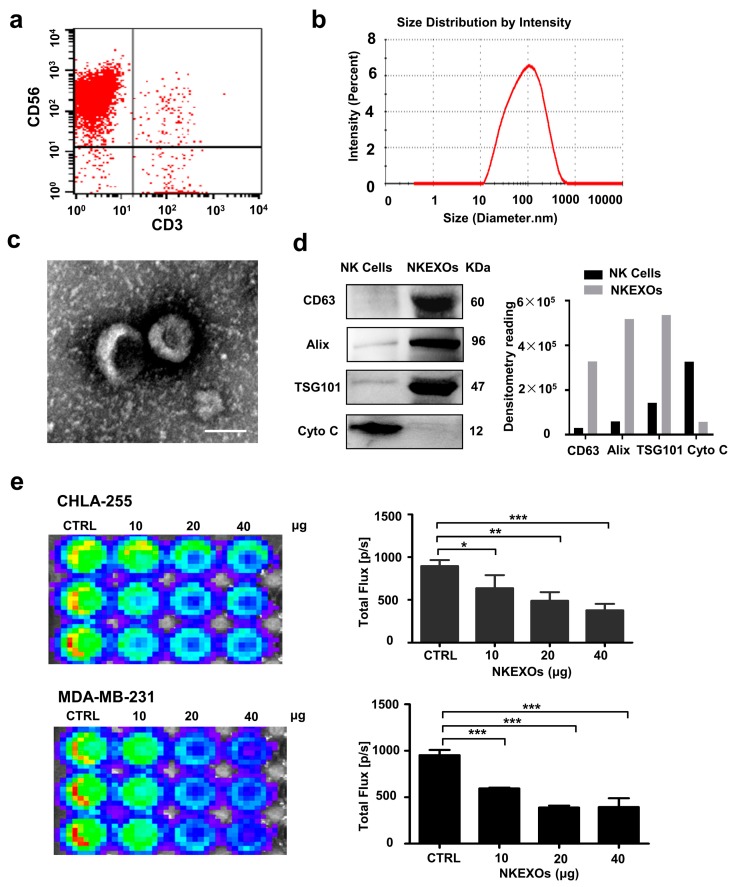
Figure 1
Characterization and function of isolated NKEXOs. ( a ) Flow cytometric analysis of NK cells (CD3u2212 and CD56+) grown for a total of 21 days. ( b ) Size distributions of NKEXO measured by dynamic li...
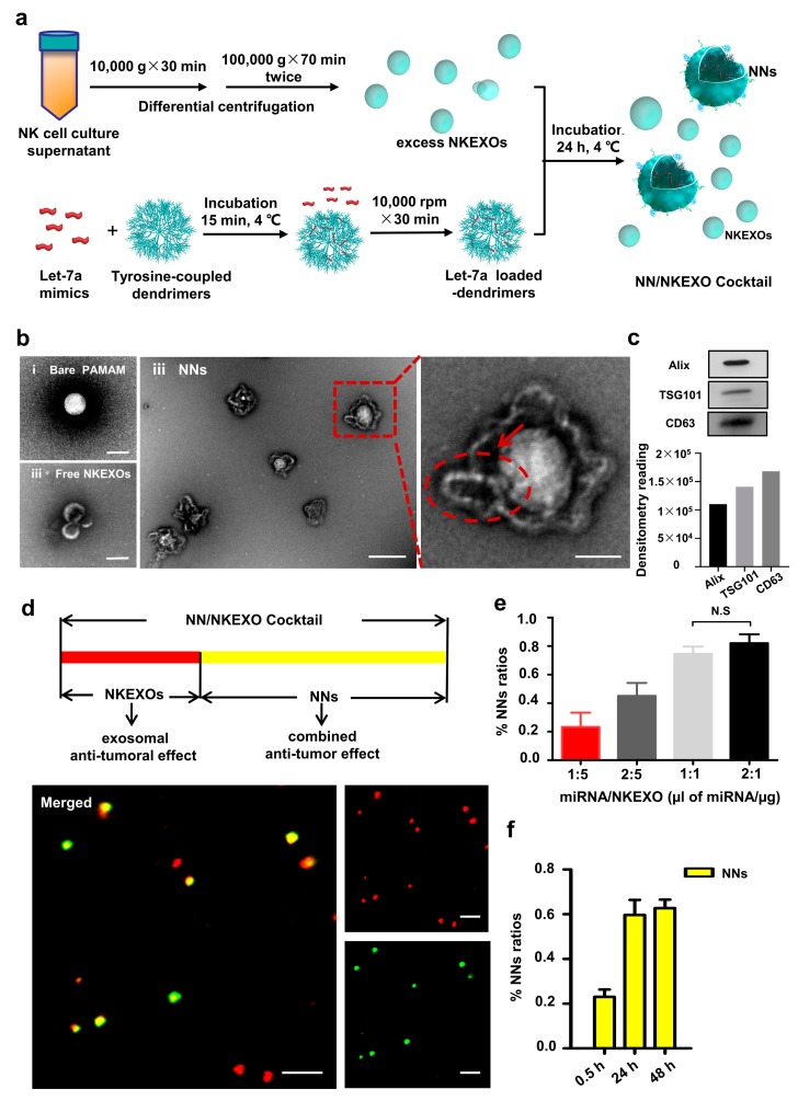
Figure 2
Fabrication and characterization of the NN/NKEXO cocktail. ( a ) Schematic design of the NN/NKEXO cocktail. ( b ) TEM images of bare PAMAM dendrimers (i), NNs (ii) and free NKEXO in supernatant (iii)....
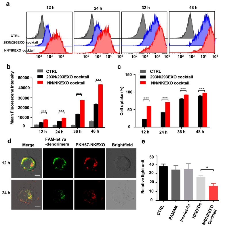
Figure 3
Binding and cytotoxicity of the NN/NKEXO cocktail to a human neuroblastoma cell line in vitro. ( a ) Representative confocal fluorescence images of CHLA-255 cells after treatment with NN/NKEXO cocktai...
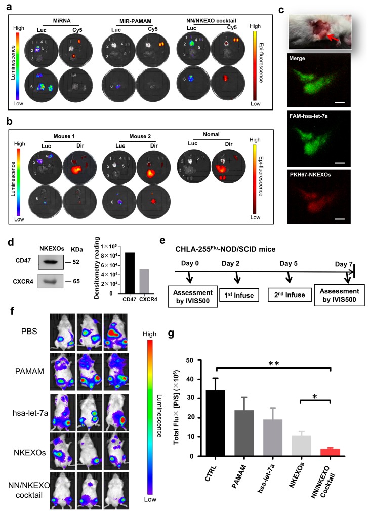
Figure 4
In vivo biodistribution and anti-tumor effect of the NN/NKEXO cocktail. ( a ) Bioluminescence (Luc) and miRNA fluorescence (Cy5) images of major organs of the NOD/SCID mice bearing systemic CHLA-255-l...
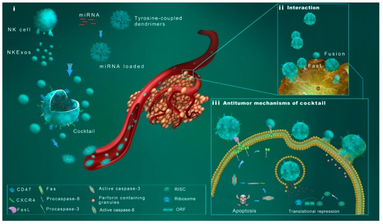
Figure 5
Schematic design of the NN/NKEXO cocktail for tumor targeting and drug delivery. ( i ) Main components of the cocktail: exosomes derived from NK cells (NKEXOs); exosome camouflaged coreu2013shell nano...
Figure images are served from the NIH/NLM PubMed Central Open Access Subset or Europe PMC; copyright remains with the publishers and authors.
💬 Discussion
0 commentsNo comments yet. Be the first to start a discussion!
Leave a Comment