Abstract
Five endosomal sorting complexes required for transport (ESCRTs) mediate the degradation of ubiquitinated membrane proteins via multivesicular bodies (MVBs) in lysosomes. ESCRT-0, -I, and -II interact with cargo on endosomes. ESCRT-II also initiates the assembly of a ringlike ESCRT-III filament consisting of Vps20, Snf7, Vps24, and Vps2. The AAA-adenosine triphosphatase Vps4 disassembles and recycles the ESCRT-III complex, thereby terminating the ESCRT pathway. A mechanistic role for Vps4 in intraluminal vesicle (ILV) formation has been unclear. By combining yeast genetics, biochemistry, and electron tomography, we find that ESCRT-III assembly on endosomes is required to induce or stabilize the necks of growing MVB ILVs. Yet, ESCRT-III alone is not sufficient to complete ILV biogenesis. Rather, binding of Vps4 to ESCRT-III, coordinated by interactions with Vps2 and Snf7, is coupled to membrane neck constriction during ILV formation. Thus, Vps4 not only recycles ESCRT-III subunits but also cooperates with ESCRT-III to drive distinct membrane-remodeling steps, which lead to efficient membrane scission at the end of ILV biogenesis in vivo.
🔬 Techniques
🔭 Microscopes
🧬 Organisms
💻 Software
✨ Fluorophores
🧪 Sample Preparation
🔬 Cell Lines
🏭 Microscope Brands
🧪 Reagent Suppliers
💻 Software Details
📋 Protocols
💾 Data Repositories
🏛️ Research Organizations (ROR)
Affiliated research institutions:
📋 Methods
Antibodies and reagents Rabbit polyclonal antisera against
Vps2 were produced by immunizing rabbits (Eurogentec). 1 mg H6-Vps2, purified from Escherichia coli , was excised from SDS-PAGE and used for immunization. The antiserum (serum agglutination test 455 α) was purified using GST-Vps2 transferred to a polyvinylidene difluoride membrane, eluted with 100 mM glycine, pH 2.5, and diluted 1:100 for Western blotting. The specificity of the antibody was tested using recombinant proteins and yeast extracts from vps2Δ mutants. Monoclonal anti-HA affinity agarose was obtained from Sigma-Aldrich. The mouse monoclonal anti-HA antibody (12CA5) was a gift from L. Hengst (Biocenter, Innsbruck Medical University, Innsbruck, Austria). Rabbit polyclonal antisera against Vps4, Snf7, and Vps24 were a gift from S.D. Emr (Weill Institute for Cell and Molecular Biology, Cornell University, Ithaca, NY) and have been previously described ( Babst et al., 1998 ). Anti-PGK (phosphoglycerate kinase 1) mouse monoclonal antibody was purchased from Invitrogen. Cycloheximide was purchased from Sigma-Aldrich. Strains, plasmids, and DNA manipulation Information on Saccharomyces cerevisiae strains, plasmids, and oligonucleotides is provided in Tables S3 , S4 , and S5 .
Protein expression and purification
Purification of recombinant proteins was performed as previously described ( Babst et al., 1998 ; Teis et al., 2008 ). Recombinant proteins were expressed in E. Coli C41(DE3)pLysS (Lucigen). GST-tagged proteins were purified with glutathione–Sepharose 4B (GE Healthcare), washed, and either eluted with glutathione or cleaved with thrombin. H6-tagged proteins were purified with Ni–nitrilotriacetic acid agarose, washed, and eluted with imidazole. H6-Vps2 was directly eluted with SDS sample buffer (2% SDS, 0.1 M Tris, pH 6.8, 10% glycerol, 0.01% bromophenol blue, and 5% β-mercaptoethanol). GST pull-down assay using GST–ESCRT-III GST pull-down assays were performed as previously described ( Shestakova et al., 2010 ). GST–ESCRT-III subunits were bound to glutathione–Sepharose 4B (GE Healthcare), washed, and incubated with Vps4-E233Q and 1 mM ATP for 10 min at RT. Bound proteins were eluted by boiling for 5 min in SDS-PAGE sample buffer and analyzed by SDS-PAGE. Immunoprecipitation experiments 30 OD 600 equivalents of the yeast cells grown to mid–log phase (OD = 0.6) were lysed by bead beating in immunoprecipitation buffer (100 mM KAc, 5 mM MgCl 2 , 0.2% NP-40, and 100 mM NaCl). Solubilized proteins were subjected to immunoprecipitation for 2 h at 4°C with 30 µl anti-HA agarose. Beads were washed four times in immunoprecipitation buffer. Bound proteins were eluted with SDS sample buffer.
Show full methods section
Antibodies and reagents Rabbit polyclonal antisera against
Vps2 were produced by immunizing rabbits (Eurogentec). 1 mg H6-Vps2, purified from Escherichia coli , was excised from SDS-PAGE and used for immunization. The antiserum (serum agglutination test 455 α) was purified using GST-Vps2 transferred to a polyvinylidene difluoride membrane, eluted with 100 mM glycine, pH 2.5, and diluted 1:100 for Western blotting. The specificity of the antibody was tested using recombinant proteins and yeast extracts from vps2Δ mutants. Monoclonal anti-HA affinity agarose was obtained from Sigma-Aldrich. The mouse monoclonal anti-HA antibody (12CA5) was a gift from L. Hengst (Biocenter, Innsbruck Medical University, Innsbruck, Austria). Rabbit polyclonal antisera against Vps4, Snf7, and Vps24 were a gift from S.D. Emr (Weill Institute for Cell and Molecular Biology, Cornell University, Ithaca, NY) and have been previously described ( Babst et al., 1998 ). Anti-PGK (phosphoglycerate kinase 1) mouse monoclonal antibody was purchased from Invitrogen. Cycloheximide was purchased from Sigma-Aldrich. Strains, plasmids, and DNA manipulation Information on Saccharomyces cerevisiae strains, plasmids, and oligonucleotides is provided in Tables S3 , S4 , and S5 .
Protein expression and purification
Purification of recombinant proteins was performed as previously described ( Babst et al., 1998 ; Teis et al., 2008 ). Recombinant proteins were expressed in E. Coli C41(DE3)pLysS (Lucigen). GST-tagged proteins were purified with glutathione–Sepharose 4B (GE Healthcare), washed, and either eluted with glutathione or cleaved with thrombin. H6-tagged proteins were purified with Ni–nitrilotriacetic acid agarose, washed, and eluted with imidazole. H6-Vps2 was directly eluted with SDS sample buffer (2% SDS, 0.1 M Tris, pH 6.8, 10% glycerol, 0.01% bromophenol blue, and 5% β-mercaptoethanol). GST pull-down assay using GST–ESCRT-III GST pull-down assays were performed as previously described ( Shestakova et al., 2010 ). GST–ESCRT-III subunits were bound to glutathione–Sepharose 4B (GE Healthcare), washed, and incubated with Vps4-E233Q and 1 mM ATP for 10 min at RT. Bound proteins were eluted by boiling for 5 min in SDS-PAGE sample buffer and analyzed by SDS-PAGE. Immunoprecipitation experiments 30 OD 600 equivalents of the yeast cells grown to mid–log phase (OD = 0.6) were lysed by bead beating in immunoprecipitation buffer (100 mM KAc, 5 mM MgCl 2 , 0.2% NP-40, and 100 mM NaCl). Solubilized proteins were subjected to immunoprecipitation for 2 h at 4°C with 30 µl anti-HA agarose. Beads were washed four times in immunoprecipitation buffer. Bound proteins were eluted with SDS sample buffer.
Subcellular fractionation assays
Subcellular fractionation of proteins into membrane-associated pellets and soluble cytoplasmic fractions was performed as previously described ( Babst et al., 1997 ). 30 OD 600 equivalents of the yeast cells grown to mid–log phase (OD = 0.6) were spheroblasted and homogenized by douncing. After a clearing spin (500 g for 5 min), supernatants were centrifuged (15,000 g for 10 min) to separate heavy membranes from cytosol.
Semi–in vitro ESCRT-III disassembly assay
ESCRT-III disassembly assays were performed as previously described ( Davies et al., 2010 ). 100 OD 600 equivalents of the yeast cells grown to mid–log phase (OD = 0.6) were spheroblasted and homogenized by douncing in chilled lysis buffer (0.2 M sorbitol, 50 mM KOAc, 2 mM EDTA, 20 mM Hepes, pH 6.8, and protease inhibitors). After a clearing spin (500 g for 5 min), supernatants were centrifuged (15,000 g for 10 min) to separate heavy membranes from cytosol. The membrane pellet was washed in 1 ml ATPase reaction buffer (100 mM KOAc, 20 mM Hepes, pH 7.4, and 5 mM MgOAc) with 1 mM sorbitol and protease inhibitors (buffer A). The repelleted membranes were resuspended in buffer A and passed through an 18-gauge needle three times and a 30-gauge needle five times for homogenization. Homogenized membranes were stored at −80°C. 100-µl reactions containing 2 OD 600 equivalent membranes, 100 nM purified Vps4, and an ATP regeneration system (10 mM phosphocreatine, 20 U/ml creatine phosphokinase, and 0.8 mM ATP) were incubated at 26°C for 30 and 60 s. The disassembly reaction was stopped by centrifugation, and membrane-bound and -released proteins were separated by centrifugation at 13,000 rpm at 4°C.
Fluorescence microscopy
For live-cell microscopy, cells were grown to midlog (OD 600 = 0.6) at RT in yeast nitrogen base (fluorochromes used in this study were GFP and mCherry) and labeled with FM4-64 (Invitrogen) as previously described ( Vida and Emr, 1995 ; Teis et al., 2008 ). Fluorescence microscopy was performed with a microscope (Axio Imager.M1; Carl Zeiss) and an α-Plan Fluar 100×, 1.45 NA oil objective (Carl Zeiss). Images were taken with a charge-coupled device camera (Sport Explorer; Visitron Systems). Acquisition software used was VisiView 2.0.3 (Visitron Systems). Image brightness and contrast was enhanced in the RGB channel using Photoshop CS4 Extended (version 11.0.2; Adobe). Cryofixation, EM, and tomography Yeast cultures (0.6 OD 600 ) were subjected to high-pressure freezing with an HMP 010 (obtained from Bal-Tec) followed by freeze substitution, epoxy resin embedding, and section poststaining essentially as described previously for plants ( Hess, 2007 ), except that freeze-substitution media consisted of acetone plus 2.5% (vol/vol) glutaraldehyde, 2% (wt/vol) uranyl acetate, 10% vol/vol methanol, 0.05% (wt/vol) OsO 4 , and 1.5% water. Thin and semithin sections (100 and 400 nm, respectively) were poststained with hot ethanolic phosphotungstic acid (0.5% wt/vol in 95% ethanol for 5 min at 60°C; Locke and Krishnan, 1971 ). Thin sections were viewed at 80 kV with a microscope (Philips CM120; FEI) equipped with a digital camera (Morada; Olympus). Electron tomography from 400-nm semithin sections (coated with 10-nm fiducial gold) was performed on a camera (Tecnai T20-G2; FEI) at 200 kV using a dual-tilt series. Images were recorded at a binning of 2 with a 4,000 × 4,000–pixel digital camera (Eagle; FEI) from 55 to −55° with 1° increments using Inspect3D automated tomography software (obtained from FEI). Tomograms were reconstructed and modeled using IMOD software ( Kremer et al., 1996 ). For ILV size and ILV membrane neck analysis, single slices of reconstructed tomograms were analyzed using 3dmod. For morphometry, we analyzed >30 MVBs, >150 ILVs, and >33 ILV budding profiles per cell type in 2D slices from tomographic reconstructions from seven different WT cells, four different snf7* , five different vps2* , and nine different snf7*, vps2* mutants. Dual-luciferase reporter assay (LUCID) Dual-luciferase assays of Sna3-FLuc trafficking to the lysosome were performed as previously described ( Nickerson et al., 2012 ). In brief, luciferase activity of Sna3-FLuc and Renilla luciferase (RLuc) was analyzed with the dual-luciferase assay system (DLR; Promega). Cells in mid–log phase (OD = 0.6) were treated with cycloheximide (50 µg/ml final) for 30 min, harvested by centrifugation, and lysed by incubation in 500 µl lysis buffer by vortexing with glass beads at RT. 5-µl aliquots of the lysate were analyzed in 96-well plate format using a luminometer (TriStar LB941; Berthold Technologies). After background subtraction, luciferase activity of Sna3-FLuc was normalized to the activity of RLuc. Block and recovery of MVB biogenesis in vps4 -ts mutants Yeast cells were grown to logarithmic phase at 26°C and then shifted to 37°C for 4 h. 50 µg/ml cycloheximide was added to the growth media 15 min before shifting them back to 26°C for 4 h. 30 OD 0.6 equivalents grown at 37°C or recovering at 26°C (with cycloheximide) were subjected to subcellular fractionation, cryofixation, and electron tomography as described in this work, except that cells grown at 37°C were also spheroblasted at 37°C. SILAC and mass spectrometry analysis vps2* mutants were grown in SILAC “light” medium, and WT cells were grown in SILAC “heavy” medium containing ( 13 C 6 / 15 N 2 ) l -lysine (20 µg/ml; Sigma-Aldrich) for 10 generations to mid–log phase (OD 600 = 0.6). Cells were washed once with ice-cold double-distilled H 2 O and used for immunoprecipitation. Immunoprecipitated proteins from WT cells (labeled with heavy lysine) and from vps2* mutants were eluted in SDS sample buffer (without bromophenol blue), mixed, and subjected to SDS-PAGE. Proteins were digested in gel with LysC. Peptides were analyzed using an HPLC system (UltiMate 3000 Nano-LC; Dionex) coupled to a mass spectrometer (LTQ Orbitrap XL; Thermo Fisher Scientific). MaxQuant version 1.2.0.18 with search engine Andromeda and Proteome Discoverer version 1.2.0.208 (Thermo Fisher Scientific) with search engine Mascot version 2.2.07 (Matrix Science) were used for data analysis. Raw data obtained by liquid chromatography–electrophoresis electrospray ionization–mass spectrometry were searched against the yeast protein database downloaded from http://downloads.yeastgenome.org/sequence/S288C_reference/orf_protein/ .
Statistical analysis
Statistical analysis was performed using robust one-way analysis of variance (ILV diameter) and Student’s t test, unpaired and two tailed (ILV diameter and LUCID). *, P < 0.05; **, P < 0.01; ***, P < 0.001. Online supplemental material Fig. S1 shows analysis of the MIT–MIM interaction. Fig. S2 presents subcellular fractionation and velocity sedimentation centrifugation to further characterize chimeric ESCRT-III complexes. Fig S3 shows Western blot analysis (full films from Fig. 4 A ) of subcellular fractionation and GPF-CPS sorting in vps20 -MIM1 and vps2 -MIM2 mutants. Fig. S4 contains further analysis of MVB and ILV morphology using electron tomography of cryofixed mutants: snf7*, vps2* double mutants without VPS21 overexpression, Vps4-MIT mutants, and in vma4Δ mutants. Fig. S5 shows analysis of snf7*, vps2* double mutants. Tables S1 and S2 show the SILAC-based quantification of Vps4-HA immunoprecipitation using MaxQuant and Proteome Discoverer, respectively, and relate to Fig. 1 D .
Table
S3 contains all yeast strains used in this study.
Table
S4and plasmids used in this study.
Table
S5 shows primers used in this study. Video 1 shows electron tomography and 3D modeling of a cryofixed WT yeast cell overexpressing Vps21. Video 2 shows electron tomography and 3D modeling of a cryofixed snf7* mutant overexpressing Vps21. Video 3 shows electron tomography and 3D modeling of a cryofixed vps2* mutant overexpressing Vps21. Video 4 shows electron tomography and 3D modeling of a cryofixed snf7*, vps2* mutant overexpressing Vps21. Video 5 shows electron tomography and 3D modeling of a cryo-fixed snf7*, vps2*, vps25 T150K mutant overexpressing Vps21. Online supplemental material is available at http://www.jcb.org/cgi/content/full/jcb.201310114/DC1 . Additional data are available in the JCB DataViewer at http://dx.doi.org/10.1083/jcb.201310114.dv .
Online supplemental material Fig. S1 shows analysis of the MIT–MIM interaction. Fig. S2 presents subcellular fractionation and velocity sedimentation centrifugation to further characterize chimeric ESCRT-III complexes. Fig S3 shows Western blot analysis (full films from Fig. 4 A ) of subcellular fractionation and GPF-CPS sorting in vps20 -MIM1 and vps2 -MIM2 mutants. Fig. S4 contains further analysis of MVB and ILV morphology using electron tomography of cryofixed mutants: snf7*, vps2* double mutants without VPS21 overexpression, Vps4-MIT mutants, and in vma4Δ mutants. Fig. S5 shows analysis of snf7*, vps2* double mutants. Tables S1 and S2 show the SILAC-based quantification of Vps4-HA immunoprecipitation using MaxQuant and Proteome Discoverer, respectively, and relate to Fig. 1 D .
Table
S3 contains all yeast strains used in this study.
Table
S4and plasmids used in this study.
Table
S5 shows primers used in this study. Video 1 shows electron tomography and 3D modeling of a cryofixed WT yeast cell overexpressing Vps21. Video 2 shows electron tomography and 3D modeling of a cryofixed snf7* mutant overexpressing Vps21. Video 3 shows electron tomography and 3D modeling of a cryofixed vps2* mutant overexpressing Vps21. Video 4 shows electron tomography and 3D modeling of a cryofixed snf7*, vps2* mutant overexpressing Vps21. Video 5 shows electron tomography and 3D modeling of a cryo-fixed snf7*, vps2*, vps25 T150K mutant overexpressing Vps21. Online supplemental material is available at http://www.jcb.org/cgi/content/full/jcb.201310114/DC1 . Additional data are available in the JCB DataViewer at http://dx.doi.org/10.1083/jcb.201310114.dv .
📊 Figures
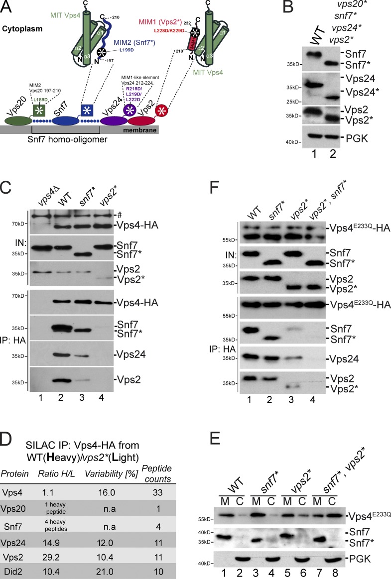
Figure 1.
Binding of Vps4 to ESCRT-III is mainly mediated by Snf7 and Vps2. (A) The schematic representation shows ESCRT-III, the Snf7 homooligomer (blue dots), the MIM2 of Vps20 (green square), the MIM2 of Snf...
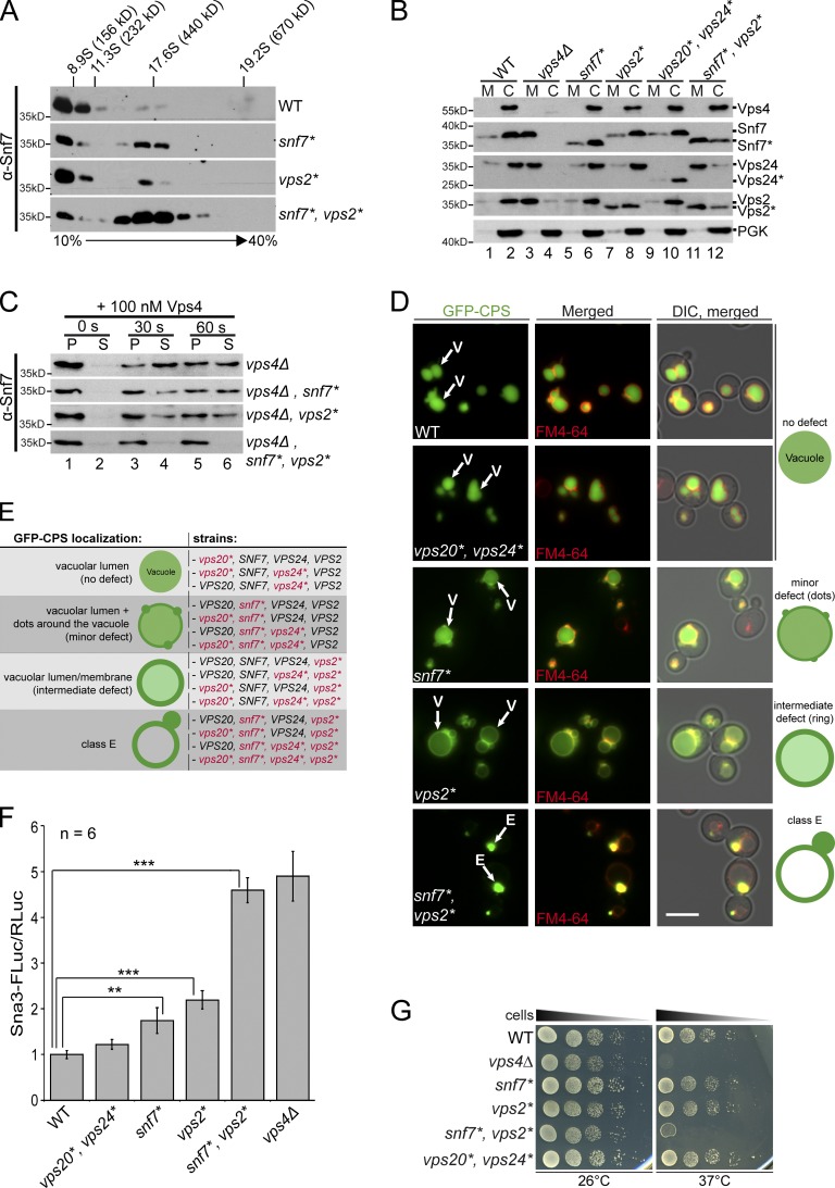
Figure 2.
The interaction of Vps4 with Snf7 and Vps2 is essential for efficient ESCRT-III disassembly and MVB sorting. (Au2013C) Experiments were analyzed by SDS-PAGE and Western blotting. (A) Solubilized membr...
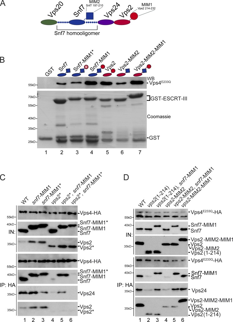
Figure 3.
Rearranging the binding of Vps4 to the ESCRT-III complex. (A) Schematic representation of the ESCRT-III complex. (B) In vitro pull-down assay with ESCRT-III chimeras. Their domain organization is show...
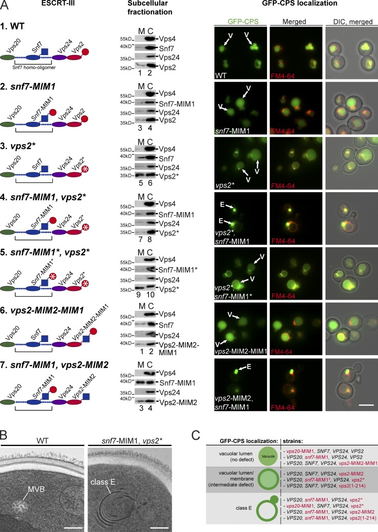
Figure 4.
The MIMs of Vps2 and Snf7 couple ESCRT-III disassembly to ILV biogenesis. (A) Sections 1u20137 show schematic representation of ESCRT-III complexes, the corresponding subcellular fractionation, and li...
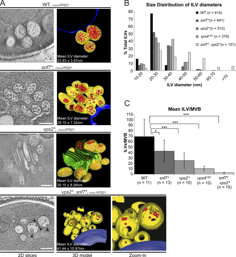
Figure 5.
Vps4-mediated ESCRT-III disassembly controls ILV biogenesis in vivo. (A) Electron tomography of cryofixed WT cells overexpressing Vps21 (TDH3- VPS21 ) and the indicated mutants. 2D slices from tomogra...
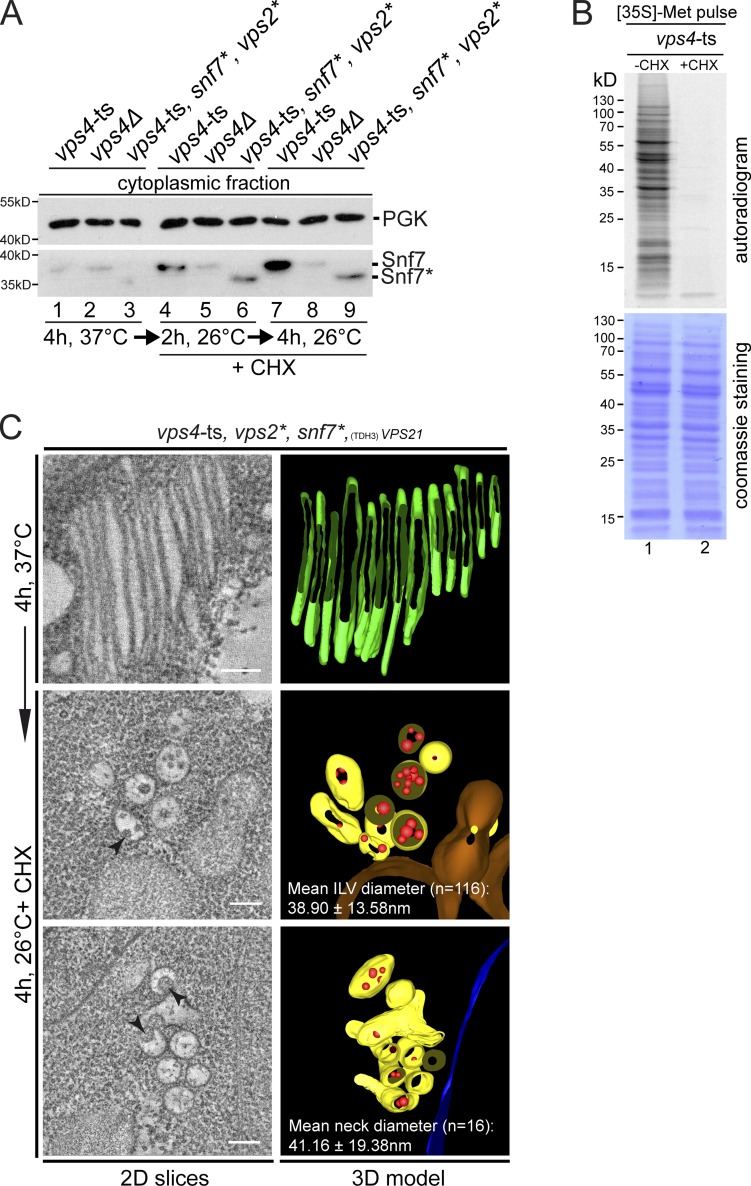
Figure 6.
Slow and continuous ESCRT-III recycling and ILV biogenesis in snf7*, vps2* double mutants. (A) vps4 -ts mutants, vps4u0394 mutants and snf7*, vps2*, vps4 -ts mutants were shifted to the nonpermissive ...
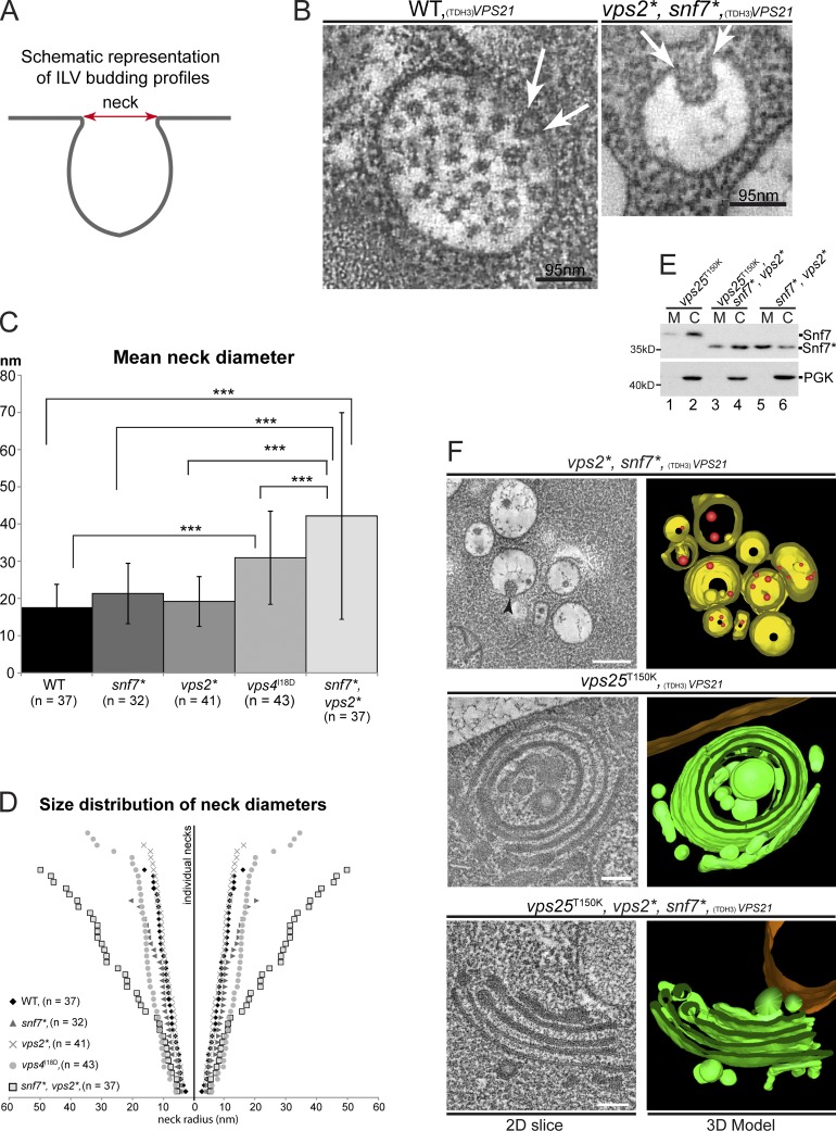
Figure 7.
Binding of Vps4 to Snf7 and Vps2 is required for ILV neck constriction. (A) Schematic representation of ILV budding profiles as detected in tomographic reconstructions. (B) MVB of WT cells and MVB-lik...
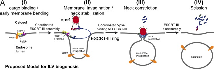
Figure 8.
Proposed model for ILV biogenesis. PI(3)P, phosphatidylinositol 3-phosphate.
Figure images are served from the NIH/NLM PubMed Central Open Access Subset or Europe PMC; copyright remains with the publishers and authors.
💬 Discussion
0 commentsNo comments yet. Be the first to start a discussion!
Leave a Comment