Abstract
Abstract Specimens below 50 kDa have generally been considered too small to be analyzed by single-particle cryo-electron microscopy (cryo-EM). The high flexibility of pure RNAs makes it difficult to obtain high-resolution structures by cryo-EM. In bacteria, riboswitches regulate sulfur metabolism through binding to the S-adenosylmethionine (SAM) ligand and offer compelling targets for new antibiotics. SAM-I, SAM-I/IV, and SAM-IV are the three most commonly found SAM riboswitches, but the structure of SAM-IV is still unknown. Here, we report the structures of apo and SAM-bound SAM-IV riboswitches (119-nt, ~40 kDa) to 3.7 Å and 4.1 Å resolution, respectively, using cryo-EM. The structures illustrate homologies in the ligand-binding core but distinct peripheral tertiary contacts in SAM-IV compared to SAM-I and SAM-I/IV. Our results demonstrate the feasibility of resolving small RNAs with enough detail to enable detection of their ligand-binding pockets and suggest that cryo-EM could play a role in structure-assisted drug design for RNA.
🔬 Techniques
💻 Software
🧪 Sample Preparation
🏭 Microscope Brands
🧪 Reagent Suppliers
📷 Detectors
💻 Software Details
💻 Code & Software
💾 Data Repositories
🏛️ Research Organizations (ROR)
Affiliated research institutions:
📋 Methods
Sample preparation
RNA was prepared as previously described 13 . Briefly, DNA templates were constructed through PCR assembly using primers listed in the Supplementary Table 2 and then purified with AMPureXP beads (Beckman Coulter). RNA was transcribed with T7 RNA polymerase at 37 °C for 4 h in a buffer containing 0.2 μM DNA template, 40 mM Tris·HCl (pH 8.1), 25 mM MgCl 2 , 3.5 mM spermidine, 0.01% TritonX-100, 40 mM DTT, 4% PEG 8000, 3 mM NTPs, and 5 U/μL T7 RNA polymerase (NEB). RNA was then purified with Zymo RNA Clean and Concentrator columns (Zymo Research) following the manufacturer’s instructions.
Cryo-EM data collection
Three microliters of the SAM-IV riboswitch RNAs at 40 μM were applied onto glow-discharged 200-mesh R2/1 Quantifoil grids. The grids were blotted for 2–4 s and rapidly cryocooled in liquid ethane using a Vitrobot Mark IV (Thermo Fisher Scientific) at room temperature and ~100% humidity. The samples were screened using a Talos Arctica cryo-electron microscope (Thermo Fisher Scientific) operated at 200 kV. To test the feasibility of cryo-EM for determining RNA structures, the apo SAM-IV riboswitch was first imaged in a Talos Arctica cryo-electron microscope with and without phase plate, respectively. For the high-resolution study, both of the apo and SAM-bound SAM-IV riboswitches were imaged in a Titan Krios cryo-electron microscope (Thermo Fisher Scientific) with GIF energy filter (Gatan) at a magnification of ×130,000 (corresponding to a calibrated sampling of 1.06 Å per pixel). Micrographs were recorded by EPU software (Thermo Fisher Scientific) with a Gatan K2 Summit direct electron detector, where each image was composed of 30 individual frames with an exposure time of 6 s and a dose rate of 7.6 electrons per second per Å 2 . Finally, a total of 7200 movie stacks for the apo state and 6030 movie stacks for the bound state were collected with a defocus range of −1.5 to −3.5 μm.
Show full methods section
Sample preparation
RNA was prepared as previously described 13 . Briefly, DNA templates were constructed through PCR assembly using primers listed in the Supplementary Table 2 and then purified with AMPureXP beads (Beckman Coulter). RNA was transcribed with T7 RNA polymerase at 37 °C for 4 h in a buffer containing 0.2 μM DNA template, 40 mM Tris·HCl (pH 8.1), 25 mM MgCl 2 , 3.5 mM spermidine, 0.01% TritonX-100, 40 mM DTT, 4% PEG 8000, 3 mM NTPs, and 5 U/μL T7 RNA polymerase (NEB). RNA was then purified with Zymo RNA Clean and Concentrator columns (Zymo Research) following the manufacturer’s instructions.
Cryo-EM data collection
Three microliters of the SAM-IV riboswitch RNAs at 40 μM were applied onto glow-discharged 200-mesh R2/1 Quantifoil grids. The grids were blotted for 2–4 s and rapidly cryocooled in liquid ethane using a Vitrobot Mark IV (Thermo Fisher Scientific) at room temperature and ~100% humidity. The samples were screened using a Talos Arctica cryo-electron microscope (Thermo Fisher Scientific) operated at 200 kV. To test the feasibility of cryo-EM for determining RNA structures, the apo SAM-IV riboswitch was first imaged in a Talos Arctica cryo-electron microscope with and without phase plate, respectively. For the high-resolution study, both of the apo and SAM-bound SAM-IV riboswitches were imaged in a Titan Krios cryo-electron microscope (Thermo Fisher Scientific) with GIF energy filter (Gatan) at a magnification of ×130,000 (corresponding to a calibrated sampling of 1.06 Å per pixel). Micrographs were recorded by EPU software (Thermo Fisher Scientific) with a Gatan K2 Summit direct electron detector, where each image was composed of 30 individual frames with an exposure time of 6 s and a dose rate of 7.6 electrons per second per Å 2 . Finally, a total of 7200 movie stacks for the apo state and 6030 movie stacks for the bound state were collected with a defocus range of −1.5 to −3.5 μm.
Image processing
All micrographs were motion-corrected using MotionCor2 32 and the contrast transfer function (CTF) was determined using CTFFIND4 33 . All particles were autopicked using the NeuralNet option in EMAN2 34 and further checked manually. The resulting number of boxed particles were 2,102,569 for the apo state and 1,830,706 for the SAM-bound state. Then, particle coordinates were imported to Relion 35 , where three rounds of 2D classification were performed to remove 2D class averages with less resolved features. Using 1,137,602 particles of the apo state, we first generated an ab-initio map using cryoSPARC 36 and refined it with Relion, yielding a 3.9-Å map. Then starting with the same ab-initio map, we reprocessed the same set of particles using cryoSPARC and yielded a final map with 3.7-Å resolution from 796,923 particles. For the SAM-bound state, we selected 984,150 particles by Relion and processed them using cryoSPARC to yield a 4.1-Å map from 588,580 particle images. Both map determinations used the 3D non-uniform refinement and local refinement options of cryoSPARC. The cited resolutions for the final maps were estimated by the 0.143 criterion of FSC curve in cryoSPARC. The 3.7-Å and 4.1-Å low-pass filters were applied, respectively, to the final 3D maps for better display (See more information in Supplementary Fig. 2 and Supplementary Table 1 ). Model building The initial SAM-IV riboswitch models were built with auto-DRRAFTER 13 . We built both fully automated models and models using information from the previously determined crystal structure of the homologous SAM-I riboswitch (PDB code: 3GX5) for the apo and SAM-bound states. For the fully automated models, 2000 models were built per round, and a total of three and four rounds of modeling were performed for the apo and SAM-bound states, respectively. For the models built with information from the previously solved SAM-I structure, P5 was first approximately fit into the density maps for the apo and SAM-bound states. We modeled the conformation of the SAM binding pocket by taking all residues that were within 5 Å of SAM in the SAM-I crystal structure (residues 6–8, 11–12, 44–47, 57–59, 87 in SAM-I numbering, corresponding to residues 2–4, 7–8, 63–65, 77 in SAM-IV numbering) and mutating the nucleotides to the SAM-IV sequence. These nucleotides were kept fixed as a rigid body in the first round of modeling for the SAM-IV apo state, and in all but the final round of modeling for the SAM-bound state. Three rounds of modeling were performed for the apo and SAM-bound states, with 2000 models built per round. These models agree closely with the fully automated models. All final models were built into half maps to prevent overfitting (Fig. S5 ). Next, the top-scoring models of the apo and SAM-bound states built with information from the SAM-I crystal structure were selected for further refinement by ERRASER and Phenix 37 to optimize the geometry while improving the fit to the cryo-EM density. As the current resolution was not sufficient to determine the conformation of the SAM binding pocket, residues 2–4, 7–8, 63–65, 77 and SAM derived from SAM-I crystal structure were fixed during the optimizations. The final models were evaluated by MolProbity and Q-score 20 . All figures were prepared using PyMol 38 or Chimera 24 . Secondary structure diagrams were prepared with RiboDraw [ https://github.com/ribokit/RiboDraw ]. Reporting summary Further information on research design is available in the Nature Research Reporting Summary linked to this article.
Supplementary information Supplementary Information Peer Review Reporting Summary Description of Additional Supplementary Files Supplementary Movie 1
📊 Figures
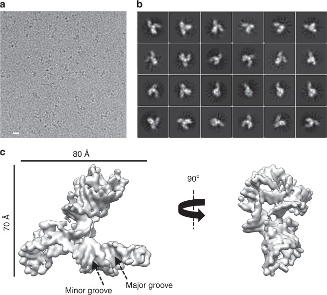
Fig. 1
Single-particle cryo-EM analysis of the apo SAM-IV riboswitch collected on Titan Krios. a Representative motion-corrected cryo-EM micrograph. Scale bar represents 100u2009u00c5. b Reference-free 2D cl...
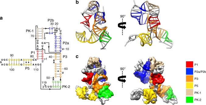
Fig. 2
The secondary and tertiary structures of the apo SAM-IV riboswitch. a The secondary structure with the domains colored differently. The black arrow indicates the direction of the backbone. b - c The m...
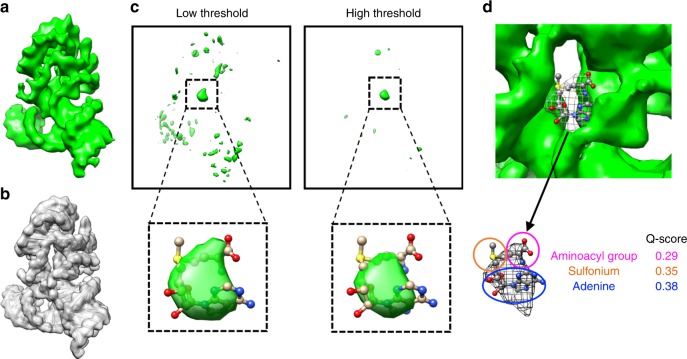
Fig. 3
Determination of the ligand location in the ligand-bound SAM-IV cryo-EM map. a Ligand-bound SAM-IV cryo-EM map. b Map computed from the SAM-IV RNA-only model from a . c Difference map between a and b ...
Figure images are served from the NIH/NLM PubMed Central Open Access Subset or Europe PMC; copyright remains with the publishers and authors.
💬 Discussion
0 commentsNo comments yet. Be the first to start a discussion!
Leave a Comment