Abstract
The epithelial anion transporter SLC26A9 contributes to airway surface hydration and gastric acid production. Colocalizing with CFTR, SLC26A9 has been proposed as a target for the treatment of cystic fibrosis. To provide molecular details of its transport mechanism, we present cryo-EM structures and a functional characterization of murine Slc26a9. These structures define the general architecture of eukaryotic SLC26 family members and reveal an unusual mode of oligomerization which relies predominantly on the cytosolic STAS domain. Our data illustrates conformational transitions of Slc26a9, supporting a rapid alternate-access mechanism which mediates uncoupled chloride transport with negligible bicarbonate or sulfate permeability. The characterization of structure-guided mutants illuminates the properties of the ion transport path, including a selective anion binding site located in the center of a mobile module within the transmembrane domain. This study thus provides a structural foundation for the understanding of the entire SLC26 family and potentially facilitates their therapeutic exploitation.
🔬 Techniques
🧬 Organisms
✨ Fluorophores
🧪 Sample Preparation
🔬 Cell Lines
🏭 Microscope Brands
🧪 Reagent Suppliers
💻 Software Details
💾 Data Repositories
🏛️ Research Organizations (ROR)
Affiliated research institutions:
📋 Methods
Key resources table
Reagent type (species) or resource Designation Source or reference Identifier Additional information Chemical compound, drug Pro293S-CDM medium Lonza Cat#BE02-025Q Chemical compound, drug HyClone HyCell TransFx-H medium GE Healthcare Cat#SH30939.02 Chemical compound, drug L-glutamine Millipore Sigma Cat#G7513 Chemical compound, drug Penicillin-streptomycin Millipore Sigma Cat#P0781 Chemical compound, drug Fetal bovine serum Millipore Sigma Cat#F7524 Chemical compound, drug Pluronic F-68 ThermoFisher Scientific Cat#24040032 Chemical compound, drug Polyethylenimine 25 K MW, linear Polysciences Cat#23966–1 Chemical compound, drug Dulbecco’s Modified Eagle’s Medium (DMEM) Millipore Sigma Cat#D5546 Chemical compound, drug Valproic acid Millipore Sigma Cat#P4543 Chemical compound, drug cOmplete, EDTA-free Protease Inhibitor Cocktail Roche Cat#5056489001 Chemical compound, drug Digitonin AppliChem Cat#A1905 Chemical compound, drug Glyco-diosgenin Anatrace Cat#GDN101 Chemical compound, drug D-desthiobiotin Millipore Sigma Cat#D1411 Chemical compound, drug n -dodecyl-β-D-maltoside (DDM) Anatrace Cat#D310 Chemical compound, drug Cholesteryl hemisuccinate, tris salt (CHS) Anatrace Cat#CH210 Chemical compound, drug 1-palmitoyl-2-oleoyl-sn-glycero-3-phosphoethanolamine (POPE) Avanti Polar Lipids, Inc Cat#850757 Chemical compound, drug 1-palmitoyl-2-oleoyl-sn-glycero-3-phospho-(1'-rac-glycerol) (POPG) Avanti Polar Lipids, Inc Cat#840457 Chemical compound, drug Diethyl ether Millipore Sigma Cat#296082 Chemical compound, drug Triton X-100 Millipore Sigma Cat#T9284 Chemical compound, drug Biotin Millipore Sigma Cat#B4501 Chemical compound, drug 9-amino-6-chloro-2-methoxyacridine (ACMA) Thermo Fisher Scientific Cat#A1324 Chemical compound, drug carbonyl cyanide 3-chloropheny lhydrazone (CCCP) Merck Millipore Cat#C2759 Chemical compound, drug 4,4’-Diisothiocyanatostilbene-2,2’-disulfonic acid (DIDS) Millipore Sigma Cat#D3514 Commercial assay or kit QuikChange site-directed mutagenesis kit Agilent Cat#200523 Commercial assay or kit NucleoBond Xtra Maxi kit Macherey-Nagel Cat#740416 Commercial assay or kit StrepTactin Superflow affinity resin slurry IBA Lifesciences Cat#2-1206-002 Commercial assay or kit Superose 6 10/300 GL GE Healthcare Cat#17-5172-01 Commercial assay or kit Zorbax GF-450 Agilent Cat#884973–902 Commercial assay or kit Superose 6 5/150 GE Healthcare Cat#29091597 Commercial assay or kit Pierce Streptavidin Plus UltraLink Resin Thermo Fisher Scientific Cat#53117 Commercial assay or kit Bio-Beads SM-2 Bio-Rad Cat# 1523920 Commercial assay or kit Avestin LiposoFast Liposome Factory Basic Millipore Sigma Cat#Z373400 Commercial assay or kit 400 nm polycarbonate filters for LiposoFast Millipore Sigma Cat#Z373435 Commercial assay or kit 96-well black-walled microplate Thermo Fisher Scientific Cat# M33089 Commercial assay or kit 200 mesh Au 1.2/1.3 cryo-EM grids Quantifoil Cat#N1-C14nAu20-01 Commercial assay or kit Amicon 100 kDa MWCO centrifugal filter EMD Millipore Cat#UFC910008 Commercial assay or kit 0.22 µm Ultrafree-MC Centrifugal Filter EMD Millipore Cat#UFC30GV Commercial assay or kit Borosilicate glass capillary with filament Sutter Instrument Cat#BF150-86-10HP Cell line (human) HEK293S GnTI- ATCC CRL-3022 Cell line (human) HEK-293T ATCC CRL-1573 Recombinant DNA Mus musculus Slc26a9 ORF shuttle clone Source BioScience ORFeome# OCACo5052B0115D; GenBank BC160193 Recombinant DNA pcDNA 3.1 (+) vector, Invitrogen Thermo Fisher Scientific Cat# V79020 Recombinant DNA Modified pcDNA 3.1 vector with C-terminal 3C protease cleavage site, Venus and Myc tags and streptavidin binding peptide Raimund Dutzler laboratory N/A Recombinant DNA Modified pcDNA 3.1 vector with C-terminal 3C protease cleavage site, Myc tag and streptavidin binding peptide Raimund Dutzler laboratory N/A Recombinant DNA Expression vector encoding membrane scaffold protein (MSP) E3D1, pMSP1E3D1 Denisov et al., 2007 Addgene, Cat#20066 Software,algorithm SerialEM 3.5.0 Mastronarde, 2005 http://bio3d.colorado.edu/SerialEM/ Software, algorithm RELION-3.0 Scheres, 2012 https://www2.mrc-lmb.cam.ac.uk/relion/ Software, algorithm CTFFIND4.1 Rohou and Grigorieff, 2015 http://grigoriefflab.jan elia.org/ctf Software, algorithm Bsoft 1.9.5 Heymann and Belnap, 2007 https://lsbr.niams.nih.gov/bsoft/ Software, algorithm Coot 0.8.8 Emsley and Cowtan, 2004 https://www2.mrc-lmb.cam.ac.uk/person al/pemsley/coot/ Software, algorithm PHENIX 1.14 Adams et al., 2002 http://phenix-online.org/ Software, algorithm REFMAC5 Murshudov et al., 2011 http://www.ccpem.ac.uk/ Software, algorithm MSMS Sanner et al., 1996 http://mgltools.scripps.edu/packages/MSMS/ Software, algorithm DINO 0.9.4 http://www.dino3d.org http://www.dino3d.org Software, algorithm PyMOL 2.3.0 DeLano, 2002 https://pymol.org/2/ Software, algorithm Chimera 1.13.1 Pettersen et al., 2004 http://www.cgl.ucsf.edu/chimera/ Software, algorithm ChimeraX 0.7 Goddard et al., 2018 https://www.cgl.ucsf.edu/chimerax/ Software, algorithm CHARMM Brooks et al., 1983 https://www.charmm.org/charmm/ Software, algorithm SWISS-MODEL Biasini et al., 2014 https://swissmodel.expasy.org/ Software, algorithm Axon Clampex 10.6 Molecular Devices N/A Software, algorithm Axon Clampfit 10.6 Molecular Devices N/A Software, algorithm Prism 7 GraphPad https://www.graphpad.com/ Cell lines GnTI - cells used for protein expression and purification were obtaiend from ATTC (ATCC CRL-3022). Adherent HEK293T cells used for electrophysiology were obtained from ATTC (ATCC CRL-1573). Both cell-lines were tested negative for mycoplasma contamination. Suspension-adapted HEK293S GnTI - cells expressing murine Slc26a9 were grown at 37°C and 5% CO2 in either Pro293S-CDM or HyClone TransFx-H media, supplemented with 2 mM L-glutamine, 100 U ml –1 penicillin/streptomycin, 1% FBS, and 1% Pluronic F-68. Adherent HEK293T cells were grown in DMEM media supplemented with 1 mM L-glutamine, 100 U ml –1 penicillin/streptomycin, 10% FBS and 1 mM sodium pyruvate.
Show full methods section
Key resources table
Reagent type (species) or resource Designation Source or reference Identifier Additional information Chemical compound, drug Pro293S-CDM medium Lonza Cat#BE02-025Q Chemical compound, drug HyClone HyCell TransFx-H medium GE Healthcare Cat#SH30939.02 Chemical compound, drug L-glutamine Millipore Sigma Cat#G7513 Chemical compound, drug Penicillin-streptomycin Millipore Sigma Cat#P0781 Chemical compound, drug Fetal bovine serum Millipore Sigma Cat#F7524 Chemical compound, drug Pluronic F-68 ThermoFisher Scientific Cat#24040032 Chemical compound, drug Polyethylenimine 25 K MW, linear Polysciences Cat#23966–1 Chemical compound, drug Dulbecco’s Modified Eagle’s Medium (DMEM) Millipore Sigma Cat#D5546 Chemical compound, drug Valproic acid Millipore Sigma Cat#P4543 Chemical compound, drug cOmplete, EDTA-free Protease Inhibitor Cocktail Roche Cat#5056489001 Chemical compound, drug Digitonin AppliChem Cat#A1905 Chemical compound, drug Glyco-diosgenin Anatrace Cat#GDN101 Chemical compound, drug D-desthiobiotin Millipore Sigma Cat#D1411 Chemical compound, drug n -dodecyl-β-D-maltoside (DDM) Anatrace Cat#D310 Chemical compound, drug Cholesteryl hemisuccinate, tris salt (CHS) Anatrace Cat#CH210 Chemical compound, drug 1-palmitoyl-2-oleoyl-sn-glycero-3-phosphoethanolamine (POPE) Avanti Polar Lipids, Inc Cat#850757 Chemical compound, drug 1-palmitoyl-2-oleoyl-sn-glycero-3-phospho-(1'-rac-glycerol) (POPG) Avanti Polar Lipids, Inc Cat#840457 Chemical compound, drug Diethyl ether Millipore Sigma Cat#296082 Chemical compound, drug Triton X-100 Millipore Sigma Cat#T9284 Chemical compound, drug Biotin Millipore Sigma Cat#B4501 Chemical compound, drug 9-amino-6-chloro-2-methoxyacridine (ACMA) Thermo Fisher Scientific Cat#A1324 Chemical compound, drug carbonyl cyanide 3-chloropheny lhydrazone (CCCP) Merck Millipore Cat#C2759 Chemical compound, drug 4,4’-Diisothiocyanatostilbene-2,2’-disulfonic acid (DIDS) Millipore Sigma Cat#D3514 Commercial assay or kit QuikChange site-directed mutagenesis kit Agilent Cat#200523 Commercial assay or kit NucleoBond Xtra Maxi kit Macherey-Nagel Cat#740416 Commercial assay or kit StrepTactin Superflow affinity resin slurry IBA Lifesciences Cat#2-1206-002 Commercial assay or kit Superose 6 10/300 GL GE Healthcare Cat#17-5172-01 Commercial assay or kit Zorbax GF-450 Agilent Cat#884973–902 Commercial assay or kit Superose 6 5/150 GE Healthcare Cat#29091597 Commercial assay or kit Pierce Streptavidin Plus UltraLink Resin Thermo Fisher Scientific Cat#53117 Commercial assay or kit Bio-Beads SM-2 Bio-Rad Cat# 1523920 Commercial assay or kit Avestin LiposoFast Liposome Factory Basic Millipore Sigma Cat#Z373400 Commercial assay or kit 400 nm polycarbonate filters for LiposoFast Millipore Sigma Cat#Z373435 Commercial assay or kit 96-well black-walled microplate Thermo Fisher Scientific Cat# M33089 Commercial assay or kit 200 mesh Au 1.2/1.3 cryo-EM grids Quantifoil Cat#N1-C14nAu20-01 Commercial assay or kit Amicon 100 kDa MWCO centrifugal filter EMD Millipore Cat#UFC910008 Commercial assay or kit 0.22 µm Ultrafree-MC Centrifugal Filter EMD Millipore Cat#UFC30GV Commercial assay or kit Borosilicate glass capillary with filament Sutter Instrument Cat#BF150-86-10HP Cell line (human) HEK293S GnTI- ATCC CRL-3022 Cell line (human) HEK-293T ATCC CRL-1573 Recombinant DNA Mus musculus Slc26a9 ORF shuttle clone Source BioScience ORFeome# OCACo5052B0115D; GenBank BC160193 Recombinant DNA pcDNA 3.1 (+) vector, Invitrogen Thermo Fisher Scientific Cat# V79020 Recombinant DNA Modified pcDNA 3.1 vector with C-terminal 3C protease cleavage site, Venus and Myc tags and streptavidin binding peptide Raimund Dutzler laboratory N/A Recombinant DNA Modified pcDNA 3.1 vector with C-terminal 3C protease cleavage site, Myc tag and streptavidin binding peptide Raimund Dutzler laboratory N/A Recombinant DNA Expression vector encoding membrane scaffold protein (MSP) E3D1, pMSP1E3D1 Denisov et al., 2007 Addgene, Cat#20066 Software,algorithm SerialEM 3.5.0 Mastronarde, 2005 http://bio3d.colorado.edu/SerialEM/ Software, algorithm RELION-3.0 Scheres, 2012 https://www2.mrc-lmb.cam.ac.uk/relion/ Software, algorithm CTFFIND4.1 Rohou and Grigorieff, 2015 http://grigoriefflab.jan elia.org/ctf Software, algorithm Bsoft 1.9.5 Heymann and Belnap, 2007 https://lsbr.niams.nih.gov/bsoft/ Software, algorithm Coot 0.8.8 Emsley and Cowtan, 2004 https://www2.mrc-lmb.cam.ac.uk/person al/pemsley/coot/ Software, algorithm PHENIX 1.14 Adams et al., 2002 http://phenix-online.org/ Software, algorithm REFMAC5 Murshudov et al., 2011 http://www.ccpem.ac.uk/ Software, algorithm MSMS Sanner et al., 1996 http://mgltools.scripps.edu/packages/MSMS/ Software, algorithm DINO 0.9.4 http://www.dino3d.org http://www.dino3d.org Software, algorithm PyMOL 2.3.0 DeLano, 2002 https://pymol.org/2/ Software, algorithm Chimera 1.13.1 Pettersen et al., 2004 http://www.cgl.ucsf.edu/chimera/ Software, algorithm ChimeraX 0.7 Goddard et al., 2018 https://www.cgl.ucsf.edu/chimerax/ Software, algorithm CHARMM Brooks et al., 1983 https://www.charmm.org/charmm/ Software, algorithm SWISS-MODEL Biasini et al., 2014 https://swissmodel.expasy.org/ Software, algorithm Axon Clampex 10.6 Molecular Devices N/A Software, algorithm Axon Clampfit 10.6 Molecular Devices N/A Software, algorithm Prism 7 GraphPad https://www.graphpad.com/ Cell lines GnTI - cells used for protein expression and purification were obtaiend from ATTC (ATCC CRL-3022). Adherent HEK293T cells used for electrophysiology were obtained from ATTC (ATCC CRL-1573). Both cell-lines were tested negative for mycoplasma contamination. Suspension-adapted HEK293S GnTI - cells expressing murine Slc26a9 were grown at 37°C and 5% CO2 in either Pro293S-CDM or HyClone TransFx-H media, supplemented with 2 mM L-glutamine, 100 U ml –1 penicillin/streptomycin, 1% FBS, and 1% Pluronic F-68. Adherent HEK293T cells were grown in DMEM media supplemented with 1 mM L-glutamine, 100 U ml –1 penicillin/streptomycin, 10% FBS and 1 mM sodium pyruvate.
Construct generation
DNA encoding the open reading frame
(ORF) for mouse Slc26a9 (GenBank accession: BC160193 ) was PCR-amplified from a cDNA clone (Source BioScience) and shuttled into a pcDNA 3.1 vector (Invitrogen) which was modified to be compatible with FX cloning technology ( Geertsma and Dutzler, 2011 ). Unless stated otherwise, all expression constructs also encoded a C-terminal Rhinovirus 3C protease cleavage site followed by venus YFP (vYFP), a myc epitope tag and a streptavidin binding peptide (SBP), giving the general construct scheme ORF-3C-vYFP-myc-SBP. Assembly of the Slc26a9 T dual-truncation construct entailed removal of the STAS IVS region, via replacement of residues Pro 558 –Val 660 with a Gly-Ser linker, and deletion of C-terminal residues Pro 745 –Leu 790 , akin to a described procedure for the isolated STAS domain from rat Prestin ( Pasqualetto et al., 2010 ). Further deletion of N-terminal residues Met 1 –Ala 30 resulted in the construct Slc26a9(Δ1-30) T . Mutations Q88A, Q88E, F92A, T127A, F128A, L391A, S392A, R205E, K221E, K270E, K431E, and K441E were introduced into Slc26a9 T using the QuikChange site-directed mutagenesis method (Agilent). Protein expression and purification Suspension-adapted HEK293S GnTI - cells (ATCC CRL-3022) were grown in either Pro293S-CDM (Lonza) or HyClone TransFx-H (GE Healthcare) media, supplemented with 2 mM L-glutamine (Sigma), 100 U ml –1 penicillin/streptomycin (Sigma), 1% FBS, and 1% Pluronic F-68. Cultures were maintained in TubeSpin Bioreactor 600 vessels (TPP), shaken at 185 rpm with an orbital radius of 50 mm, and incubated at 37°C and 5% CO 2 . For protein expression, a transient transfection protocol relying on 25 kDa linear polyethylenimine (PEI, Polysciences) was employed. One day prior to transfection, cells at high density (3–5 × 10 6 ml –1 ) were diluted into fresh media to a density of 0.6–0.8 × 10 6 ml –1 . Transfection-grade plasmid DNA was purified from MC1061 E. coli culture using the NucleoBond Xtra Maxi kit (Macherey-Nagel), and a ratio of 1.3 μg DNA per 10 6 of HEK293S-GnTI - cells was used for transfection. Plasmid DNA was diluted into non-supplemented DMEM media (Sigma) at a concentration of 0.015 μg μl −1 , and PEI was added to a concentration of 0.038 μg μl –1 from a 1 mg ml –1 , pH 7 stock solution. After 10–15 min, the DNA-PEI mixtures were diluted 10-fold directly into the cultured cells, and valproic acid (Sigma) was added to a final concentration of 3 mM. After 40–48 hr, cells were harvested by centrifugation at 500 g for 10 min, washed with PBS, and then either directly used for protein extraction and purification or flash frozen in liquid nitrogen and stored at –80°C. All subsequent protein extraction and purification procedures were carried out at 4°C. Cell pellets from 4–liter expression batches were resuspended in 60 ml of resuspension buffer (25 mM HEPES, pH 7.4, 200 mM NaCl, 5% glycerol, 2 mM CaCl 2 , and 2 mM MgCl2, 10 μg ml –1 DNase, and protease inhibitors (cOmplete EDTA-free, Roche). For cryo-EM analysis of detergent-solubilized Slc26a9 T , the protein was extracted in digitonin (AppliChem), and subsequently purified in the presence of the synthetic digitonin substitute glyco-diosgenin (GDN, Anatrace). To extract membrane proteins, 2% (w/v) digitonin powder was directly dissolved in the cell resuspension, and the mixture was incubated for 1.5 hr under gentle agitation. Insoluble material was removed via ultracentrifugation for 40 min at 150,000 g and the supernatant was passed through a 5 μm syringe filter (Sartorius). The clarified extract was applied to 12 ml of StrepTactin Superflow affinity resin slurry (IBA Lifesciences), which was pre-equilibrated in wash buffer composed of 25 mM HEPES, pH 7.4, 200 mM NaCl, 5% glycerol, and 0.02% GDN. The affinity resin was washed with 20 CV of wash buffer, before elution with 15 ml of wash buffer supplemented with 10 mM D-desthiobiotin (Sigma). The protein was concentrated using a 100 kDa molecular weight cut-off (MWCO) centrifugal filter (Amicon) to 500 μl, typically resulting in a concentration of 1–2 mg ml –1 fusion protein, and 3C protease was added at a protein:protease mass ratio of 1:2. After a 1 hr incubation, the sample was centrifuged at 10,000 g for 3 min to pellet aggregated material, and the supernatant was passed through a 0.22 μm centrifugal filter (Millipore), before being injected onto a Superose 6 10/300 column (GE Healthcare) equilibrated in SEC buffer, 10 mM HEPES, pH 7.4, 200 mM NaCl, 0.02% GDN. Protein from peak fractions containing cleaved Slc26a9 T protein was pooled, concentrated to 3 mg ml –1 , and immediately used for cryo-EM grid preparation. For the preparation of Slc26a9 T protein to be reconstituted into either liposome or lipid nanodiscs, a similar extraction and purification procedure was performed, with the following modifications. Resuspended cells were extracted with a mixture of 1.5% n -dodecyl-β-D-maltoside (DDM, Anatrace) and 0.15% cholesteryl hemisuccinate (CHS, Anatrace), and protein was purified in the presence of 0.03% DDM and 0.003% CHS. For protein which was designated for liposome reconstitution, all other purification procedures were equivalent to the protocol used for cryo-EM sample preparation. However, for nanodisc preparations, an uncleaved variant of Slc26a9 T , lacking a fluorescent fusion protein and therefore consisting of Slc26a9 T -3C-myc-SBP (henceforth abbreviated as Slc26a9 T -SBP), was purified and used for nanodisc assembly. The behavior of all vYFP-tagged Slc26a9 constructs in detergent extracts was assessed with an HPLC system equipped for fluorescence-coupled size exclusion chromatography (FSEC, Agilent), using a Zorbax GF-450 column (Agilent). The same system was also employed to monitor the quality of purified proteins, using UV detection with a Superose 6 5/150 column (GE Healthcare). All protein samples were purified to ≥95% homogeneity, as assessed by SDS-PAGE. Liposome and nanodisc reconstitution For reconstitution of Slc26a9 T into liposomes, a procedure relying on detergent-destabilization of preformed liposomes was employed, as previously described ( Geertsma et al., 2008 ). Synthetic POPE and POPG lipids (Avanti Polar Lipids) at a mass ratio of 3:1 POPE:POPG were washed in diethyl ether, dried under N 2 in a glass round-bottom flask, and hydrated via gentle sonication in liposome buffer, 10 mM HEPES, pH 7.4, 100 mM KCl, to a concentration of 20 mg ml –1 . The lipid mixture was subjected to three freeze-thaw cycles, followed by extrusion through two 400 nm polycarbonate filters (LiposoFast Basic, Avestin) to form large unilamellar vesicles (LUVs). LUVs were diluted in liposome buffer to 4 mg ml –1 , and 10% (w/v) Triton X-100 was added dropwise until the solution absorbance at 540 nm value reached a maximum, indicating suitable destabilization of liposomes for incorporation of membrane protein. Purified Slc26a9 T (in DDM-CHS) was added to the destabilized LUVs at a protein:lipid ratio of 1:80 (w/w). After 20 min of incubation at RT, the sample was cooled to 4°C and detergent was removed via sequential additions of 250 mg SM-2 Bio-Beads (Bio-Rad) per 5 ml, every 24 hr for three days. Bio-Beads were removed via gravity filtration, and Slc26a9 T proteoliposomes were pelleted via ultracentrifugation at 150,000 g for 30 min, resuspended in liposome buffer to 20 mg ml –1 , frozen in liquid N 2 , and stored at – 80°C. Identical procedures were used to produce mock liposomes lacking any reconstituted membrane protein. Slc26a9 T -SBP was reconstituted into lipid nanodiscs as described ( Ritchie et al., 2009 ), with subtle modifications. We utilized the engineered membrane scaffold protein (MSP) MSP1-E3D1 because the estimated resultant nanodisc dimeter of 12 nm is ideal for the incorporation of Slc26a9 T , which our preliminary cryo-EM analysis had suggested to possess a length of 10–11 nm when solubilized in detergent. MSP1-E3D1 was purified as described ( Ritchie et al., 2009 ) and a 3:1 (w/w) mixture of the synthetic lipids POPC:POPG was dried and hydrated in 10 mM HEPES, pH 7.4, 100 mM KCl, as described above for proteoliposome preparations, except the final lipid concentration was 10 mM, and DDM was added to the stock lipid mixture to a final concentration of 27.5 mM. To assemble nanodiscs, purified Slc26a9 T -SBP was diluted into 10 mM HEPES, 200 mM NaCl, 0.5 mM DDM, to a final protein concentration of 6 μM. Lipids were added to a concentration of 1.9 mM, and the mixture was incubated on ice for 30 min. Next, MSP1-E3D1 was added to a concentration of 20 μM, giving a final protein:lipid:MSP molar ratio of 1:450:20, at a final volume of 750 μl. After an additional 30 min of incubation on ice, 200 mg of SM2 Bio-Beads was added to remove detergent, and the reaction was allowed to incubate overnight under slow rotation at 4°C. Bio-beads were removed via gravity filtration, and Slc26a9 T -SBP-reconstituted nanodiscs were isolated from empty nanodiscs via secondary purification with 3 ml of Streptavidin Plus UltraLink affinity resin (ThermoFisher) using detergent-free nanodisc buffer, 10 mM HEPES, pH 7.4, 150 mM NaCl, and elution with 5 mM biotin. Finally, nanodiscs were injected onto a Superose 6 5/300 column equilibrated in nanodisc buffer, and a peak containing Slc26a9 T -SBP nanodiscs was concentrated to 1 mg ml –1 and immediately used for cryo-EM grid preparation.
Liposome anion transport assay
To measure electrogenic anion transport in Slc26a9 T proteoliposomes, we used a method based on a previously described fluorometric assay ( Kane Dickson et al., 2014 ) in which the internal liposome buffer ideally contains no permeant ions. Since Slc26a9 has low permeability to cations and sulfate, we exchanged the internal buffer of Slc26a9 T proteoliposomes and mock liposomes to 10 mM HEPES, 50 mM Na 2 SO 4 by pelleting the proteoliposomes (150,000 g, 30 min), resuspending in internal sulfate buffer to a concentration of 1 mg ml –1 , and subjecting the sample to three freeze-thaw cycles. Finally, liposomes were again pelleted and resuspended to 20 mg ml –1 in internal sulfate buffer. All subsequent procedures were carried out at RT to restrict formation of multilamellar vesicles, and mock liposomes were always assayed in parallel as a negative control. To form small unilamellar vesicles, 20 μl aliquots of liposomes in 0.2 ml conical tubes were placed in a bath sonicator for 5–10 s, until the opaque solution became translucent. Liposomes were diluted 100-fold into flux buffer, consisting of 10 mM HEPES, pH 7.4, 75 mM NaCl, and 2 μM of the fluorophore 9-amino-6-chloro-2-methoxyacridine (ACMA, ThermoFisher), and 100 μl aliquots were transferred to a 96-well black-walled microplate (ThermoFisher). ACMA fluorescence was monitored with an Infinite M1000 spectrofluorometer (Tecan) in 5 s intervals, using excitation and emission wavelengths of 412 nm and 482 nm, respectively. After recording baseline fluorescence for 60 s, data collection was paused, 300 nM of the proton ionophore carbonyl cyanide 3-chlorophenylhydrazone (CCCP, Sigma) was added, and fluorescence measurements were immediately continued. Fluorescence intensity for each experimental condition was normalized to the initial value directly following addition of CCCP. For inhibition experiments, diluted proteoliposomes (0.2 mg ml –1 ) were pre-incubated for 5 min with 0–250 µM 4,4’−2,2’-disulfonic acid (DIDS, Sigma) and dimethyl sulfoxide (DMSO) was added to all samples to a final concentration of 1% to maintain solubility of DIDS.
Cryo-EM sample preparation and data collection
For structure determination of Slc26a9 T by cryo-EM, 2.5 μl samples of GDN-purified protein at a concentration of 3 mg ml –1 were applied to glow-discharged holey carbon grids (Quantifoil R1.2/1.3 Au 200 mesh). For the structural characterization of the protein in a membrane-like environment, 2.5 μl samples of Slc26a9 T reconstituted in E3D1 lipid nanodiscs at a concentration of 0.5–1 mg ml –1 were applied in a similar manner. Excess liquid was removed in a controlled environment (4°C and 100% relative humidity) by blotting grids for 3–6 s. Grids were subsequently flash frozen in liquid propane-ethane mix using a Vitrobot Mark IV (Thermo Fisher Scientific). Slc26a9 T in detergent (dataset 1) was imaged in a 300 kV Titan Krios (Thermo Fisher Scientific) with a 100 μm objective aperture. Slc26a9 T in nanodiscs (dataset 2) was recorded on a 300 kV Tecnai G 2 Polara (FEI) with a 100 μm objective aperture. All data were collected using a post-column quantum energy filter (Gatan) with a 20 eV slit and a K2 Summit direct detector (Gatan) operating in super-resolution (for dataset 1) and counting (for dataset 2) modes. Dose-fractionated micrographs were recorded in an automated manner using SerialEM ( Mastronarde, 2005 ) with a defocus range of –0.5 to –3.0 μm. Dataset one was recorded at a nominal magnification of 46,511 corresponding to a pixel size of 1.075 Å/pixel (0.5375 Å/pixel in super-resolution) with a total exposure time of 13 s (65 individual frames) and a dose of approximately 1.1 e – /Å 2 /frame. Dataset two was recorded at a nominal magnification of 37,313 corresponding to a pixel size of 1.34 Å/pixel with a total exposure time of 12.5 s (50 individual frames) and a dose of approximately 1.2 e – /Å 2 /frame. The total electron dose on the specimen level for dataset 1 and 2 was approximately 70 e – /Å 2 and 60 e – /Å 2 , respectively.
Cryo-EM image processing
The recorded super-resolution images of Slc26a9 T in detergent (dataset 1) were down-sampled twice by Fourier cropping and all individual frames were used for correction of beam-induced movement using a dose-weighting scheme in RELION’s own implementation of MotionCor2 algorithm available in version 3.0 ( Zivanov et al., 2018a ). The CTF parameters were estimated on summed movie frames using CTFFIND4.1 ( Rohou and Grigorieff, 2015 ). All individual frames of Slc26a9 T reconstituted in nanodiscs (dataset 2) were pre-processed in the same manner. Low-quality micrographs showing a significant drift or poor CTF estimates were discarded resulting in datasets of 2838 images of Slc26a9 T in detergent and 3134 images of Slc26a9 T in nanodiscs, which were subjected to further data processing in RELION ( Scheres, 2012 ). From dataset one 416,164 particles were picked automatically using low-pass filtered 2D templates generated from an initial reference-free 2D classification. The particles were extracted with a box size of 232 pixels, down-scaled twice and subjected to a couple of rounds of 2D classification. Having discarded false positives and particles of poor quality, the dataset was reduced to 241,438 particles. The initial 3D reconstruction, which was generated from 3600 randomly chosen cleaned particles, was low-pass filtered to 60 Å and used as a template in a subsequent 3D classification. Multiple independent rounds of non-symmetrized 3D classification with a different number of classes were performed in order to isolate the most homogeneous subset of particles. One out of five classes, contained almost two-thirds of all the particles and showed clear two-fold symmetry. 157,644 particles belonging to this class were subjected to auto-refinement with imposed C2 symmetry. The particles were then unbinned to a pixel size of 1.075 Å/pixel and refined to 4.3 Å using C2 symmetry and a soft mask around the protein-detergent micelle density. The reconstruction was further improved to 4.09 Å by performing per-particle defocus and beam tilt corrections followed by Bayesian polishing ( Zivanov et al., 2018a ; Zivanov et al., 2018b ). To enhance features of the peripheral helices α6 and α7, which were of lower local resolution than the core of the transmembrane domain, the density representing the detergent belt was subtracted from individual particles. In silico modified particles were used as an input in a subsequent masked 3D classification ( Scheres, 2016 ). To separate any remaining heterogeneity particle angles were kept the same as the orientations from the consensus model. The final class containing 112,930 homogeneous particles was further auto-refined with C2 symmetry and in the presence of a soft mask around the protein density. The final map at 3.96 Å was sharpened using an isotropic b-factor of –205 Å 2 . The initial dataset of Slc26a9 T in E3D1 lipid nanodiscs contained 711,032 particles, which were subjected to 2D classification in order to discard particles of poor quality, small aggregates and any remaining empty nanodiscs. After a stringent selection 308,872 particles were used in a subsequent non-symmetrized 3D classification. Similarly to the dataset of Slc26a9 T in detergent, multiple independent runs were performed to optimally separate the heterogeneity in the dataset. Here, 3D classes showed more apparent heterogeneity compared to the detergent dataset. One out of seven classes containing 64,670 particles displayed well-resolved two-fold symmetric STAS domains although the extracellular loops located between the helices α3 and α4 of each subunit showed poorer density indicating increased flexibility of this region. As at this level of 3D classification the quality of the reconstruction did not allow to observe whether this flexibility is further extended to the TM domain or whether is affecting one or two monomers, further refinement was continued, side-by-side, without imposing any symmetry as well as with imposing C2 symmetry. Within the auto-refined non-symmetrized class one monomer was better resolved comparing to the second monomer and it also showed similar arrangement as either monomer in the structure of the protein in detergent. On the other hand, the second monomer showed increased flexibility only in the TM domain and the extracellular region as its STAS domain was identical as in the first monomer and as the STAS domain in the detergent structure. These results have suggested that despite eliminating particles with large heterogeneous differences during previous rounds of 2D and 3D classification, small conformational changes accounted for the remaining flexibility. In order to separate these changes into discrete classes a focused 3D classification was performed ( Scheres, 2016 ). In this approach the mask around the flexible monomer was applied and the angles were kept fixed at the orientations from the auto-refined model. Given the classification focused only on a small region of the protein, the regularization parameter T was increased to 40. Approximately one-fourth of the remaining particles from both non-symmetrized and C2-symmetrized consensus models contributed to a class that recovered the density representing the extracellular loop. After identification of these homogeneous subsets that represent the stable monomer, final 3D auto-refinement of the whole dimeric particles was performed either with C1 or C2 symmetry imposed, yielding in both cases two-fold symmetric reconstructions of 8.4 Å and 7.77 Å, respectively. In all cases resolution was estimated in the presence of a soft solvent mask and based on the gold standard Fourier Shell Correlation (FSC) 0.143 criterion ( Chen et al., 2013 ; Rosenthal and Henderson, 2003 ; Scheres, 2012 ; Scheres and Chen, 2012 ). High-resolution noise substitution was applied to correct FSC curves for the effect of soft masking in real space ( Chen et al., 2013 ). The local resolution was estimated using BlocRes from the Bsoft package ( Cardone et al., 2013 ; Heymann and Belnap, 2007 ).
Model building and refinement
The model of Slc26a9 T in the inward-facing state was built in Coot ( Emsley and Cowtan, 2004 ) using the transmembrane domain structure of SLC26Dg (PDBID: 5DA0) and the chicken Prestin STAS domain (PDBID: 5EZB) as templates. The cryo-EM density of Slc26a9 T in detergent was of sufficiently high resolution to unambiguously assign residues 5–27, 42–559 and 661–740. The model was improved iteratively by cycles of real-space refinement in PHENIX ( Adams et al., 2002 ) with secondary structure and 2-fold NCS constraints applied, reciprocal-space refinement in REFMAC5 ( Brown et al., 2015 ; Murshudov et al., 2011 ) and manual corrections in Coot. Validation of the refinement of the model was performed in REFMAC5, distributed as part of the CCP-EM suite ( Burnley et al., 2017 ), and represented as Fourier Shell Correlation (FSC sum ) between the refined model and the corresponding final cryo-EM density map. For cross-validation the detergent cryo-EM dataset was split into two subsets which were used to calculate two independent maps (half map one and half map 2). To detect possible overfitting and hence over-estimation of the resolution, random shifts, up to 0.5 Å, were applied to the coordinates of the model of the inward-facing state, which was then refined against the unfiltered half map 1. The cross-validation was done by comparing the FSC work (estimated for the shaken-refined model and half map 1) and the FSC free (estimated for the shaken-refined model and half map 2, which was not used in the refinement). The model of the intermediate state of Slc26a9 T in nanodiscs was assembled by splitting the model of the inward-facing state into six independent entities, the STAS, gate and core domains, and refining them in PHENIX as rigid-bodies. In the next stage, the STAS, gate and core domain were linked into a single polypeptide chain and refined as described above with global minimization applied. The secondary structure and NCS constrains were maintained at all times. As the protein side-chains were not defined in our 7.77 Å cryo-EM reconstruction of the intermediate state, we have truncated all the side-chains to alanine. The validation of the refinement was performed as described above and represented as FSC sum . Owing to the low-resolution of the 3D reconstruction from the dataset in nanodiscs, FSC work and FSC free were not calculated. Surfaces were calculated with MSMS ( Sanner et al., 1996 ). Figures and videos containing molecular structures and densities were prepared with DINO ( http://www.dino3d.org ), PyMOL ( DeLano, 2002 ), Coot ( Emsley and Cowtan, 2004 ),Chimera ( Pettersen et al., 2004 ) and ChimeraX ( Goddard et al., 2018 ). Modeling and Poisson-Boltzmann calculations The electrostatic potential in the intracellular vestibule leading to the Cl – -binding site was calculated by solving the linearized Poisson–Boltzmann equation in CHARMM ( Brooks et al., 1983 ; Im et al., 1998 ) on a 150 Å ×170 Å × 200 Å grid (1 Å grid spacing) followed by focusing on a 120 Å x 130 Å x 160 Å grid (0.5 Å grid spacing). Partial protein charges were derived from the CHARMM36 all-hydrogen atom force field. Hydrogen positions were generated in CHARMM. Histidines were protonated. The protein was assigned a dielectric constant ( ϵ ) of 2. Its transmembrane region was embedded in a 30 Å-thick slab ( ϵ = 2) representing the hydrophobic core of the membrane and two adjacent 15 Å-thick regions ( ϵ = 30) representing the headgroups. The membrane region contained a 22 Å-high and 30 Å-wide aqueous cylinder ( ϵ = 80) covering the intracellular vestibule of the protein and was surrounded by an aqueous environment ( ϵ = 80). Calculations were carried out in either 150 mM of monovalent mobile ions in the aqueous regions (except for the membrane-inserted cylinder). The electrostatic surface potential shown in Figure 3—figure supplement 2F was calculated and displayed with COOT ( Emsley and Cowtan, 2004 ). Homology models of murine SLC26 paralogs were prepared with the SWISS-MODEL homology modelling server ( Biasini et al., 2014 ). Electrophysiology Adherent HEK293T cells (ATCC CRL-1573) were grown in DMEM media supplemented with 1 mM L-glutamine (Sigma), 100 U ml –1 penicillin/streptomycin (Sigma), 1 mM sodium pyruvate (Sigma), and 10% FBS, at 37°C and 5% CO 2 , in 6 cm culture dishes. For transfection, 2.5–5 μg plasmid DNA encoding the construct of interest was mixed with 25 kDa linear polyethylenimine (Polysciences) in a ratio of 1:2.5 (w/w) in 0.3 ml PBS, incubated for 10 min at room temperature, and added dropwise to cells. Transfected cells were used within 24 hr for whole-cell recordings, or within 40 hr for excised patch recordings. Borosilicate glass capillaries (OD = 1.5 mm, ID = 0.86 mm, Sutter) were pulled and fire-polished, giving patch pipettes with resistances of 3–8 MΩ when backfilled with 150 mM NaCl pipette solution. All voltage-clamp signals were recorded using an Axopatch 200B amplifier (Molecular Devices) digitized with a Digidata 1440A A/D converter (Molecular Devices), filtered at 5 kHz, sampled at 20 kHz, and collected with Clampex 10.6 (Molecular Devices). For full-length Slc26a9, membrane seal resistance was typically 2–10 GΩ. However, for Slc26a9 T , membrane seal resistance was frequently
📊 Figures
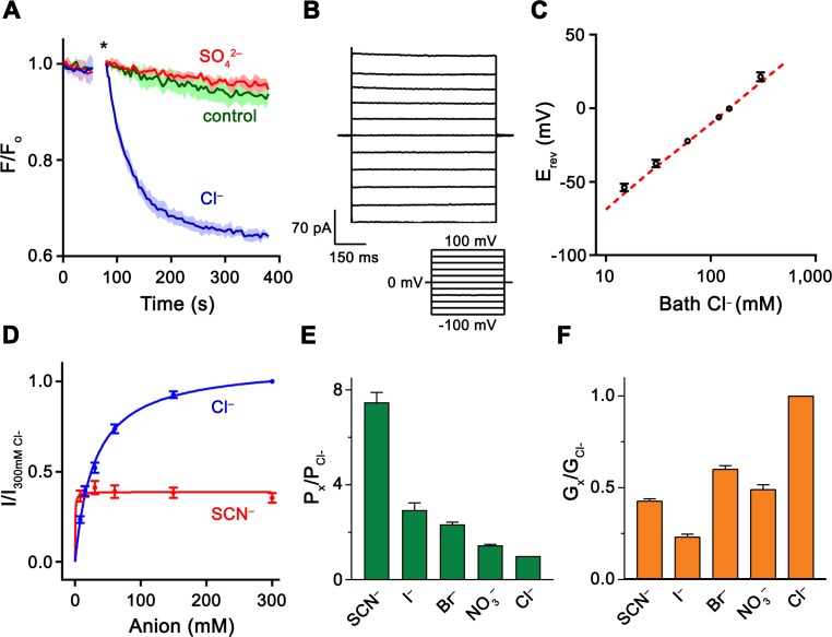
Figure 1.
Functional properties of Slc26a9.
( A ) Cl u2013 transport of Slc26a9 T reconstituted into proteoliposomes, monitored by the fluorescence change of the pH gradient-sensitive fluorophore ACMA. (*) Indicates addition of the H + ionophor...
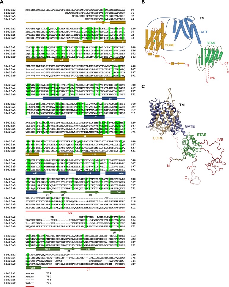
Figure 1u2014figure supplement 1.
Sequence alignment and topology.
( A ) Sequence alignment of the murine Slc26 paralogs Slc26a2 ( NP_031911.1 ), Slc26a4 ( NP_035997.1 ), Slc26a5 ( NP_109652.3 ), and Slc26a9 ( NP_796217.2 ). Identical residues are highlighted in gree...
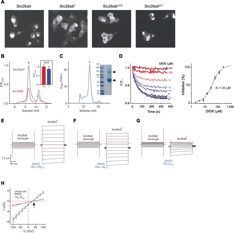
Figure 1u2014figure supplement 2.
Expression, purification and functional characterization of Slc26a9 T .
( A ) Fluorescence-microscopy images of HEK293T cells transfected with different Slc26a9 constructs (From left to right: full-length Slc26a9 , the u0394IVSu0394CT truncation construct Slc26a9 T , the ...
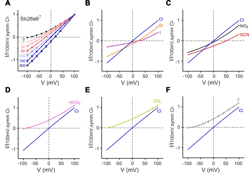
Figure 1u2014figure supplement 3.
Functional properties of Slc26a9 T .
( A ) Iu2013V relationships for excised inside-out patches from HEK293T cells expressing Slc26a9 T -vYFP, under varying NaCl gradients. The perfusate (intracellular) NaCl concentration was varied from...
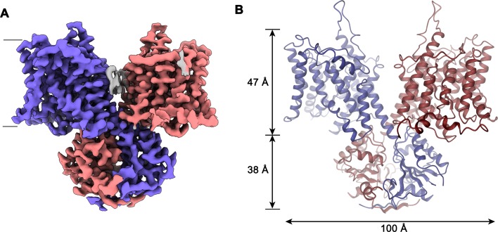
Figure 2.
Slc26a9 T structure.
( A ) Cryo-EM density of Slc26a9 T in the detergent GDN at 3.96 u00c5 contoured at 5u03c3. Density corresponding to distinct subunits in the dimeric protein is colored in blue and red respectively. Re...
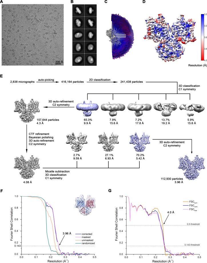
Figure 2u2014figure supplement 1.
Cryo-EM reconstruction of Slc26a9 T in detergent at 3.96 u00c5.
( A ) Representative cryo-EM micrograph acquired with Titan Krios microscope. ( B ) 2D class averages of Slc26a9 T . ( C ) Angular distribution plot of all particles included in the final C2-symmetriz...
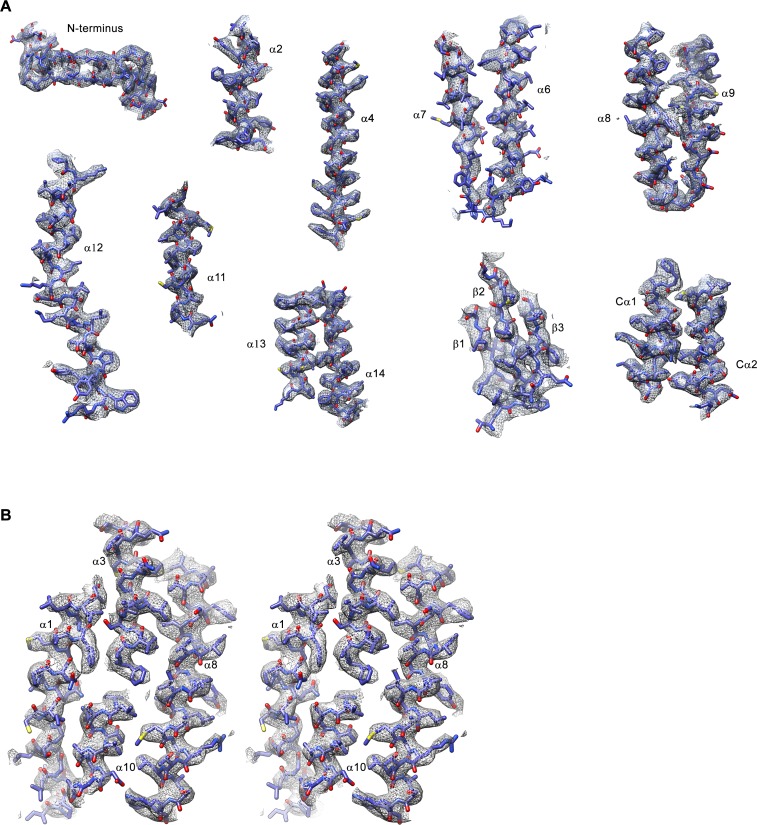
Figure 2u2014figure supplement 2.
Cryo-EM density of the Slc26a9 T structure in detergent.
( A ) Sections of the cryo-EM density (gray, 7u03c3) superimposed on the refined structure of Slc26a9 T . Secondary structure elements are labeled. ( B ) Stereo view of cryo-EM density (7u03c3) of the...
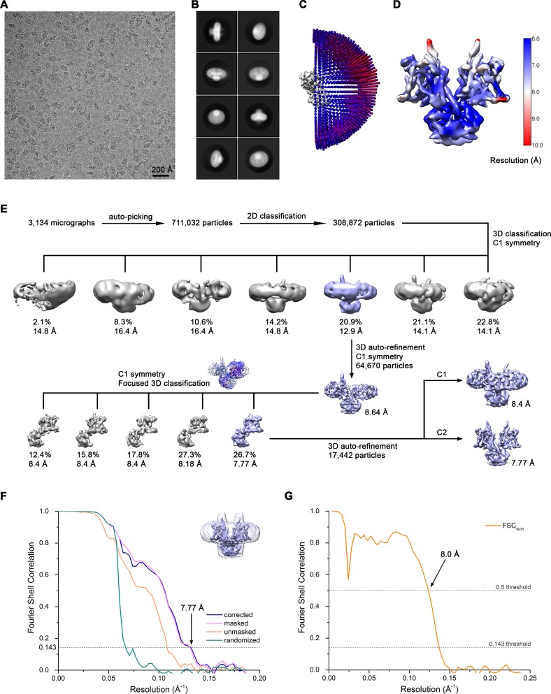
Figure 2u2014figure supplement 3.
Cryo-EM reconstruction of Slc26a9 T in nanodiscs at 7.8 u00c5.
( A ) Representative cryo-EM micrograph acquired with Tecnai G 2 Polara microscope. ( B ) 2D class averages of Slc26a9 T . ( C ) Angular distribution plot of all particles included in the final C2-sym...
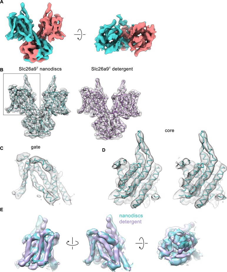
Figure 2u2014figure supplement 4.
Cryo-EM density of the Slc26a9 T structure in nanodiscs.
( A ) Cryo-EM density (6u03c3) of Slc26a9 T in lipid nanodiscs at 7.77 u00c5. Density corresponding to distinct subunits is colored in cyan and salmon respectively. ( B ) Left, Cu03b1-representation o...
Video 1.
Cryo-EM density map of the dimeric Slc26a9 T .
Shown is the cryo-EM map of the protein in detergent with the refined model of the inward-facing state superimposed.
Video 2.
Cryo-EM density map of the dimeric Slc26a9 T in nanodiscs.
Shown is the cryo-EM map of the protein in nanodiscs with the modeled structure of the intermediate state superimposed.
Video 3.
The structure of Slc26a9 T .
Shown are unique features of a mammalian SLC26 transporter. The oligomerization interface is minimal between the transmembrane domains and is predominantly mediated by the swapped STAS domains. The vi...
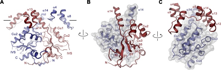
Figure 3.
STAS domain and dimer interface.
( A ) Ribbon representation of the STAS domain dimer and interacting parts of the N-terminus and the TMD. The view is as in Figure 2B . ( B ) Elements of mutual STAS domain interactions and contacts w...
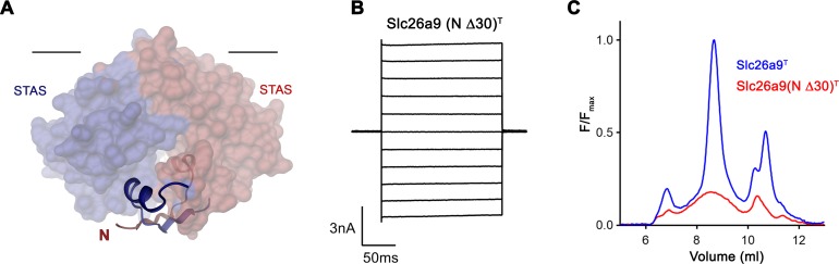
Figure 3u2014figure supplement 1.
Effect of N-terminal truncation of Slc26a9 T on function and stability.
( A ) Depiction of the interaction of residues 5u201325 of both subunits with the STAS domain dimer. The N-termini are displayed as ribbon interacting with the STAS domain depicted by its molecular su...
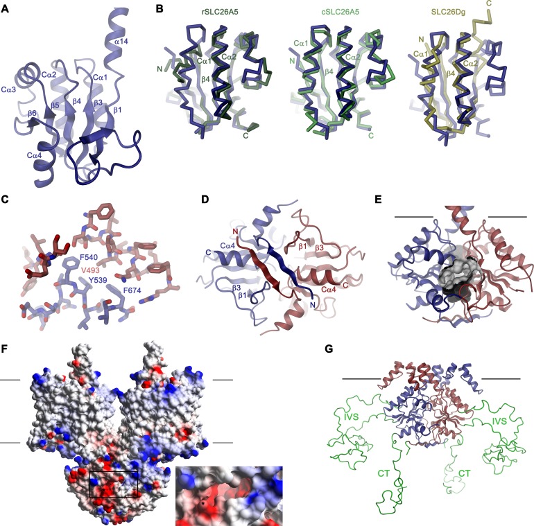
Figure 3u2014figure supplement 2.
Structural features of the Slc26a9 STAS domain.
( A ) Ribbon representation of the STAS domain of a single subunit of Slc26a9 T with secondary structure elements labeled. ( B ) Superposition of the STAS domain of Slc26a9 T with the X-ray structures...
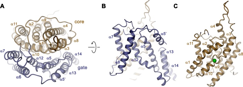
Figure 4.
Transmembrane domain.
( A ) Ribbon representation of the TMD of a single subunit of Slc26a9 T viewed from the extracellular side. ( B ) View of the gate and ( C ) the core module from within the membrane. Green sphere indi...
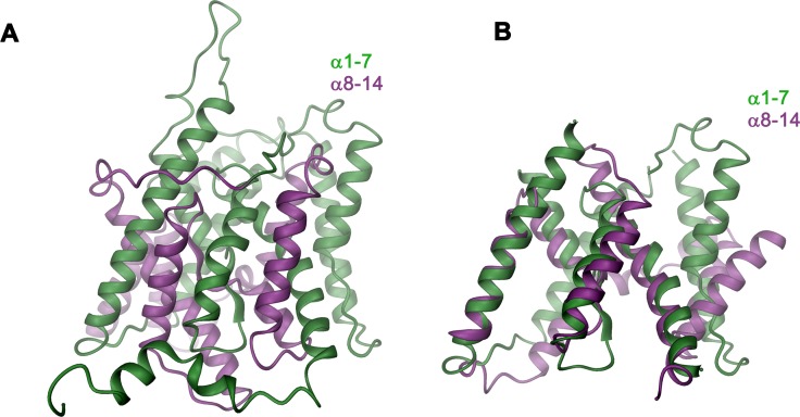
Figure 4u2014figure supplement 1.
Structural features of the TMD.
( A ) Ribbon representation of the TMD of a single subunit of Slc26a9 T viewed from within the membrane. The N-terminal (u03b11-u03b17) and C-terminal (u03b18-u03b114) halves are colored in green and ...
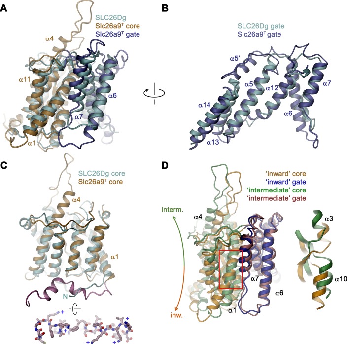
Figure 5.
Comparison of the TMD.
( A ) Structural superposition of the TMDs of Slc26a9 T (beige and blue) and SLC26Dg (PDBID: 5DA0, cyan). ( B ) Superposition of the gate domains and ( C ) core domains of Slc26a9 T and SLC26Dg. N-ter...
Video 4.
Conformational transition from the inward-facing to the intermediate state.
Ribbon representation of the transmembrane domain as a morph between the inward-facing and intermediate states. The structure is viewed from the peripheral. Residues 276 to 312 are removed for clarity...
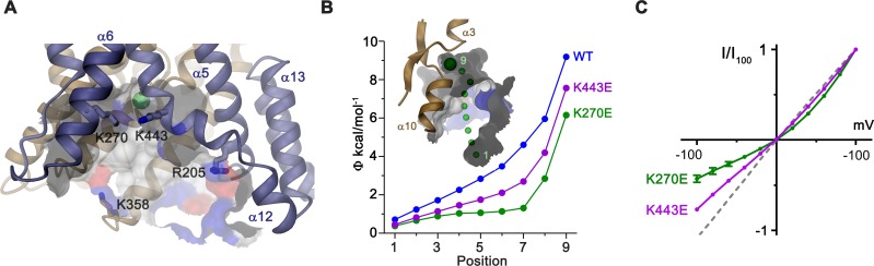
Figure 6.
Electrostatic properties of the intracellular cavity.
( A ) View of the intracellular cavity leading to the Cl u2013 -binding site. Section of the molecular surface is shown with contact region of acidic residues colored in red and basic residues in blue...
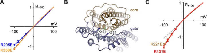
Figure 6u2014figure supplement 1.
Functional properties of mutants of basic residues.
( A ) Introduction of negative charges at positions of basic residues distal to the putative anion-binding site show negligible effects on the linear Iu2013V relation. Shown are Iu2013V relationships ...
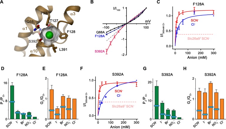
Figure 7.
Substrate binding site.
( A ) Structure of the Cl u2013 binding site.u00a0The molecular surface of the binding pocket is shown. Selected residues are displayed as sticks. Bound Cl u2013 is shown as a green sphere. ( B ) I-V ...
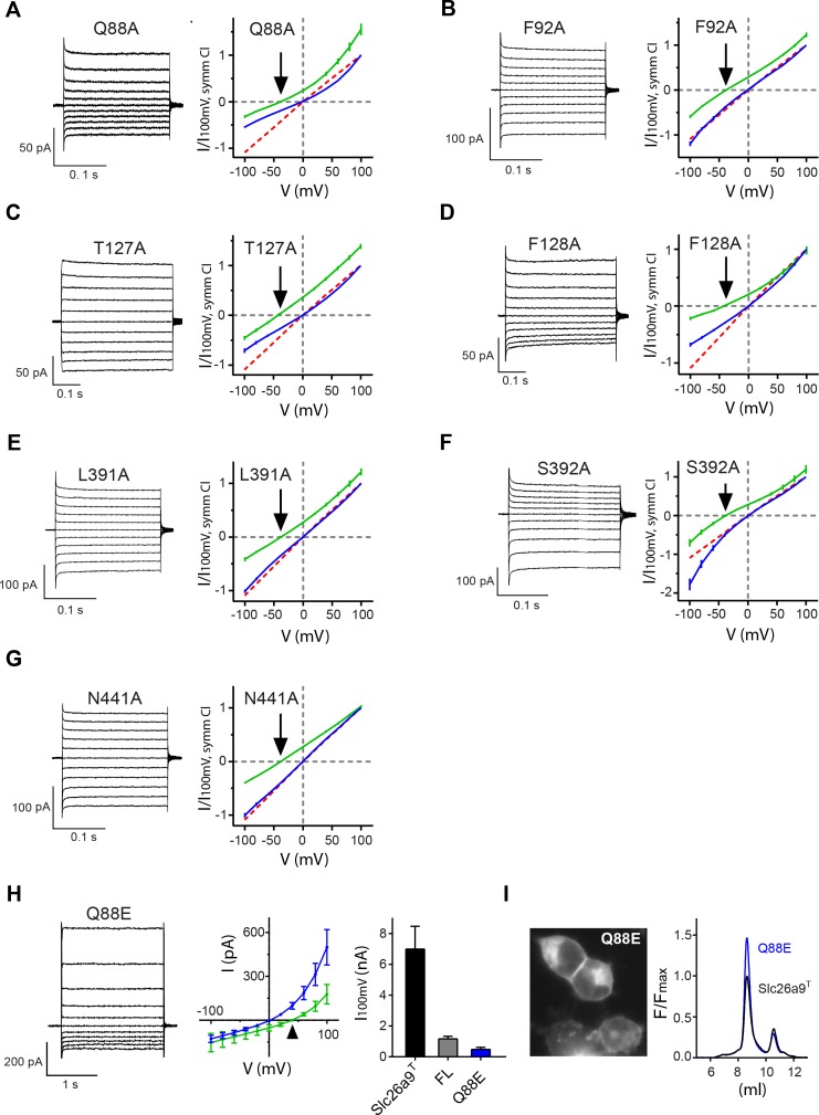
Figure 7u2014figure supplement 1.
I-V-relationships of anion binding site mutants.
( Au2013G ) Alanine mutants investigated by inside-out patch-clamp electrophysiology of HEK392T cells expressing Slc26a9 T anion binding-site mutations. Binding-site mutations do not alter Cl u2013 se...
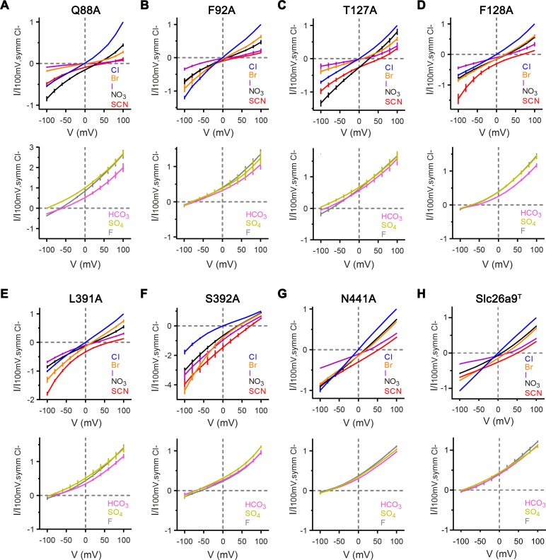
Figure 7u2014figure supplement 2.
Anion selectivity of binding site mutants.
Currents were recorded by inside-out patch-clamp electrophysiology at asymmetric conditions containing 150 mM extracellular Cl u2013 and 150 mM of the indicated anion. ( Au2013G ) Top, Bi-ionic Iu2013...
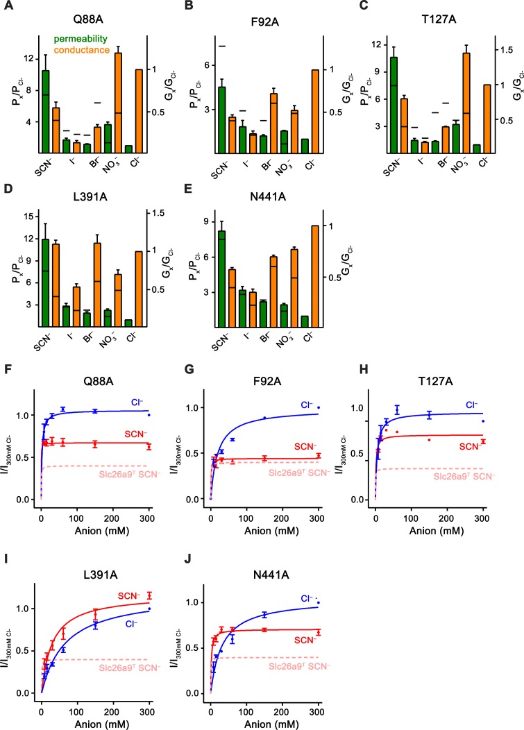
Figure 7u2014figure supplement 3.
Kinetic properties of anion binding site mutants.
Data was recorded from excised patches. ( Au2013E ) Relative permeabilities (P x /P Cl, green) and macroscopic conductivities (G x /G Cl , orange) for ( A ) Q88A, ( B ) F92A, ( C ) T127A, ( D ) L391A ...
Video 5.
Structure of the presumed ion binding site.
Ribbon representation of the anion binding site with side-chains of interacting residues displayed as sticks. Surface of the binding pocket around a modeled chloride (green sphere) is shown. View is a...
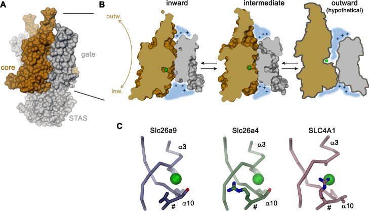
Figure 8.
Transport mechanism.
( A ) Molecular surface of Slc26a9 T viewed towards the long dimension of the molecule. ( B ) Sections of the TMD in the inward-facing conformation defined by the Slc26a9 T detergent structure (left),...
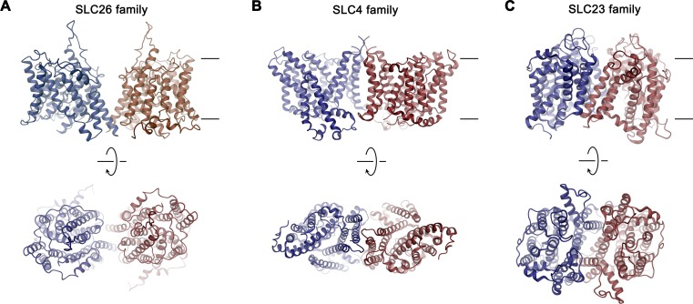
Figure 8u2014figure supplement 1.
Dimer architecture of transport proteins sharing the 7u00a0+u00a07 inverted repeat topology.
The TMDs of selected transporters are shown as representatives for their respective family as ribbon. ( A ), SLC26 family: Slc26a9, ( B ), SLC4 family: SLC4A1/Band 3 (PDBID: 4YZF), ( C ), SLC23 family...
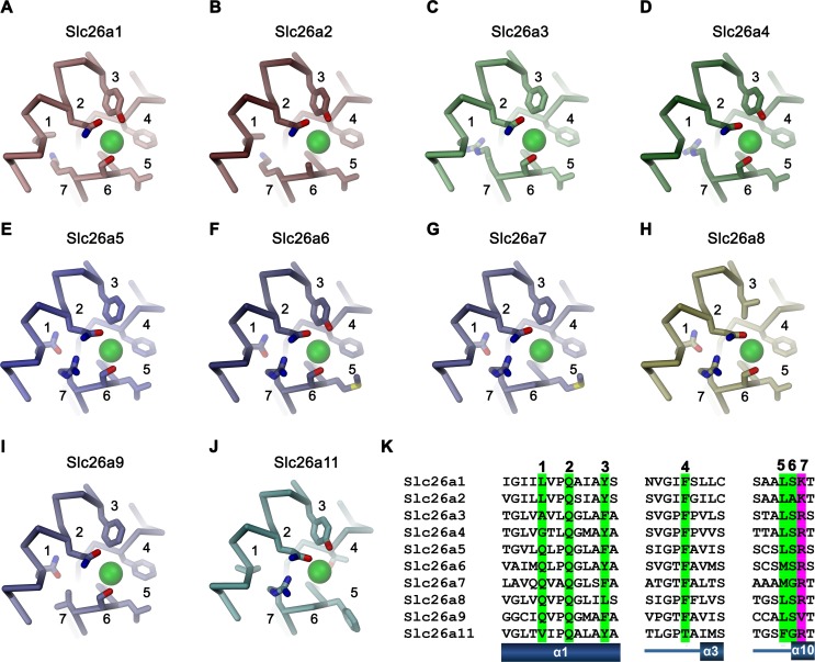
Figure 8u2014figure supplement 2.
Ion binding by SLC26 proteins.
Putative anion binding region of murine Slc26 paralogs are shown as Cu03b1-trace with selected side-chains in interaction distance with the bound anion displayed as sticks. Numbering corresponds to th...
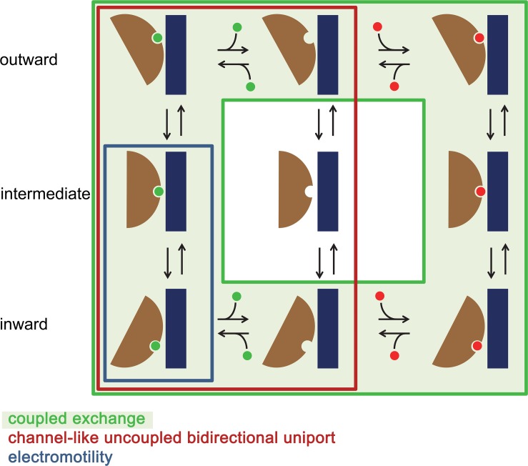
Figure 8u2014figure supplement 3.
Mechanistic relationships within the SLC26 family.
Schematic depiction of plausibleu00a0kinetic cycles underlying different transport modes observed in the SLC26 family. States occupied in a channel-like uncoupled bidirectional uniport as observed in ...
Figure images are served from the NIH/NLM PubMed Central Open Access Subset or Europe PMC; copyright remains with the publishers and authors.
💬 Discussion
0 commentsNo comments yet. Be the first to start a discussion!
Leave a Comment