Abstract
The plasma membrane adenosine triphosphate (ATP) release channel pannexin 1 (PANX1) has been implicated in many physiological and pathophysiological processes associated with purinergic signaling, including cancer progression, apoptotic cell clearance, inflammation, blood pressure regulation, oocyte development, epilepsy and neuropathic pain. Here we present near-atomic-resolution structures of human and frog PANX1 determined by cryo-electron microscopy that revealed a heptameric channel architecture. Compatible with ATP permeation, the transmembrane pore and cytoplasmic vestibule were exceptionally wide. An extracellular tryptophan ring located at the outer pore created a constriction site, potentially functioning as a molecular sieve that restricts the size of permeable substrates. The amino and carboxyl termini, not resolved in the density map, appeared to be structurally dynamic and might contribute to narrowing of the pore during channel gating. In combination with functional characterization, this work elucidates the previously unknown architecture of pannexin channels and establishes a foundation for understanding their unique channel properties.
🔬 Techniques
🧬 Organisms
✨ Fluorophores
🧪 Sample Preparation
🔬 Cell Lines
🏭 Microscope Brands
💾 Data Repositories
🏛️ Research Organizations (ROR)
Affiliated research institutions:
📊 Figures
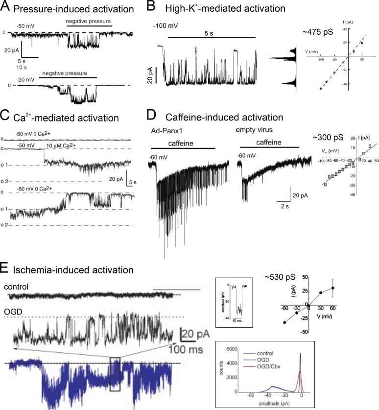
Figure 1.
Panx1 channels of large and linear unitary conductance. (A) Pressure-induced single-channel activity obtained from a Xenopus oocyte heterologously expressing human PANX1 (adapted from Bao et al., 2004...
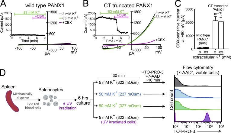
Figure 2.
Raising extracellular K does not activate recombinant or native Panx1 channels. (A and B) Whole-cell currents were obtained from HEK293T cells expressing either wild-type PANX1 (A) or C-terminally tru...
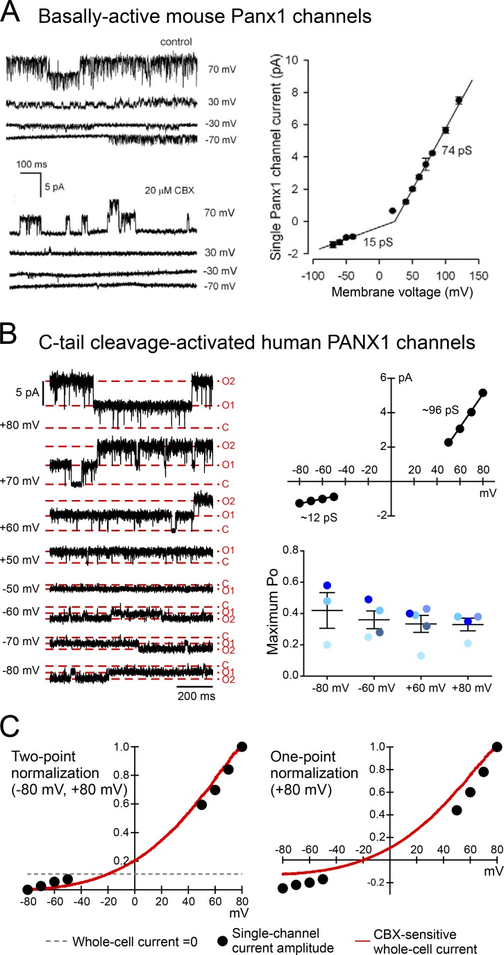
Figure 3.
Panx1 channels of u2264100 pS and outwardly rectifying unitary conductance. (A) Single-channel activity and outwardly rectifying unitary conductance of basally active mouse Panx1 heterologously expres...
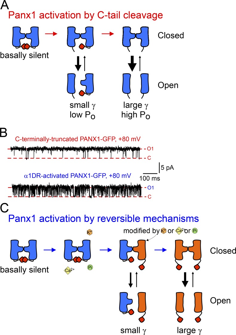
Figure 4.
A general model for Panx1 activation by progressive displacement of autoinhibitory C-tails. (A) Diagram depicts an irreversible activation of Panx1 channels by progressive removal of C-tails. (B) C-ta...
Figure images are served from the NIH/NLM PubMed Central Open Access Subset or Europe PMC; copyright remains with the publishers and authors.
💬 Discussion
0 commentsNo comments yet. Be the first to start a discussion!
Leave a Comment