Abstract
INTRODUCTION: Organ-specific composition of extracellular matrix proteins (ECM) is a determinant of metastatic host organ involvement. The chemokine CXCL12 and its receptor CXCR4 play important roles in the colonization of human breast cancer cells to their metastatic target organs. In this study, we investigated the effects of chemokine stimulation on adhesion and migration of different human breast cancer cell lines in vivo and in vitro with particular focus on the liver as a major metastatic site in breast cancer. METHODS: Time lapse microscopy, in vitro adhesion and migration assays were performed under CXCL12 stimulation. Activation of small GTPases showed chemokine receptor signalling dependence from ECM components. The initial events of hepatic colonisation of MDA-MB-231 and MDA-MB-468 cells were investigated by intravital microscopy of the liver in a rat model and under shRNA inhibition of CXCR4. RESULTS: In vitro, stimulation with CXCL12 induced increased chemotactic cell motility (p<0.05). This effect was dependent on adhesive substrates (type I collagen, fibronectin and laminin) and induced different responses in small GTPases, such as RhoA and Rac-1 activation, and changes in cell morphology. In addition, binding to various ECM components caused redistribution of chemokine receptors at tumour cell surfaces. In vivo, blocking CXCR4 decreased extravasation of highly metastatic MDA-MB-231 cells (p<0.05), but initial cell adhesion within the liver sinusoids was not affected. In contrast, the less metastatic MDA-MB-468 cells showed reduced cell adhesion but similar migration within the hepatic microcirculation. CONCLUSION: Chemokine-induced extravasation of breast cancer cells along specific ECM components appears to be an important regulator but not a rate-limiting factor of their metastatic organ colonization.
🔬 Techniques
✨ Fluorophores
🧪 Sample Preparation
🔬 Cell Lines
🏭 Microscope Brands
🧪 Reagent Suppliers
💻 Software Details
🏛️ Research Organizations (ROR)
Affiliated research institutions:
📋 Methods
Time lapse microscopy, in vitro adhesion and migration assays were performed under CXCL12 stimulation. Activation of small GTPases showed chemokine receptor signalling dependence from ECM components. The initial events of hepatic colonisation of MDA-MB-231 and MDA-MB-468 cells were investigated by intravital microscopy of the liver in a rat model and under shRNA inhibition of CXCR4.
Materials and Methods Reagents Phycoerythrin (PE)-conjugated anti-human CXCR4 antibodies were obtained from R&D Systems (Wiesbaden, Germany). Unlabelled, function-blocking anti-human CXCR4 antibody was a kind gift from A. Müller, Düsseldorf, Germany. Anti-human RhoA antibody was purchased from Santa Cruz (Santa Cruz, CA, USA), anti-human Rac1 from BD Pharmingen (San Jose, CA, USA) and anti-human Cdc42 antibody was obtained from Cell Signaling (Danvers, MA, USA). For integrin subunits the following antibodies were used: β1, β4, α2, α3 (all from Chemicon, Hofheim, Germany), α1 (upstate biotechnology, Hamburg, Germany), α5 (Serotec, Eching, Germany) and α6 (gift from J. Eble, Münster, Germany). Alexa Fluor 488 and 546 labelled secondary antibodies, phalloidin, CalceinAM and Hoechst 33342 were purchased from Molecular Probes/Invitrogen (Karlsruhe, Germany).
Human recombinant chemokine
SDF1α/CXCL12 was obtained from R&D systems (Wiesbaden, Germany). Glutathione-Agarose Rac/Cdc41 PAK-1 PBD beads and Rho Rhotekin RBD beads were obtained from Upstate/Millipore (Eching, Germany). ECM components C I, FN and LN were purchased from Sigma-Aldrich (Saint Louis, Missouri, USA). All other chemicals were purchased from Sigma or Roth (Karlsruhe, Germany).
Show full methods section
Time lapse microscopy, in vitro adhesion and migration assays were performed under CXCL12 stimulation. Activation of small GTPases showed chemokine receptor signalling dependence from ECM components. The initial events of hepatic colonisation of MDA-MB-231 and MDA-MB-468 cells were investigated by intravital microscopy of the liver in a rat model and under shRNA inhibition of CXCR4.
Materials and Methods Reagents Phycoerythrin (PE)-conjugated anti-human CXCR4 antibodies were obtained from R&D Systems (Wiesbaden, Germany). Unlabelled, function-blocking anti-human CXCR4 antibody was a kind gift from A. Müller, Düsseldorf, Germany. Anti-human RhoA antibody was purchased from Santa Cruz (Santa Cruz, CA, USA), anti-human Rac1 from BD Pharmingen (San Jose, CA, USA) and anti-human Cdc42 antibody was obtained from Cell Signaling (Danvers, MA, USA). For integrin subunits the following antibodies were used: β1, β4, α2, α3 (all from Chemicon, Hofheim, Germany), α1 (upstate biotechnology, Hamburg, Germany), α5 (Serotec, Eching, Germany) and α6 (gift from J. Eble, Münster, Germany). Alexa Fluor 488 and 546 labelled secondary antibodies, phalloidin, CalceinAM and Hoechst 33342 were purchased from Molecular Probes/Invitrogen (Karlsruhe, Germany).
Human recombinant chemokine
SDF1α/CXCL12 was obtained from R&D systems (Wiesbaden, Germany). Glutathione-Agarose Rac/Cdc41 PAK-1 PBD beads and Rho Rhotekin RBD beads were obtained from Upstate/Millipore (Eching, Germany). ECM components C I, FN and LN were purchased from Sigma-Aldrich (Saint Louis, Missouri, USA). All other chemicals were purchased from Sigma or Roth (Karlsruhe, Germany).
Cell lines and culture conditions
Subclones of the MDA-MB-231 cells (originally obtained from the ATCC) were provided by M. Kochetkowa, Adelaide, Australia. These cells were originally derived from a 51 years old Caucasian female and are invasive and tumorigenic in nude mice. MDA-MB-468 cell line was purchased from ATCC (Manassas, VA) and was originally derived from a pleural effusion of a 51-year old black female patient with metastatic adenocarcinoma of the breast. MDA-MB-231 cell line was maintained in RPMI 1640 or DMEM medium (Gibco/Invitrogen, Karlsruhe, Germany) containing 10% foetal bovine serum (FBS, Gibco) without antibiotics. Two clones with shRNA-mediated CXCR4 reduction were also provided by M. Kochetkowa. These clones (MDA-MB-231-19 and MDA-MB-231-27) were obtained using CXCR4 shRNA-expressing constructs 5-gatctGGTGGTCTATGTTGGCGTCTGttcaagaGACAGACG CCAACATAGACCACCtttttta-3 and 5-agcttaaaaaaGGTGGTCTATGTTGGCGTCTG tctcttgaacagacgccaacatagaccacca-3 (21-nucleotide CXCR4 at position 470–490 of human CXCR4 cDNA). [21] , [27] MDA-MB-468 cells were cultured in DMEM medium (Lonza, Verviers, Belgium) containing 10% FBS and L-glutamine (Gibco/Invitrogen, Karlsruhe, Germany) without antibiotics. The cells were starved overnight in serum-free media before their application in experiments. After trypsinization, the cells were resuspended in serum-free adhesion medium (containing 1% bovine serum albumin) for reconstitution of surface proteins prior to experimentation.
Transwell assay
The breast cancer cells were added to FN, LN or C I-coated transwell inserts with 8 µm-pore-size (Nunc/Thermo Fisher Scientific, Rockford, IL, USA). The cells were suspended into the upper chamber at a final concentration of 0,7×10 5 cells/ml in 500 µl adhesion medium. After 60 min adhesion time, CXCL12 in concentrations 25, 50 or 100 ng/ml were added to the lower chamber. Unstimulated cells served as negative control. For diffuse stimulation CXCL12 at equal concentrations was added in the lower and upper chamber. After 4 or 16 h of incubation, the cells on the upper surface of the filter were removed by wiping with Q-tips, and the migrated cells on the lower side were fixed with formalin and stained with crystal violet and haematoxylin. Cellular transmigration was enumerated in 16 standardized microscopic fields per membrane. To test the specific chemotactic response of the cells to CXCL12 the CXCR4 receptor was blocked using a neutralizing mouse-anti-human CXCR4 antibody (kind gift of A. Müller, Düsseldorf, Germany). The antibody was added to the upper chamber at concentrations of 5 and 10 µg/ml and migration of the cells was observed for 4 or 16 h.
Static adhesion assays
Microtiter plates (96 wells, Greiner BioOne, Frickenhausen, Germany) were coated with C I (10 µg/ml), FN (10 µg/ml), LN (10 µg/ml) or 1% BSA (negative control). Blocking of nonspecific binding sites was performed with 1% BSA for 30 min. After reconstitution of cell surface proteins in adhesion medium for 45 min, cells were resuspended in adhesion medium at a final concentration of 1×10 6 cells/ml and seeded to the coated wells. The cells were stimulated with CXCL12 with concentrations of 25, 50 and 100 ng/ml. After 30 or 60 min adhesion time cells were washed, fixed with formalin and then stained with crystal violet for 15 min. The absorbance was measured at 630nm using a spectrophotometer. All experiments were performed in triplicates and repeated at least three times.
Time lapse microscopy
The cells were added to ECM-coated culture dishes for 20 min. After washing adhesion medium containing CXCL12 at different concentrations was added. The cells were observed for 60 min under a time lapse video microscope (Nikon, Düsseldorf, Germany). For quantification of cell motility cell tracking was done using software packages Cell∧D.
Intravital microscopy
Intravital microscopy was performed as previously described [6] , [7] . The adequacy and reliability of this model in the investigation of early interactions between circulating tumour cells and the hepatic microcirculation was confirmed previously [28] . Briefly, Sprague Dawley rats (200 to 250 g) (Charles River, Sulzfeld, Germany) were cared for in accordance with standards of the German Council on Animal Care, under an approved protocol of the local animal welfare committee (Landesamt für Naturschutz, Umweltschutz und Veterinärmedizin: LANUV G84/2002). Rats were anesthetized using inhalation of isofluorane (Curamed, Karlsruhe, Germany). Permanent catheters were introduced into the left heart via the carotid artery and the right heart via the jugular vein. After a wide median laparotomy was performed, the left liver lobe was careful mobilized without disturbing hepatic microcirculation. Using a heated operating table, animals were fixed under an upright microscope and positioned on their left side. This positioning allowed a partial luxation of the mobilized left liver lobe that was placed on a specific holder to investigate its lower surface. During the experiments the liver was continuously irrigated with isotonic saline solution. An upright epifluorescence microscope (Zeiss, Oberkochen, Germany) was used with a 20-fold objective that was located over a glass slip covering the organ surfaces. The microscope was connected with a video enhancer-zoom lens system and a low-light charge-coupled device video camera (Peiper, Düsseldorf, Germany) allowing real-time imaging via a separate monitor. Fluorescence images were recorded using timer-containing S-VHS video system for further analysis. In Vivo Observation of Metastatic Tumour Cell Adhesion and Extravasation For intravital observation of adhesive interactions between circulating tumour cells and the host organ microcirculation, single cell suspensions of CalceinAM fluorescence-labelled tumour cells (1×10 6 ) were injected intra-arterially within 60 sec. Previously [6] , [7] , we have shown that the route of cell application (left heart, right heart, portal vein) did not influence the adhesive or migratory behaviour within the liver sinusoids. This technique did not interfere with cardio-circulatory or pulmonary functions of the animals. Off-line analysis was used to determine tumour cell behaviour within the target organs as previously described [24] , [29] . Various parameters were used for further investigation and semiquantitative analysis of these interactions. A semiquantitative analysis of tumour cell adhesion and extravasation was performed throughout a 30 min observation period, and the numbers of adherent cells were counted for each of the 5 min intervals. Using a standardized procedure, all fields were analysed in each observation period and average numbers of adherent cells, migrated cells, and total cells observed were counted. The numbers provided represent the total numbers of cells within 30 microscopic fields for each 5 min period. Numbers of arrested cells represent the total of adherent and extravasated cells. Relative migration rates were calculated as percentages of cells within the host organ parenchyma in relation to the numbers of arrested cells.
GTPase activation assay
Non-adherent cells (10 7 ) in suspension unstimulated or stimulated with CXCL12 (25, 50, 100 ng/ml) for 15 min were washed once with ice cold PBS and lysed with 50mM Tris-HCl pH 7.4, 150 mM NaCl, 1% NP40, 0.5% deoxycholate, 0.1% SDS, 5mM EDTA and 1 µl inhibitor cocktail (Sigma) per 1 ml lysis puffer. Alternatively, adherent cells were seeded at different ECM proteins (C I, FN and LN) for 60 min to exclude interference with initial adhesive behaviour and subsequently stimulated with CXCL12 in a similar manner. As controls poly-l-lysine (PLL for non-integrin mediated adhesion) and BSA (negative control) were used. Lysates were clarified by centrifugation at 14,000xg for 5 min and stored at −80°C. After protein quantitation and standardization 8 µl Rho Assay Reagent (Rhotekin RBD glutathione agarose beads) or 10 µl Rac/Cdc42 assay reagent (PAK-1 PBD agarose conjugate) were added to 1000 µg total protein and incubated for 45 min at 4°C. Samples were washed three times with magnesium-containing lysis puffer (25 mM HEPES pH 7.5, 150 mM NaCl, 1% Igepal CA-630, 10% Glycerol, 10mM MgCl 2 , 1mM EDTA) and 1 µl inhibitor cocktail per 1 ml lysis puffer. Agarose beads were resuspended in 4x Laemmli sample puffer and boiled for 5min. Total Rho, Rac-1 or CDC42, respectively, served as loading control in each of the experiments. The lysates were loaded on 12% polyacrylamide gels, then transferred to PVDF membranes and GTPases finally detected with rabbit anti-RhoA antibody (Santa Cruz), mouse anti-Rac antibody (BD Biosciences Pharmingen) or rabbit anti-Cdc42 antibody (Cell Signalling). Bands were visualized with enhanced chemiluminescence (Millipore, Schwalbach, Germany). Quantitative densitometry analysis was performed using ImageJ densitometry software (version 1.6, NIH, Bethesda, MD) and selected bands were semi-quantified based on their optical densities.
Immunofluorescence staining
The cells were added to C I, FN, LN or Poly(L)lysine (PLL)-coated chamber slides, incubated for 60 min and then stimulated using different chemokine concentrations (25, 50 and 100 ng/ml) for different time intervals (5, 15 and 30 min). Subsequently, cells were washed and fixed using 4% paraformaldehyde. For intracellular staining, the cells were blocked and permeabilized using PBS containing 1% BSA and 0, 1% Triton X100, whereas permeabilization was not performed to achieve cell surface staining. Cells were incubated for 30 min with anti-human CXCR4 antibodies and subsequently with Alexa Fluor conjugated secondary antibody for additionally 30 min. For actin filament staining phalloidin Alexa Fluor 488 and for nuclear staining Hoechst 33342 was used. Imaging was performed as combination of 3D-fluorescence reconstruction and phase contrast microscopy using a Nikon Eclipse TE2000 microscope. For each substrate 20 representative CXCR4 clusters were evaluated regarding their localization at the cell, their size and shape. The size of CXCR4 clusters was calculated using software packages Cell∧D (Olympus, Münster, Germany).
Flow Cytometry
Cells were fixed with 4% paraformaldehyde and then washed and resuspended in PBS containing 0.5% BSA. After this the cells were incubated for 45 min with PE-conjugated anti chemokine antibodies (R&D systems). After washing the integrin or chemokine receptor surface expression was measured using a FC500 flow cytometer (Beckman Coulter, Krefeld, Germany). After gating of the cell population, the mean fluorescence intensities (MFI) of the antibody-stained cells were detected and the relative amounts of positive cells were calculated using the flow cytometer software.
Statistical analysis
Statistical analysis was performed using the SPSS V.14 (SPSS Inc., Chicago, IL) statistical program. Data were shown as mean±SD. For comparison of different parameters between the treatment groups p-values were calculated according to the Scheffé-test (ANOVA post-hoc-test) for dependent or independent samples as appropriate. For other analyses Student's t-test has been used. Significant differences were accepted for p
📊 Figures
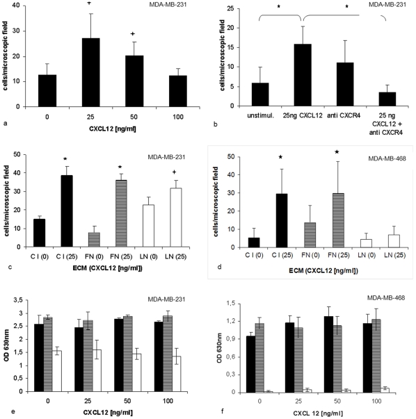
Figure 1
CXCL12 stimulated cell adhesion and migration.
Transwell migration assays were performed for 16 h using different gradients of CXCL12 (0u2013100 ng/ml). (a) Bell-shaped response is shown for MDA-MB-231 cells at C I. (b) This response was specific ...
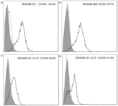
Figure 2
Flow cytometry analysis of breast cancer cells.
The cell surface expression of CXCR4 (u2043) was found in different breast cancer cell lines MDA-MB-231 (a) and MDA-MB-468 (b). Downregulation of CXCR4 expression in clones MDA-MB-231 Cl 19 (c) and MD...
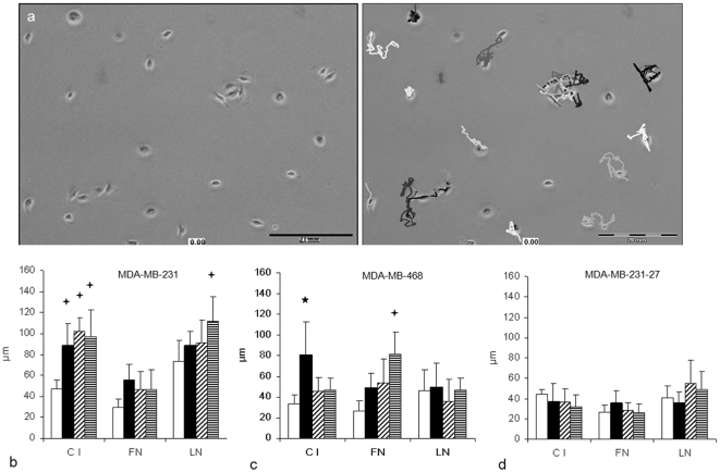
Figure 3
CXCL12 increases cell motility dependent from the ECM.
(a) Cells that were attached to different ECM components were observed for one hour using time-lapse video microscopy. The length of their moving path was determined using a cell tracking software (Ce...
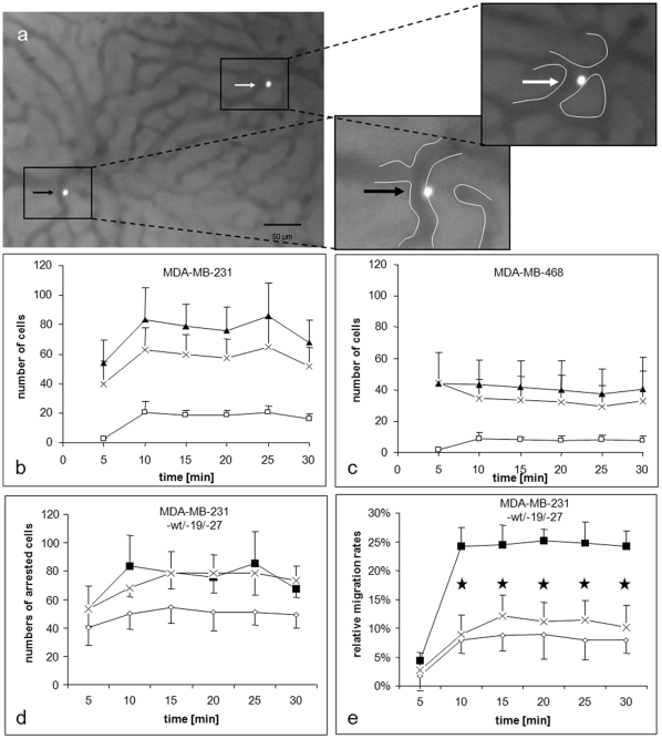
Figure 4
In vivo migration of breast cancer cells within the liver sinusoids.
Single cell suspensions of fluorescence-labelled tumour cells were injected into Spague-Dawley rats. (a) Example of an adherent cells (white arrow) and a cell starting to migrate (black arrow) into th...
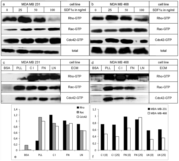
Figure 5
CXCL12 and ECM-induced activation of small Rho GTPases.
Cells were treated with different concentrations of CXCL12 in single cell suspensions (a+b) or plated at various adhesive substrates (c+d). Level of activation of RhoA, Rac1 and Cdc42 assessed by pull...
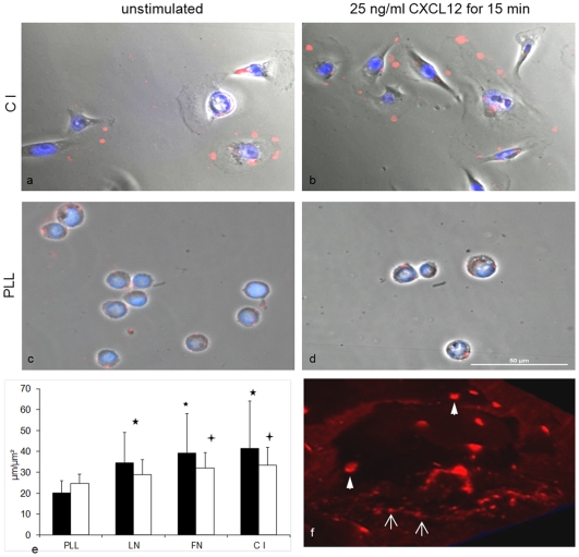
Figure 6
Clustering of CXCR4 at tumour cell surfaces.
CXCR4 was visualized in wild-type MDA-MB-231 cells using fluorescence-labelled antibodies (red). Nuclei were counterstained (blue). Scale 50 u00b5m. Figures were taken 15 min after CXCL12 stimulation ...
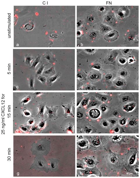
Figure 7
Kinetics of CXCR4 cell surface expression.
MDA-MB-231 cells were seeded at C I (left column) or FN (right column) and stimulated with CXCL12 for up to 30 min. Fixed cells without stimulation (a+b) or with CXCL12 stimulation (25 ng/ml) for 5 mi...
Figure images are served from the NIH/NLM PubMed Central Open Access Subset or Europe PMC; copyright remains with the publishers and authors.
💬 Discussion
0 commentsNo comments yet. Be the first to start a discussion!
Leave a Comment