Abstract
We recently reported that Inosine Monophosphate Dehydrogenase (IMPDH), a rate-limiting enzyme in de novo guanine nucleotide biosynthesis, clustered into macrostructures in response to decreased nucleotide levels and that there were differences between the IMPDH isoforms, IMPDH1 and IMPDH2. We hypothesised that the Bateman domains, which are present in both isoforms and serve as energy-sensing/allosteric modules in unrelated proteins, would contribute to isoform-specific differences and that mutations situated in and around this domain in IMPDH1 which give rise to retinitis pigmentosa (RP) would compromise regulation. We employed immuno-electron microscopy to investigate the ultrastructure of IMPDH macrostructures and live-cell imaging to follow clustering of an IMPDH2-GFP chimera in real-time. Using a series of IMPDH1/IMPDH2 chimera we demonstrated that the propensity to cluster was conferred by the N-terminal 244 amino acids, which includes the Bateman domain. A protease protection assay suggested isoform-specific purine nucleotide binding characteristics, with ATP protecting IMPDH1 and AMP protecting IMPDH2, via a mechanism involving conformational changes upon nucleotide binding to the Bateman domain without affecting IMPDH catalytic activity. ATP binding to IMPDH1 was confirmed in a nucleotide binding assay. The RP-causing mutation, R224P, abolished ATP binding and nucleotide protection and this correlated with an altered propensity to cluster. Collectively these data demonstrate that (i) the isoforms are differentially regulated by AMP and ATP by a mechanism involving the Bateman domain, (ii) communication occurs between the Bateman and catalytic domains and (iii) the RP-causing mutations compromise such regulation. These findings support the idea that the IMPDH isoforms are subject to distinct regulation and that regulatory defects contribute to human disease.
🔬 Techniques
🔭 Microscopes
🧬 Organisms
💻 Software
✨ Fluorophores
🧪 Sample Preparation
🔬 Cell Lines
🏭 Microscope Brands
🧪 Reagent Suppliers
🔎 Objectives
💻 Software Details
🏛️ Research Organizations (ROR)
Affiliated research institutions:
📋 Methods
Reagents and Materials
Reagents were from Sigma–Aldrich (Castle Hill, NSW, Australia) unless otherwise stated. Tissue culture reagents and foetal bovine serum were from Invitrogen (Mount Waverley, VIC, Australia) and Bovogen Biologicals (Essendon, VIC, Australia) respectively. The anti-panIMPDH antibody was a kind gift from Frank Collart [52] , the anti-HA antibody was from Covance (Berkley, CA, USA), the anti-tubulin antibody from Abcam (Cambridge, UK), the anti-GFP antibody [53] and isoform-specific IMPDH antibodies [9] were generated as previously described. All secondary antibodies were from Molecular Probes (Eugene, OR, USA).
Generation of IMPDH constructs
To yield pmEGFP-C1 HA-IMPDH2-GFP, HA-IMPDH2 cDNA [35] was amplified by PCR with forward 5′-GGTGGTGCTAGCGCCACCATGTACCCATACGATGTGCCAGATTACGCT-3′ and reverse 5′- GGTGGCGACCGGTCCACCAGAACCACCTGCACCAGATGCACCTGTTCCGAAAAGCCGCTTCTCATACG-3′ primers, which was inserted into pmEGFP-C1 (Clontech, Mountain View, CA, USA) on NheI/AgeI sites. IMPDH chimeras and the truncated core domain constructs were cloned with an N-terminal HA-tag using a three-step PCR method (see Table S1 for primers) employed by Nimmesgern et al. , (1999) [17] . Firstly, two distinct PCR products, A and B, were generated using template and primer pairs detailed in Table S1 . In the second round, PCR products A and B became the template for amplification with the forward primer of PCR A and reverse primer of PCR B resulting in a chimeric full-length product. This amplicon was shuttled into Blunt II TOPO (Invitrogen) prior to subcloning on HindIII/NotI site into pcDNA5/FRT/TO (Invitrogen). A hexa-histidine (His)-tag was inserted at the 5′ end of human IMPDH1 or IMPDH2 cDNA [35] by PCR prior to cloning into pET20b (+) (Novagen, Madison, WI, USA). QuikChange site-directed mutagenesis kit (Stragene, La Jolla, CA, USA) was used to introduce point mutations, CGC to CCC (R224P) and GAC to AAC (D226N).
Show full methods section
Reagents and Materials
Reagents were from Sigma–Aldrich (Castle Hill, NSW, Australia) unless otherwise stated. Tissue culture reagents and foetal bovine serum were from Invitrogen (Mount Waverley, VIC, Australia) and Bovogen Biologicals (Essendon, VIC, Australia) respectively. The anti-panIMPDH antibody was a kind gift from Frank Collart [52] , the anti-HA antibody was from Covance (Berkley, CA, USA), the anti-tubulin antibody from Abcam (Cambridge, UK), the anti-GFP antibody [53] and isoform-specific IMPDH antibodies [9] were generated as previously described. All secondary antibodies were from Molecular Probes (Eugene, OR, USA).
Generation of IMPDH constructs
To yield pmEGFP-C1 HA-IMPDH2-GFP, HA-IMPDH2 cDNA [35] was amplified by PCR with forward 5′-GGTGGTGCTAGCGCCACCATGTACCCATACGATGTGCCAGATTACGCT-3′ and reverse 5′- GGTGGCGACCGGTCCACCAGAACCACCTGCACCAGATGCACCTGTTCCGAAAAGCCGCTTCTCATACG-3′ primers, which was inserted into pmEGFP-C1 (Clontech, Mountain View, CA, USA) on NheI/AgeI sites. IMPDH chimeras and the truncated core domain constructs were cloned with an N-terminal HA-tag using a three-step PCR method (see Table S1 for primers) employed by Nimmesgern et al. , (1999) [17] . Firstly, two distinct PCR products, A and B, were generated using template and primer pairs detailed in Table S1 . In the second round, PCR products A and B became the template for amplification with the forward primer of PCR A and reverse primer of PCR B resulting in a chimeric full-length product. This amplicon was shuttled into Blunt II TOPO (Invitrogen) prior to subcloning on HindIII/NotI site into pcDNA5/FRT/TO (Invitrogen). A hexa-histidine (His)-tag was inserted at the 5′ end of human IMPDH1 or IMPDH2 cDNA [35] by PCR prior to cloning into pET20b (+) (Novagen, Madison, WI, USA). QuikChange site-directed mutagenesis kit (Stragene, La Jolla, CA, USA) was used to introduce point mutations, CGC to CCC (R224P) and GAC to AAC (D226N).
Cell Culture Chinese Hamster Ovary
(CHO) cells and HeLa cells were cultured in complete F12 HAMs and Dulbecco's Modified Eagle's medium respectively, supplemented with 10% FBS and 2 mM L-glutamine. Cells were transfected and treated as previously described [9] . HeLa cells stably expressing HA-IMPDH2-GFP were initially selected, and subsequently maintained, with geneticin (600 µg/ml) added to the media 24 h post transfection with the pmEGFP-C1 HA-IMPDH2-GFP plasmid. A population of cells with a low fluorescence, due to low expression of HA-IMPDH2-GFP, were further selected by fluorescence activated cell sorted analysis. The resulting heterogeneous stable population of cells expressed HA-IMPDH2-GFP at approximately 10% of the level of endogenous IMPDH. Cells were treated with either vehicle, 2 µM MPA for either 4 h or as indicated or 2 µM MPA for 4 h and supplemented with 100 µM guanosine for the final 2 h.
Immunofluorescence and time-lapse videomicroscopy
Indirect immunofluorescence microscopy was performed as previously described [9] with cells being imaged on a LSM510 META confocal microscope at 100× magnification (Carl Zeiss MicroImaging, Jena, Germany). For 3D time-lapse (4D) videomicroscopy, HeLa HA-IMPDH2-GFP cells were cultured on 24 mm glass bottomed dishes (Proscitech, Qld, Australia). Cells were washed with PBS and complete F12 HAMS prior to replacing with complete F12 HAMS containing the inhibitors. Cells were then imaged, on a pre-heated (37°C) stage top insert with 5% humidified CO 2 circulating, through a C-Apochromat 40×/1.20 W Korr UV-VIS-IR M27 objective using 4–6% 488 nm laser intensity on a LSM510 META confocal microscope (Zeiss). Confocal Z-series (0.9–1.1 µm increments) were acquired over time using the LSM software (Zeiss) and collected images were processed, analysed and movies constructed using Image J v1.41 software (NIH). All images and movies are maximum intensity projections of the 3D image. Electron microscopy and correlative light and electron microscopy CHO cells were fixed with 0.1% glutaraldehyde/4% PFA and processed for EM as previously described [54] . Sections were labelled with antibodies to IMPDH followed by 10 nm protein A-gold. For cryofixation and correlative light and electron microscopy, HeLa cells selected for stable low expression of HA-IMPDH2-GFP were treated with MPA and then high pressure frozen, freeze substituted and embedded in HM20 resin at low temperature as described previously [55] with modifications to the Lowicryl HM20 infiltration which was shortened to one day (50%, 75% and 100% for 1 h each followed by two 12 h 100% infiltrations all at −50°C). Sections were labelled with antibodies to GFP followed by 10 nm protein A-gold. In silico analysis UCSF Chimera (version 1.3; [56] ) was used to coordinate superimposition of protein data bank files for human IMPDH2 (1B3O; [24] ) and IMPDH1 (1JCN; [25] ), utilising the default parameters of the matchmaker function, and for the structure visualisation.
Protein purification
Escherichia coli BL21 (DE3) transformed with pET20b constructs were induced at room temperature (RT) for 12–14 h by addition of isopropyl-beta-D-thiogalactopyranoside (1 mM). Cell pellets were resuspended in either binding buffer 1 (50 mM Tris pH 8.0, 100 mM KCl, 30 mM imidazole, 1.5 M urea, 10 mM 2-mercaptoethanol) for IMPDH1 proteins and the core protein or binding buffer 2 (50 mM Tris pH 6.8, 500 mM KCl, 30 mM imidazole) for IMPDH2, containing protease inhibitors (1 µg/ml leupeptin, 1 µg/ml pepstatin, 1 µg/ml antipain, 250 µM benzamidine and 3 mM AEBSF). Lysates were sonicated, centrifuged at 17000× g for 30 min at 4°C and His-IMPDH proteins purified on a talon affinity resin (Clontech) or nickel-nitriloacetic acid column (Invitrogen) according to the manufacturer's instructions. Protein was eluted with appropriate binding buffer containing 250 mM imidazole and dialysed into activity assay buffer (100 mM KCl, 100 mM Tris-HCl pH 8.0, 1 mM DTT) with glycerol added to a final concentration of 20% for storage. Protease protection assay Purified His-IMPDH protein (0.9 µM) was incubated for 20 min at RT with 1 mM nucleotides or 1 mM MPA with 1 mM IMP and NAD in activity assay buffer prior to addition of 20 µg/ml elastase for a further 20 min. Reactions were ceased by addition of Laemmli SDS-PAGE sample buffer and heat denaturation, then analysed by SDS-PAGE. Protein bands were visualised with coomassie staining and the full-length (intact) protein quantitated using the LI-COR Odyssey Infrared Imaging System. Percentage protection was calculated using the following formula: % protection = ((full-length protein remaining after digestion in sample/undigested protein) – (full-length protein remaining with elastase only digestion/undigested protein))×100%. [ 32 P] ATP filter binding assay The ATP binding assay was based on those previously described [22] , [23] , [57] . In brief, purified His-IMPDH protein (0.9 µM), or BSA (used as a negative control), was mixed with 1 µM [α- 32 P] ATP (800 Ci/mmol; Perkin Elmer, Waltham, MA, USA) and cold ATP (5 µM or 100 µM) for 20 min at RT in binding buffer (50 µl total reaction −100 mM KCl, 100 mM Tris-HCl pH 8.0, 1 mM DTT and 2.5 µM BSA) in duplicate. Reactions were terminated by rapid filtration, loading 15 µl onto 3× pre-equilibrated (in binding buffer) MF filter membrane discs (Millipore, Billerica, MA, USA) under vacuum. Filters were washed rapidly (
📊 Figures
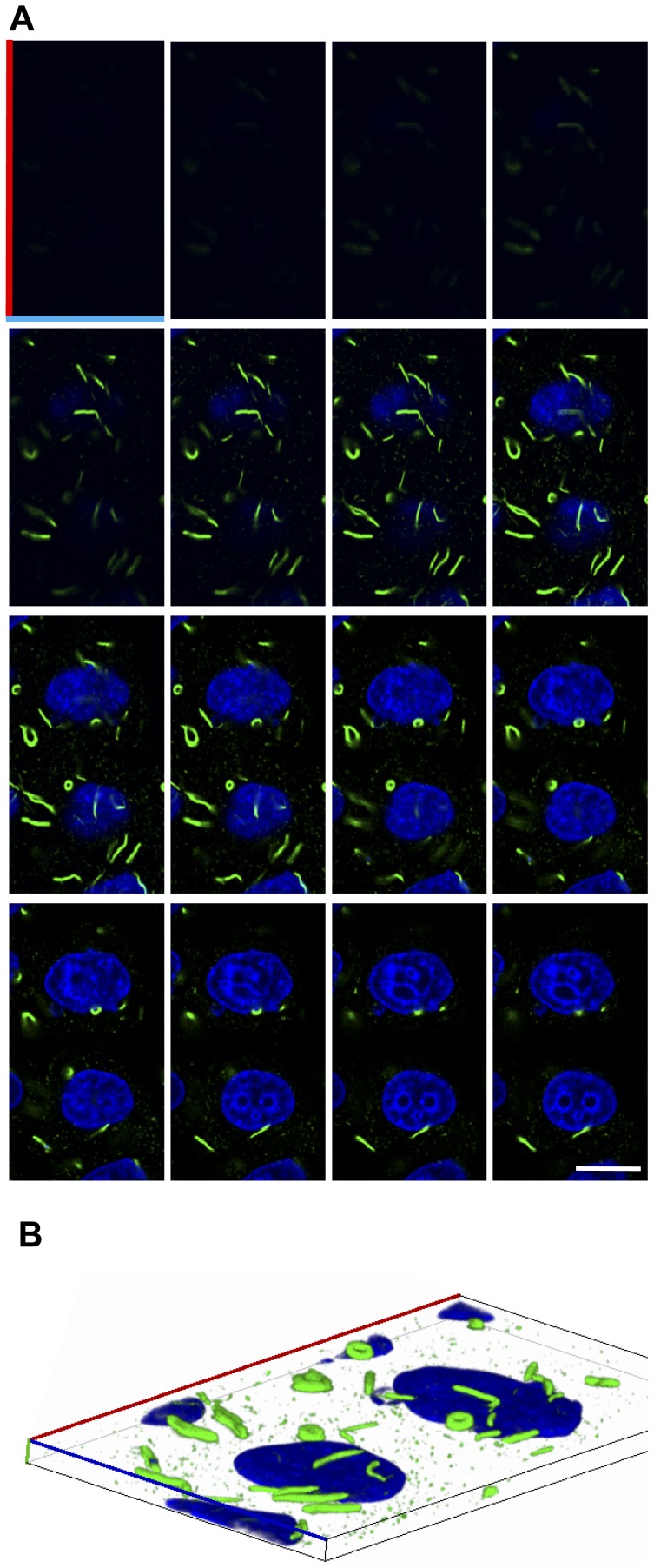
Figure 1
Optical sections of IMPDH macrostructures.
Representative confocal z-series of HeLa cells treated with 1 u00b5M MPA for 4 h. Cells were fixed, permeabilised and labelled with the anti-panIMPDH antibody (green) and nuclei were counterstained wi...
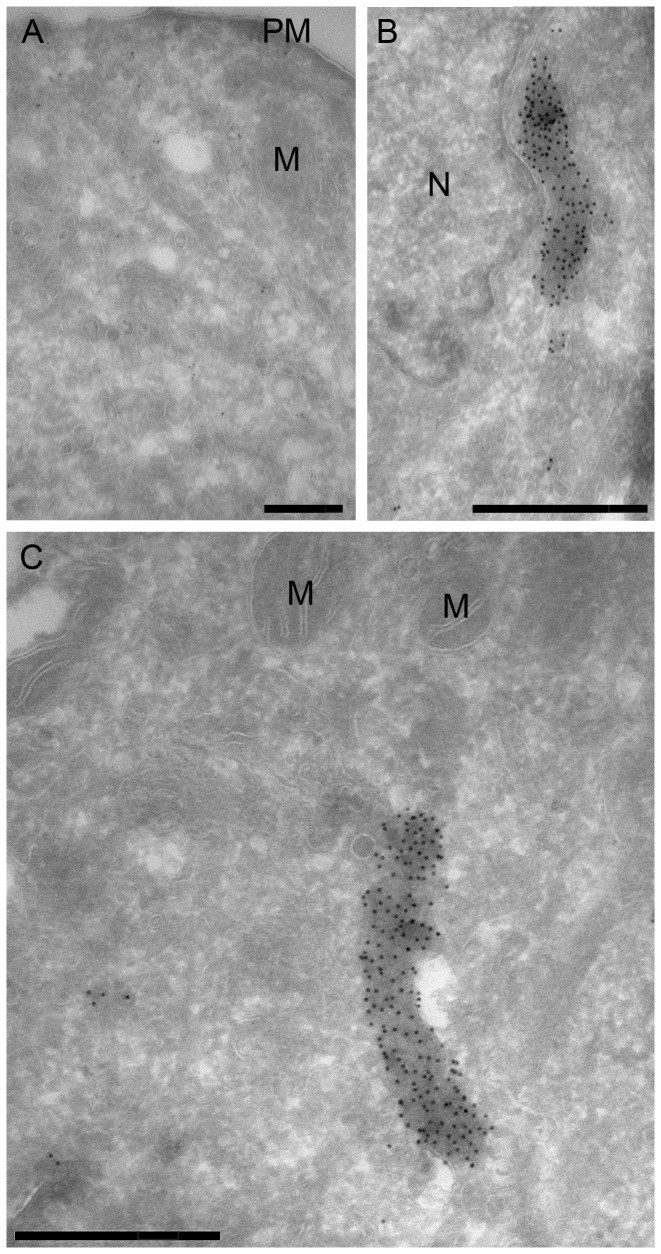
Figure 2
Immuno-EM of IMPDH localisation.
Representative electron micrographs of CHO cells treated with (A) vehicle or (B and C) 2 u00b5M MPA for 4 h. Cells were fixed, processed and labelled with anti-panIMPDH antibody and gold-labelled anti...
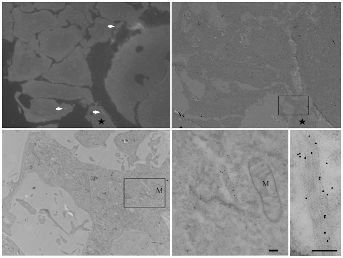
Figure 3
Analysis of IMPDH macrostructures in cryofixed material by correlative light and electron microscopy.
HeLa cells selected for stable low expression of HA-IMPDH2-GFP were treated with 2 u00b5M MPA for 4 h prior to high pressure freezing, freeze-substitution, and embedding in resin at low temperature. S...
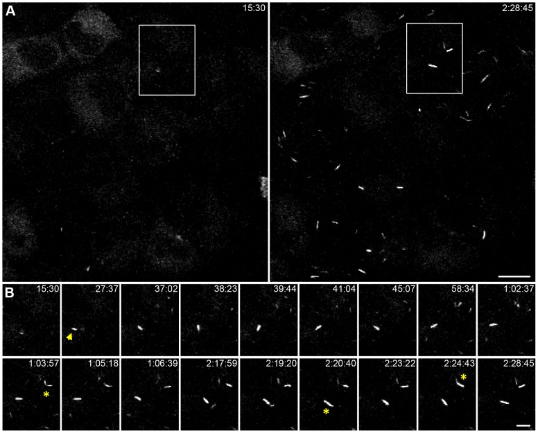
Figure 4
MPA induced clustering of HA-IMPDH2-GFP in live cells.
Images are maximum intensity projections from confocal z-series of live HeLa HA-IMPDH2-GFP cells treated with 2 u00b5M MPA. Selected frames, from Video S1 , are presented with the time captured relati...
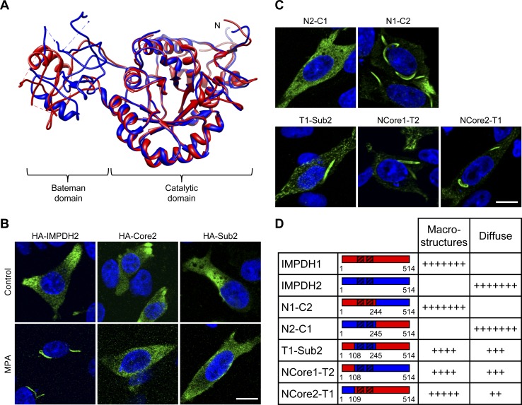
Figure 5
Investigating a role for the Bateman domain in IMPDH clustering.
(A) Superimposed structure of IMPDH2 (1B3O; yellow) with IMPDH1 (1JCN; red). Ligands have been removed for clarity. N labels the N-terminus. (B) Micrographs of CHO cells transiently expressing HA-IMPD...
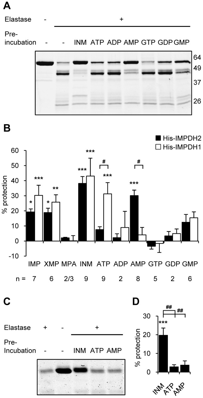
Figure 6
Nucleotides protect IMPDH in an isoform-specific manner via the Bateman domain.
(A) Representative Coomassie stained SDS-PAGE gel of a protease protection assay experiment, performed as outline in methods, with His-IMPDH2. INM stands for IMP, NAD and MPA. Molecular weight marker ...
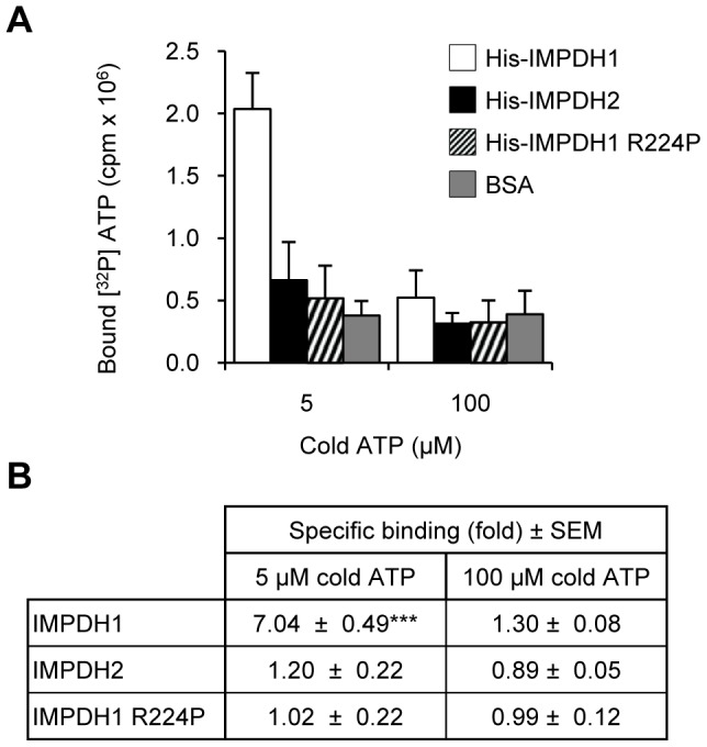
Figure 7
IMPDH1 directly binds ATP.
(A) Representative ATP binding experiment with His-IMPDH proteins, shows [ 32 P] ATP bound to His-IMPDH1 only in the presence of 5 u00b5M cold ATP. Data represents mean counts u00b1 SD. (B) Specific b...
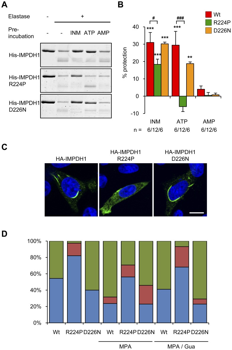
Figure 8
R224P mutation affects ATP mediated protease protection and affects spontaneous clustering.
(A) Representative Coomassie stained gel of a protease protection assay with His-IMPDH1 proteins. INM stands for IMP, NAD and MPA. (B) Quantitation of remaining full-length protein from protease prote...
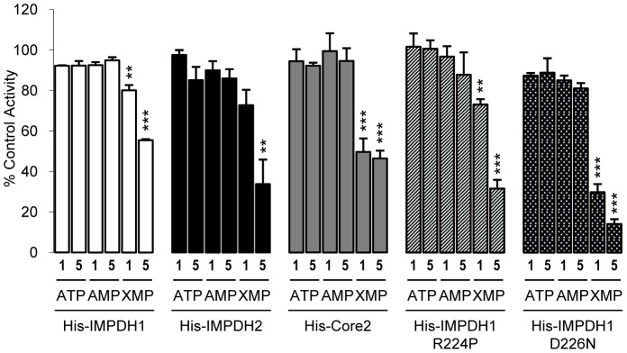
Figure 9
IMPDH activity in the presence of nucleotides.
Activity of purified His-IMPDH proteins following 20 min pre-incubation with 1 mM or 5 mM nucleotides (ATP, AMP, XMP) and normalised to control, no addition, activity. Shown is the mean u00b1 SEM (nu2...
Figure images are served from the NIH/NLM PubMed Central Open Access Subset or Europe PMC; copyright remains with the publishers and authors.
💬 Discussion
0 commentsNo comments yet. Be the first to start a discussion!
Leave a Comment