Abstract
Prokaryotic nanocompartments, also known as encapsulins, are a recently discovered proteinaceous organelle-like compartment in prokaryotes that compartmentalize cargo enzymes. While initial studies have begun to elucidate the structure and physiological roles of encapsulins, bioinformatic evidence suggests that a great diversity of encapsulin nanocompartments remains unexplored. Here, we describe a novel encapsulin in the freshwater cyanobacterium Synechococcus elongatus PCC 7942. This nanocompartment is upregulated upon sulfate starvation and encapsulates a cysteine desulfurase enzyme via an N-terminal targeting sequence. Using cryo-electron microscopy, we have determined the structure of the nanocompartment complex to 2.2 Å resolution. Lastly, biochemical characterization of the complex demonstrated that the activity of the cysteine desulfurase is enhanced upon encapsulation. Taken together, our discovery, structural analysis, and enzymatic characterization of this prokaryotic nanocompartment provide a foundation for future studies seeking to understand the physiological role of this encapsulin in various bacteria.
🔬 Techniques
🔭 Microscopes
🧬 Organisms
✨ Fluorophores
🧪 Sample Preparation
🏭 Microscope Brands
🧪 Reagent Suppliers
📷 Detectors
💻 Software Details
💾 Data Repositories
🏛️ Research Organizations (ROR)
Affiliated research institutions:
📋 Methods
Phylogenetic analysis of encapsulin genes Homologs of SrpI were compiled using NCBI BLASTp with query sequence WP_011055154.1 ( Synechococcus elongatus ). BLASTp searches were carried out in February 2020 and hits with an E-value
📊 Figures
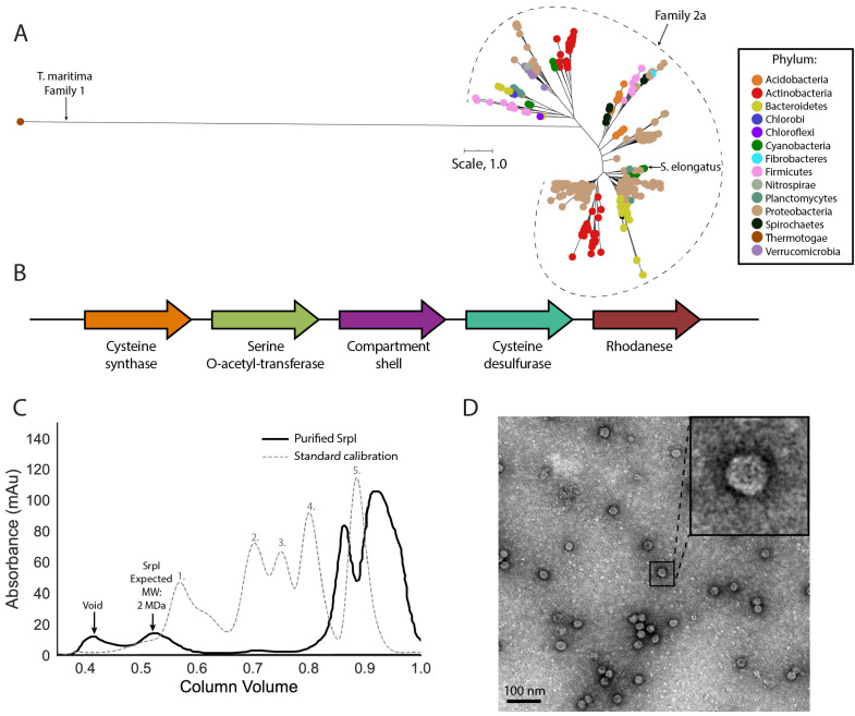
Figure 1.
SrpI is a bacterial nanocompartment that is widespread throughout bacterial phyla and found neighboring sulfur metabolism genes.
( A ) Maximum-likelihood phylogenetic tree of Family 2a encapsulin shell proteins using the T. maritima Family 1 encapsulin shell protein ( WP_004080898.1 ) as a Family 1 representative. Scale bar, on...
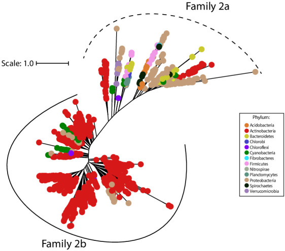
Figure 1u2014figure supplement 1.
Family 2 encapsulins can be divided into two phylogenetically distinct subfamilies.
Maximum likelihood phylogenetic tree of 1383 members of Family 2 encapsulins. Family 2a (dashed line grouping) and Family 2b (solid line grouping) represent two subfamilies. Family 2 encapsulins are f...
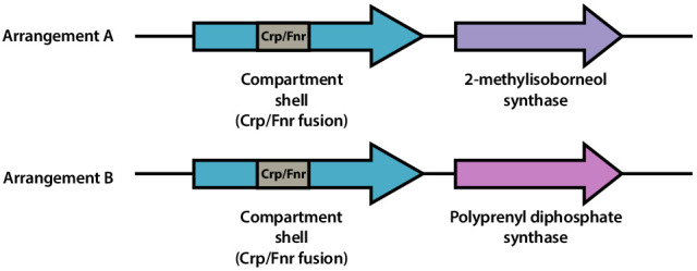
Figure 1u2014figure supplement 2.
Family 2b shell genes neighbor 2-methylisoborneol synthase or polyprenyl diphosphate synthase.
Schematic of representative genomic arrangements for Family 2b shell genes determined using the EFI-GNT web tool. One or both arrangements ( A and B ) may be found within a given genome. Family 2b she...
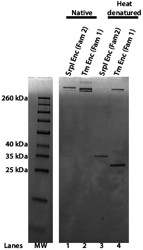
Figure 1u2014figure supplement 3.
The Family 2 encapsulin, SrpI, forms a high-molecular-weight complex similar to the Family 1encapsulin from T. maritima .
SDS-PAGE analysis of purified S. elongatus PCC 7942 Family 2 encapsulin (SrpI Enc) and T. maritima Family 1 encapsulin (Tm Enc). Lanes 1 and 2 correspond to samples that were not heat-denatured (Nativ...
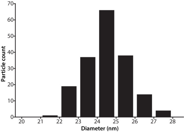
Figure 1u2014figure supplement 4.
Size distribution of SrpI encapsulin.
Diameter of purified SrpI encapsulin determined by negative stain transmission electron microscopy.u00a0Quantification of 180 particles from micrographs performed using FIJI image processing package.
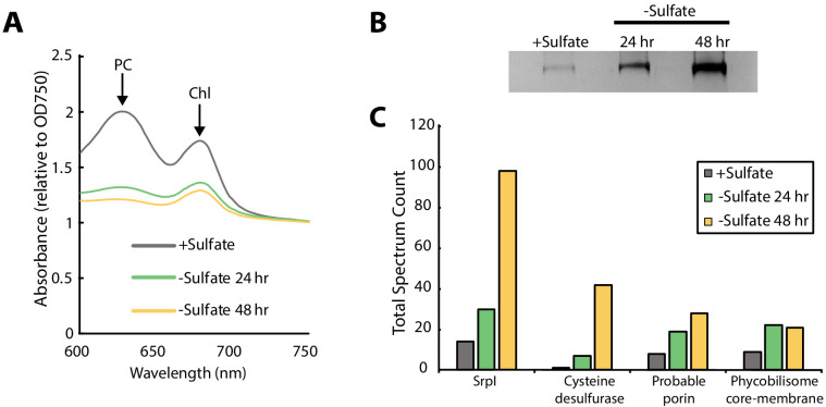
Figure 2.
SrpI encapsulin is upregulated in S. elongatus upon sulfate starvation.
( A ) Absorbance spectra of S. elongatus liquid cultures under nutrient-replete conditions (+Sulfate) and sulfur starvation (-Sulfate) for 24 and 48 hr. Absorbance maxima of phycocyanin (PC) at 620 nm...
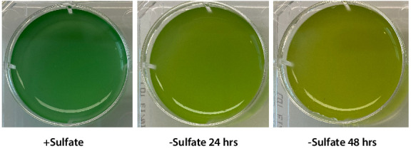
Figure 2u2014figure supplement 1.
Chlorosis phenotype of sulfur-starved S. elongatus PCC 7942.
Liquid cultures of S. elongatus PCC 7942 grown in nutrient replete medium (+Sulfate) or under sulfate starvation for 24 hr and 48 hr.
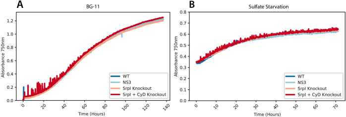
Figure 2u2014figure supplement 2.
Growth curves of S. elongatus PCC 7942 mutants under sulfur replete and sulfur starvation conditions.
Wild type, SrpI knockout-CmR, SrpI and CyD knockout-CmR, and NS3-CmR strains of S. elongatus PCC 7942 cells were ( A ) inoculated at OD750u00a0=u00a00.02 into nutrient replete BG-11 media with constan...
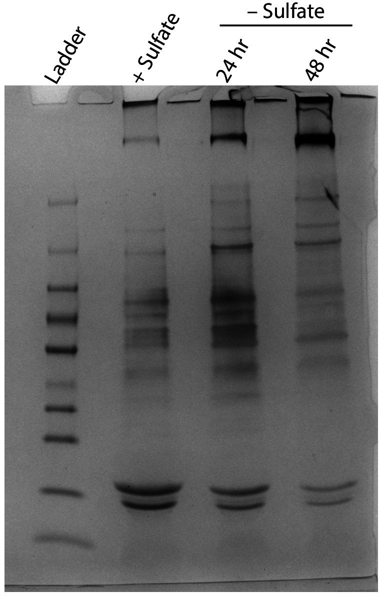
Figure 2u2014figure supplement 3.
Non-denaturing SDS-PAGE analysis of lysates from nutrient-replete and sulfur starved S. elongatus cultures visualized by silver stain.
Inputs were normalized using absorbance at 280 nm.
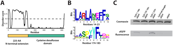
Figure 3.
An N-terminal signal sequence directs cargo loading in vivo .
( A ) Domain organization of cysteine desulfurase (CyD; Synpcc7942_B2661) and the predicted disorder scores calculated using DISOPRED3. CyD can be split into two domains u2013 a highly disordered N-te...
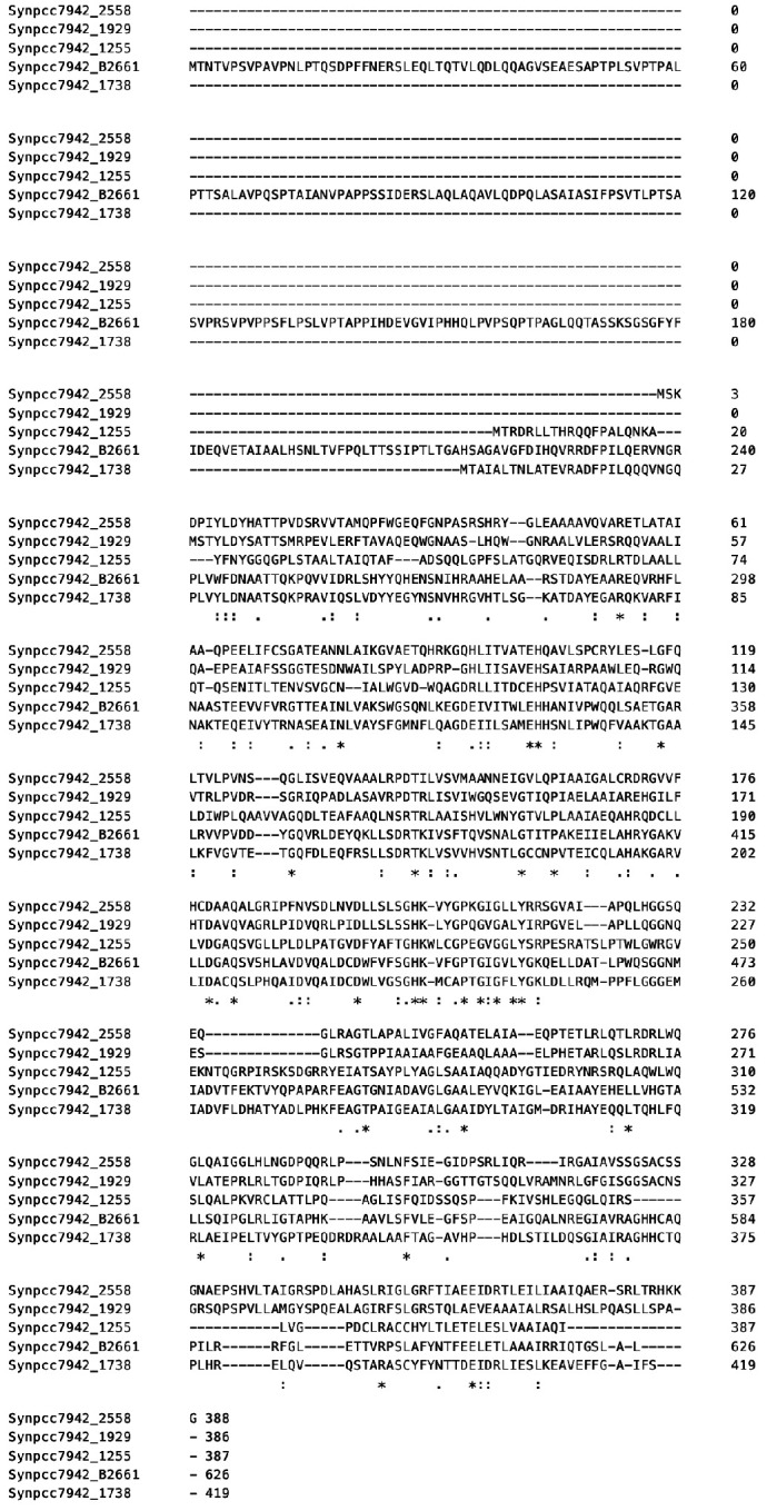
Figure 3u2014figure supplement 1.
SrpI-associated cysteine desulfurase, CyD, possesses a unique N-terminal domain that is absent in the four other cysteine desulfurase genes in the S. elongatus PCC 7942 genome.
Multiple sequence alignment of the SrpI encapsulin-associated cysteine desulfurase (Synpcc7942_B2661) and the other genomic cysteine desulfurases (Synpcc7942_2558, Synpcc7942_1929, Synpcc7942_1255, Sy...
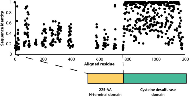
Figure 3u2014figure supplement 2.
Sequence conservation of the F2A encapsulin-associated cysteine desulfurase is sparse throughout the disordered N-terminal domain.
Percent sequence identity from multiple sequence alignment of 997 F2A encapsulin-associated cysteine desulfurase sequences.
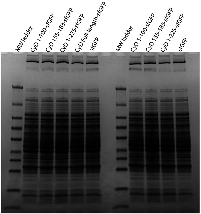
Figure 3u2014figure supplement 3.
Full gel from Coomassie stain panel of Figure 3C .
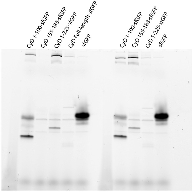
Figure 3u2014figure supplement 4.
Full gel from sfGFP fluorescence panel of Figure 3C .
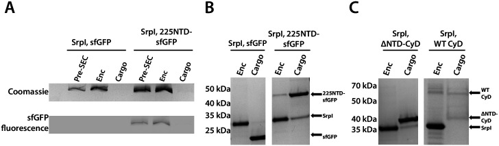
Figure 4.
The 225-NTD of CyD is necessary and sufficient for cargo loading in vitro .
( A )u00a0Non-denaturing SDS-PAGE of sfGFP or the CyD N-terminal domain-sfGFP fusion (225NTD-sfGFP) loaded in vitro into SrpI encapsulin. sfGFP fluorescence followed by Coomassie staining of the encap...
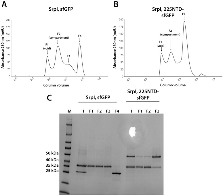
Figure 4u2014figure supplement 1.
Analysis of sfGFP and 225NTD-sfGFP loading into SrpI encapsulin.
( A and B ) size exclusion chromatogram of SrpI encapsulin shell protein (35 kDa) refolded in the presence of untagged sfGFP (27 kDa) and 225NTD-sfGFP (50 kDa) respectively using a Superose 6 Increase...
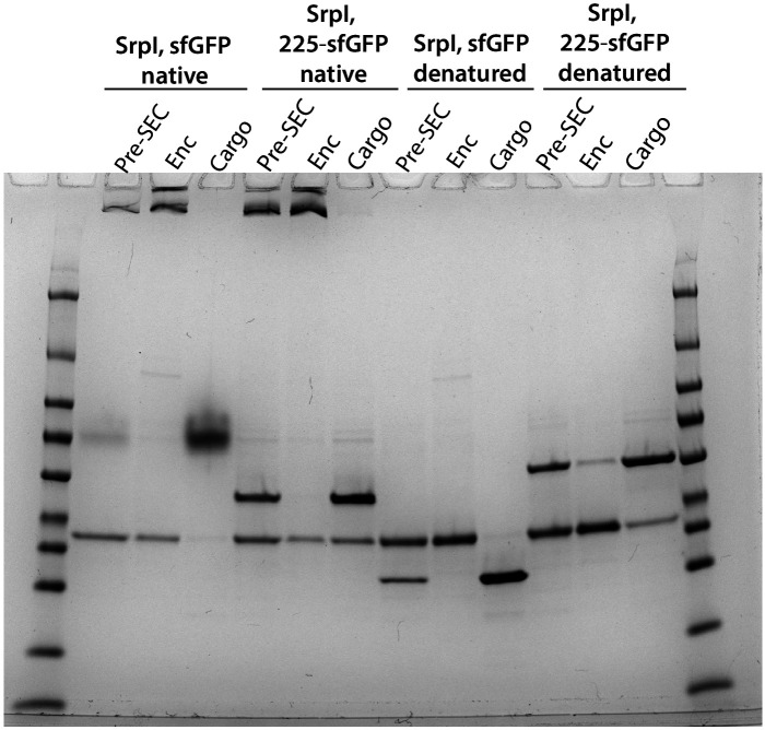
Figure 4u2014figure supplement 2.
Full Coomassie stained gel for panels A and B of Figure 4 .
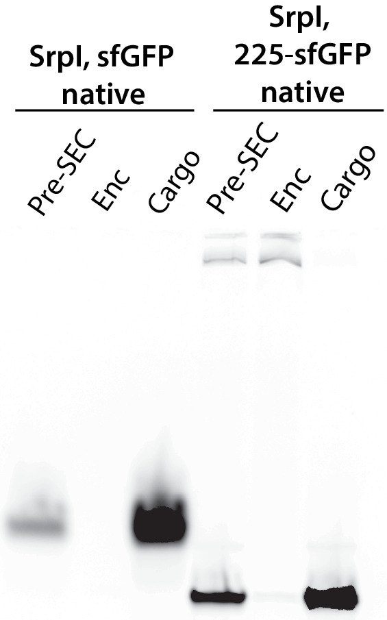
Figure 4u2014figure supplement 3.
Full gel for fluorescent panel of Figure 4A .
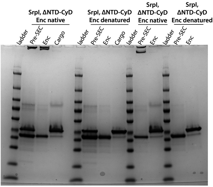
Figure 4u2014figure supplement 4.
Full gel of the SrpI, u0394NTD-CyD panel from Figure 4C .
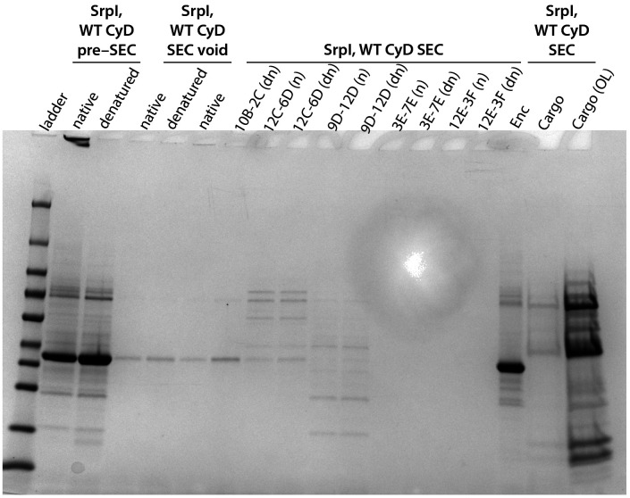
Figure 4u2014figure supplement 5.
Full gel of the SrpI, WT CyD panel from Figure 4C .
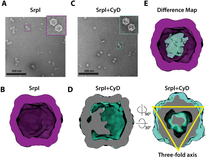
Figure 5.
Negative stain analysis indicates CyD loading into SrpI encapsulin.
( A ) Negative stain micrograph of an apo-SrpI shell in contrast with ( C ) the holo-SrpI shell that includes the CyD cargo.u00a0( B and D ) 3D reconstruction of apo-Srpl and holo-SrpI, respectively. ...
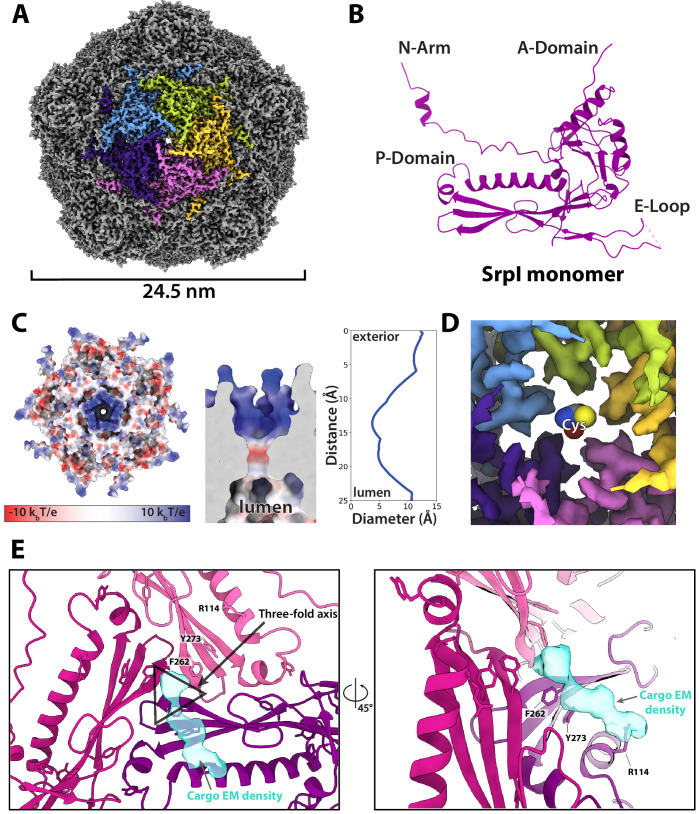
Figure 6.
CryoEM structure of the SrpI encapsulin reveals a common HK97 fold, a potential mechanism for cysteine selectivity, and a cargo binding site ( A ) The SrpI encapsulin structure at 2.2 u00c5 resolution.
This Srpl encapsulin forms a Tu00a0=u00a01 icosahedral structure 24.5 nm in diameter. Five subunits around a fivefold axis are shown in distinct colors. ( B ) SrpI monomer subunits have a HK97 fold wi...
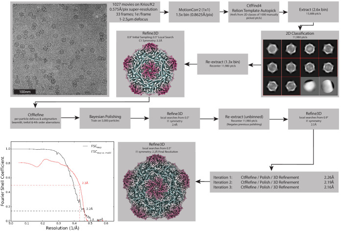
Figure 6u2014figure supplement 1.
Processing pipeline for the Srpl encapsulin.
Processing workflow within RELION that used to reconstruct the holo-Srpl structure. An identical approach was used for the apo-Srpl encapsulin. Once both these structures were determined, symmetry exp...
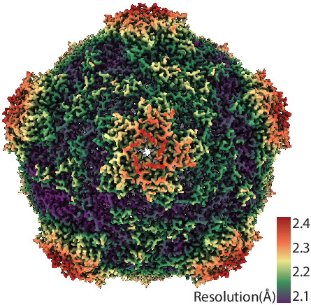
Figure 6u2014figure supplement 2.
CryoEM resolution map of SrpI encapsulin.
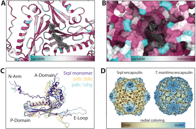
Figure 6u2014figure supplement 3.
Secondary, tertiary, and quaternary homology between Srpl and other known encapsulins.
( A and B ) Sequence conservation of Family 2a encapsulins mapped onto the atomic model and surface display for the Srpl shell respectively (conservation was calculated via ConSurf). ( C ) Single subu...
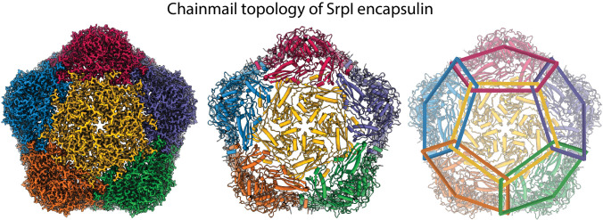
Figure 6u2014figure supplement 4.
Chainmail-like topography of Srpl.
Coloring of individual subunits highlights the chainmail overlapping topology of the Srpl encapsulin.
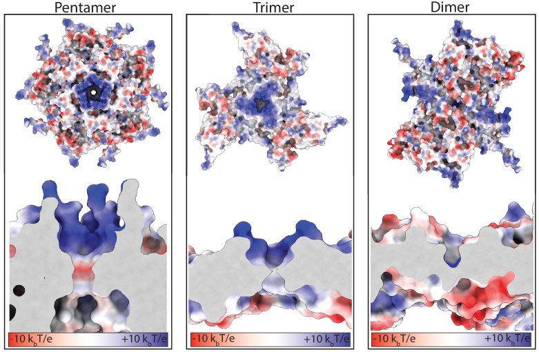
Figure 6u2014figure supplement 5.
Electrostatic surface charges at the symmetry axes of the Srpl shell.
Close-up and slice-through views of the five-fold, three-fold, and two-fold axes. Surfaces colored according to electrostatic potential (red=negative, blue=positive).
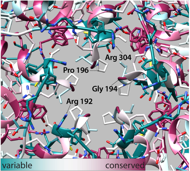
Figure 6u2014figure supplement 6.
Conservation of residues at the fivefold pore.
Ball and stick model of the fivefold pore (displayed exterior of the compartment facing out). Residues are colored by sequence conservation as calculated by the ConSurf webserver (cyan u2013 conserved...
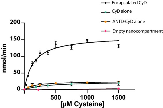
Figure 7.
Cysteine desulfurase activity is enhanced upon encapsulation.
Substrate-dependent activity of encapsulated cysteine desulfurase (encapsulated CyD), unencapsulated cysteine desulfurase (CyD alone), unencapsulated cysteine desulfurase lacking its NTD (u0394NTD-CyD...
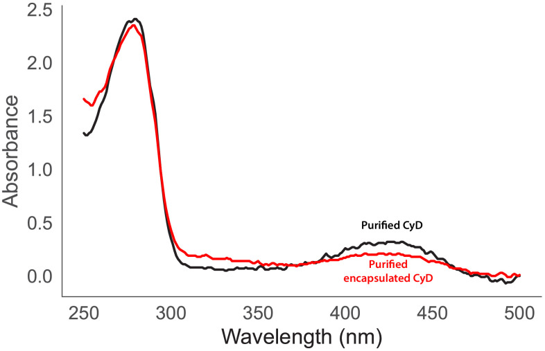
Figure 7u2014figure supplement 1.
Ultraviolet-visible absorbance spectra of purified CyD (black) and encapsulated CyD (Red).
Figure images are served from the NIH/NLM PubMed Central Open Access Subset or Europe PMC; copyright remains with the publishers and authors.
💬 Discussion
0 commentsNo comments yet. Be the first to start a discussion!
Leave a Comment