Abstract
AbstractGd‐based T 1‐weighted contrast agents have dominated the magnetic resonance imaging (MRI) contrast agent market for decades. Nevertheless, they are reported to be nephrotoxic and the U.S. Food and Drug Administration has issued a general warning concerning their use. In order to reduce the risk of nephrotoxicity, the MRI performance of the Gd‐based T 1‐weighted contrast agents needs to be improved to allow a much lower dosage. In this study, novel dotted core–shell nanoparticles (FeGd‐HN3‐RGD2) with superhigh r 1 value (70.0 mM−1 s−1) and very low r 2/r 1 ratio (1.98) are developed for high‐contrast T 1‐weighted MRI of tumors. 3‐(4,5‐Dimethylthiazol‐2‐yl)‐2,5‐diphenyltetrazolium bromide (MTT) assay and histological analyses show good biocompatibility of FeGd‐HN3‐RGD2. Laser scanning confocal microscopy images and flow cytometry demonstrate active targeting to integrin αvβ3 positive tumors. MRI of tumors shows high tumor ΔSNR for FeGd‐HN3‐RGD2 (477 ± 44%), which is about 6‐7‐fold higher than that of Magnevist (75 ± 11%). MRI and inductively coupled plasma results further confirm that the accumulation of FeGd‐HN3‐RGD2 in tumors is higher than liver and spleen due to the RGD2 targeting and small hydrodynamic particle size (8.5 nm), and FeGd‐HN3‐RGD2 is readily cleared from the body by renal excretion.
🔬 Techniques
✨ Fluorophores
🔬 Cell Lines
🏭 Microscope Brands
🧪 Reagent Suppliers
💻 Software Details
🏛️ Research Organizations (ROR)
Affiliated research institutions:
📋 Methods
Chemicals: Poly(acrylic acid) (PAA, Mw = 1800), iron (Ⅲ) chloride (FeCl 3 , ≥ 97%), gadolinium (III) nitrate hexahydrate (Gd(NO 3 ) 3 · 6H 2 O, 99.9%), N -(3-Dimethylaminopropyl)- N’ - ethylcarbodiimide (EDC, ≥ 97%), N-hydroxysuccinimide (NHS, 98 %), Rhodamine 6G (R6G), phalloidin-FITC, and Hoechst 33258 were purchased from Sigma-aldrich (USA). Iron (Ⅱ) sulfate heptahydrate (FeSO 4 ·7H 2 O) was purchased from Acros organics. Glu-{Cyclo[Arg-Gly-Asp-(D-Phe)-Lys]} 2 ( i.e. dimeric RGD peptide, or RGD2, 97.92%, Mw = 1318.51) was purchased from C S Bio Co. (CA, USA 94025). Synthesis of ES-MION/GdON hybrid nanoparticles (FeGd-HN): 40 mL of PAA (Mw = 1800) solution (4.0 mg/mL) was first purged with nitrogen (≥ 50 min) to remove oxygen. The polymer solution was then heated to reflux (100 o C). After that, a 0.8 mL mixture of iron precursors (500 mM FeCl 3 plus 250 mM FeSO 4 ) was quickly injected into the heated polymer solution, followed by addition of 12 mL of ammonia solution (28 %). The reaction was kept at 100 o C for 30 min under magnetic stirring to obtain the seeds of extremely small magnetic iron oxide nanoparticles (ES-MIONs). After that, 0.8 mL of Gd(NO 3 ) 3 (62.5 ~ 1000 mM) and 6.0 mL of ammonia solution (28 %) were added into the reaction system. The reaction was continued for 90 min under magnetic stirring at 100 o C to obtain the FeGd-HN. The ES-MIONs were synthesized by a similar method without addition of Gd precursor. [ 18 ] Finally, the solutions were cooled down to room temperature. The obtained FeGd-HN and ES-MIONs were purified by membrane dialysis (MWCO 6–8 kDa) against Milli-Q water for 5 days with a daily change of the water. The purified FeGd-HN and ES-MIONs were concentrated by centrifugal ultrafiltration (Millipore, MWCO 3 kDa). The Fe and Gd concentrations of the solutions were measured by inductively coupled plasma optical emission spectrometry (ICP-OES; Agilent 5100). The Fe or Gd recovery of the FeGd-HN was calculated from the molar ratio of Fe or Gd in the obtained FeGd-HN to the feeding Fe or Gd. In addition, 2.0 mL of the nanoparticle solutions were dried at 70 o C to calculate the mass concentration of the nanoparticles (with Fe 3 O 4 , Gd 2 O 3 and PAA). Characterization of FeGd-HN: Distributions of Fe, Gd, and O in the FeGd-HN nanoparticles were mapped using electron energy loss spectroscopic imaging (EELSI) in a scanning transmission electron microscope (STEM). [ 39 – 41 ] The EELSI data were acquired with a Tecnai TF30 transmission electron microscope (FEI, Inc.) equipped with a Quantum imaging filter (Gatan Inc.), operating at an accelerating voltage of 300 kV. STEM images containing 2048 × 2048 pixels were acquired using a high-angle annular dark-field (HAADF) detector (Fischione Instruments, Inc.). For small selected regions within the HAADF images, EELSI data were acquired with approximately 35 pixels x 35 pixels x 2048 energy channels, spanning energy losses from 445 eV to 1470 eV, with 0.5 eV/channel. The pixel size was approximately 1 nm x 1 nm, and the pixel dwell time 0.1 s. Synthesis of FeGd-HN3-RGD2: The RGD2 were conjugated onto the surface of FeGd-HN3 via the reaction between –COOH and –NH 2 in the presence of EDC/NHS. Typically, 10 μL of EDC (55 μmol) and 50 μL 13 mg/mL of NHS (5.65 μmol) were added into 5.0 mL of FeGd-HN3 solution ( C Fe = 3.10 mM, C Gd = 1.61 mM, ice cold) under magnetic stirring. After that, 100 μL of RGD2 (5.0 mg/mL, 3.8 mM) were added into the mixtures. After 16 h of reaction at room temperature under magnetic stirring, the obtained FeGd-HN3-RGD2 were washed 3 times using Milli-Q water by centrifugal ultrafiltration (Millipore, MWCO 3 kDa) to remove unreacted EDC, NHS, RGD2, and side product EDU, and finally dissolved in 5.0 mL of Milli-Q water. Synthesis of Rhodamine 6G-loaded FeGd-HN3-RGD2: To investigate the internalization of FeGd-HN3 or FeGd-HN3-RGD2 in cells by flow cytometry and laser scanning confocal microscopy (LSCM), Rhodamine 6G (R6G) was loaded onto the surface of FeGd-HN3 or FeGd-HN3-RGD2. Typically, 4.0 mL of FeGd-HN3 ( C Fe = 3.10 mM, C Gd = 1.61 mM), or FeGd-HN3-RGD2 ( C Fe = 3.10 mM, C Gd = 1.61 mM) were mixed with 0.7 mL of Rhodamine 6G (10 μM) under magnetic stirring at room temperature. After 24 h, the obtained R6G-FeGd-HN3 or R6G-FeGd-HN3-RGD2 solution was washed using Milli-Q water by centrifugal ultrafiltration (Millipore, MWCO 3 kDa) to remove free R6G. The resultant R6G-FeGd-HN3 or R6G-FeGd-HN3-RGD2 was dispersed in 4.0 mL of Milli-Q water. Cellular Uptake of the Nanoparticles Measured by ICP: 2.0 mL of U-87 MG cells in complete growth medium were seeded into each well of a 6-well culture plate with a cell density of 1.0×10⁵ cells/mL and allowed to adhere at 37 oC for 24 h. The growth medium was then replaced with fresh media (2.0 mL, without FBS) without or with FeGd-HN3-RGD2, or FeGd-HN3 (CGd = 80 μM). After further 2.0 h incubation, the cells were washed twice with PBS, treated with trypsin for 3.0 min, and then centrifuged at 500 × g for 5 min to remove the extracellular nanoparticles. The obtained cells were used for Gd measurement by ICP.
Show full methods section
Chemicals: Poly(acrylic acid) (PAA, Mw = 1800), iron (Ⅲ) chloride (FeCl 3 , ≥ 97%), gadolinium (III) nitrate hexahydrate (Gd(NO 3 ) 3 · 6H 2 O, 99.9%), N -(3-Dimethylaminopropyl)- N’ - ethylcarbodiimide (EDC, ≥ 97%), N-hydroxysuccinimide (NHS, 98 %), Rhodamine 6G (R6G), phalloidin-FITC, and Hoechst 33258 were purchased from Sigma-aldrich (USA). Iron (Ⅱ) sulfate heptahydrate (FeSO 4 ·7H 2 O) was purchased from Acros organics. Glu-{Cyclo[Arg-Gly-Asp-(D-Phe)-Lys]} 2 ( i.e. dimeric RGD peptide, or RGD2, 97.92%, Mw = 1318.51) was purchased from C S Bio Co. (CA, USA 94025). Synthesis of ES-MION/GdON hybrid nanoparticles (FeGd-HN): 40 mL of PAA (Mw = 1800) solution (4.0 mg/mL) was first purged with nitrogen (≥ 50 min) to remove oxygen. The polymer solution was then heated to reflux (100 o C). After that, a 0.8 mL mixture of iron precursors (500 mM FeCl 3 plus 250 mM FeSO 4 ) was quickly injected into the heated polymer solution, followed by addition of 12 mL of ammonia solution (28 %). The reaction was kept at 100 o C for 30 min under magnetic stirring to obtain the seeds of extremely small magnetic iron oxide nanoparticles (ES-MIONs). After that, 0.8 mL of Gd(NO 3 ) 3 (62.5 ~ 1000 mM) and 6.0 mL of ammonia solution (28 %) were added into the reaction system. The reaction was continued for 90 min under magnetic stirring at 100 o C to obtain the FeGd-HN. The ES-MIONs were synthesized by a similar method without addition of Gd precursor. [ 18 ] Finally, the solutions were cooled down to room temperature. The obtained FeGd-HN and ES-MIONs were purified by membrane dialysis (MWCO 6–8 kDa) against Milli-Q water for 5 days with a daily change of the water. The purified FeGd-HN and ES-MIONs were concentrated by centrifugal ultrafiltration (Millipore, MWCO 3 kDa). The Fe and Gd concentrations of the solutions were measured by inductively coupled plasma optical emission spectrometry (ICP-OES; Agilent 5100). The Fe or Gd recovery of the FeGd-HN was calculated from the molar ratio of Fe or Gd in the obtained FeGd-HN to the feeding Fe or Gd. In addition, 2.0 mL of the nanoparticle solutions were dried at 70 o C to calculate the mass concentration of the nanoparticles (with Fe 3 O 4 , Gd 2 O 3 and PAA). Characterization of FeGd-HN: Distributions of Fe, Gd, and O in the FeGd-HN nanoparticles were mapped using electron energy loss spectroscopic imaging (EELSI) in a scanning transmission electron microscope (STEM). [ 39 – 41 ] The EELSI data were acquired with a Tecnai TF30 transmission electron microscope (FEI, Inc.) equipped with a Quantum imaging filter (Gatan Inc.), operating at an accelerating voltage of 300 kV. STEM images containing 2048 × 2048 pixels were acquired using a high-angle annular dark-field (HAADF) detector (Fischione Instruments, Inc.). For small selected regions within the HAADF images, EELSI data were acquired with approximately 35 pixels x 35 pixels x 2048 energy channels, spanning energy losses from 445 eV to 1470 eV, with 0.5 eV/channel. The pixel size was approximately 1 nm x 1 nm, and the pixel dwell time 0.1 s. Synthesis of FeGd-HN3-RGD2: The RGD2 were conjugated onto the surface of FeGd-HN3 via the reaction between –COOH and –NH 2 in the presence of EDC/NHS. Typically, 10 μL of EDC (55 μmol) and 50 μL 13 mg/mL of NHS (5.65 μmol) were added into 5.0 mL of FeGd-HN3 solution ( C Fe = 3.10 mM, C Gd = 1.61 mM, ice cold) under magnetic stirring. After that, 100 μL of RGD2 (5.0 mg/mL, 3.8 mM) were added into the mixtures. After 16 h of reaction at room temperature under magnetic stirring, the obtained FeGd-HN3-RGD2 were washed 3 times using Milli-Q water by centrifugal ultrafiltration (Millipore, MWCO 3 kDa) to remove unreacted EDC, NHS, RGD2, and side product EDU, and finally dissolved in 5.0 mL of Milli-Q water. Synthesis of Rhodamine 6G-loaded FeGd-HN3-RGD2: To investigate the internalization of FeGd-HN3 or FeGd-HN3-RGD2 in cells by flow cytometry and laser scanning confocal microscopy (LSCM), Rhodamine 6G (R6G) was loaded onto the surface of FeGd-HN3 or FeGd-HN3-RGD2. Typically, 4.0 mL of FeGd-HN3 ( C Fe = 3.10 mM, C Gd = 1.61 mM), or FeGd-HN3-RGD2 ( C Fe = 3.10 mM, C Gd = 1.61 mM) were mixed with 0.7 mL of Rhodamine 6G (10 μM) under magnetic stirring at room temperature. After 24 h, the obtained R6G-FeGd-HN3 or R6G-FeGd-HN3-RGD2 solution was washed using Milli-Q water by centrifugal ultrafiltration (Millipore, MWCO 3 kDa) to remove free R6G. The resultant R6G-FeGd-HN3 or R6G-FeGd-HN3-RGD2 was dispersed in 4.0 mL of Milli-Q water. Cellular Uptake of the Nanoparticles Measured by ICP: 2.0 mL of U-87 MG cells in complete growth medium were seeded into each well of a 6-well culture plate with a cell density of 1.0×10⁵ cells/mL and allowed to adhere at 37 oC for 24 h. The growth medium was then replaced with fresh media (2.0 mL, without FBS) without or with FeGd-HN3-RGD2, or FeGd-HN3 (CGd = 80 μM). After further 2.0 h incubation, the cells were washed twice with PBS, treated with trypsin for 3.0 min, and then centrifuged at 500 × g for 5 min to remove the extracellular nanoparticles. The obtained cells were used for Gd measurement by ICP.
📊 Figures
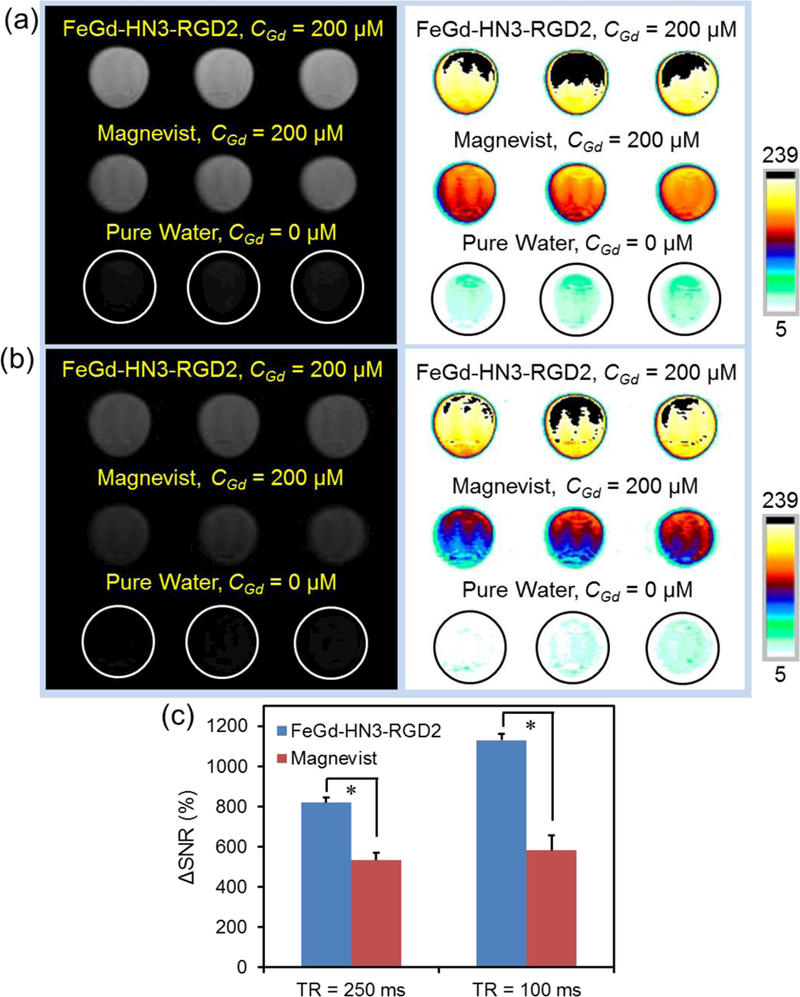
Figure 1.
T 1 -weighted MR images of our FeGd-HN3-RGD2 and Magnevist u00ae solutions ( C Gd = 200 u03bcM) compared with the pure water (control) with 250 ms of TR (a), or 100 ms of TR (b). TE = 10 ms. Magnetic ...
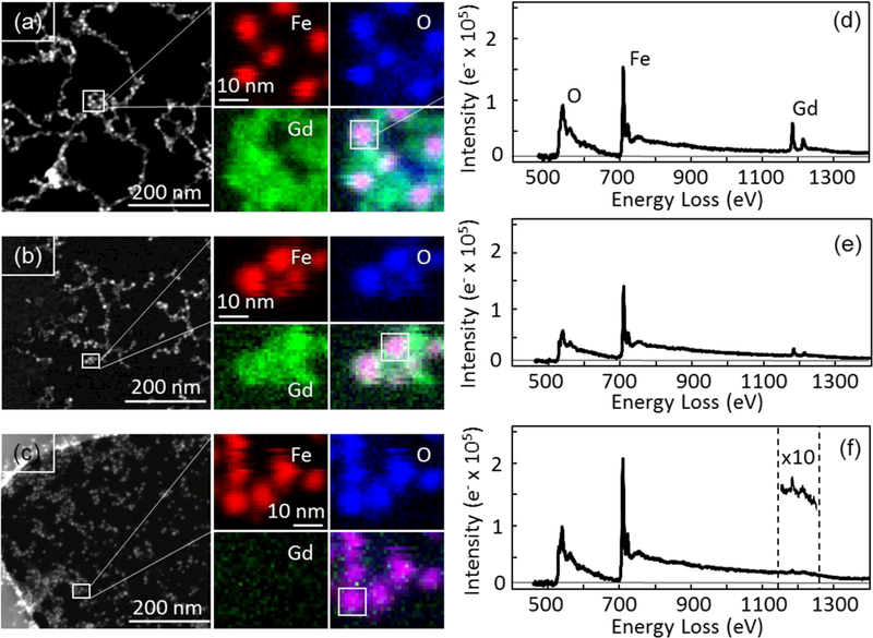
Figure 2.
Characterization of scanning transmission electron microscope (STEM), electron energy loss spectroscopic imaging (EELSI) (a-c), and EELS spectra (d-f) for the FeGd-HN1 (a, d), FeGd-HN3 (b, e), and FeG...
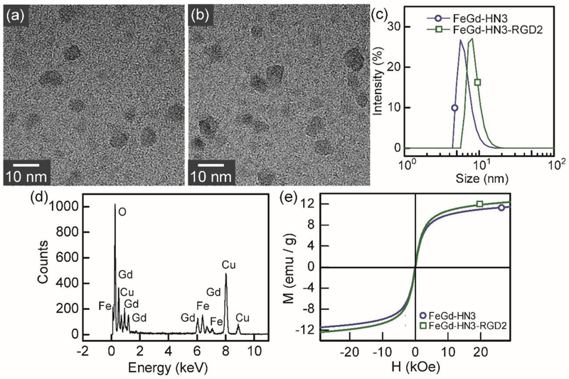
Figure 3.
Characterization of FeGd-HN3 and FeGd-HN3-RGD2. (a, b): High resolution TEM (HR-TEM) images of FeGd-HN3 (a) and FeGd-HN3-RGD2 (b). (c): Size distribution of FeGd-HN3 ( d h = 6.5 nm) and FeGd-HN3-RGD2 ...
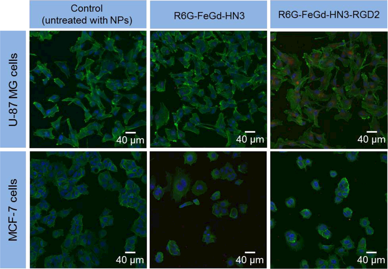
Figure 4.
LSCM images of U-87 MG or MCF-7 cells incubated with R6G-FeGd-HN3 or R6G-FeGd-HN3-RGD2. The cells untreated with nanoparticles are used as the control. The cytoskeleton stained with phalloidin-FITC is...
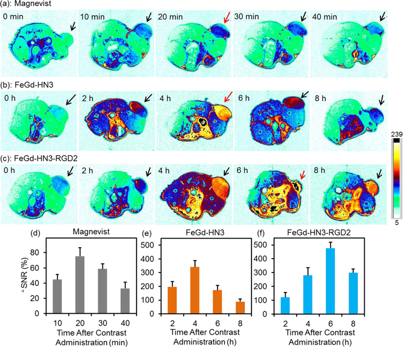
Figure 5.
In vivo T 1 -weighted MR images of U-87 MG tumor-bearing nude mice (slice orientation: axial) (a-c) and quantification of the tumors (d-f) after intravenous injection of Magnevist, FeGd-HN3, or FeGd-H...
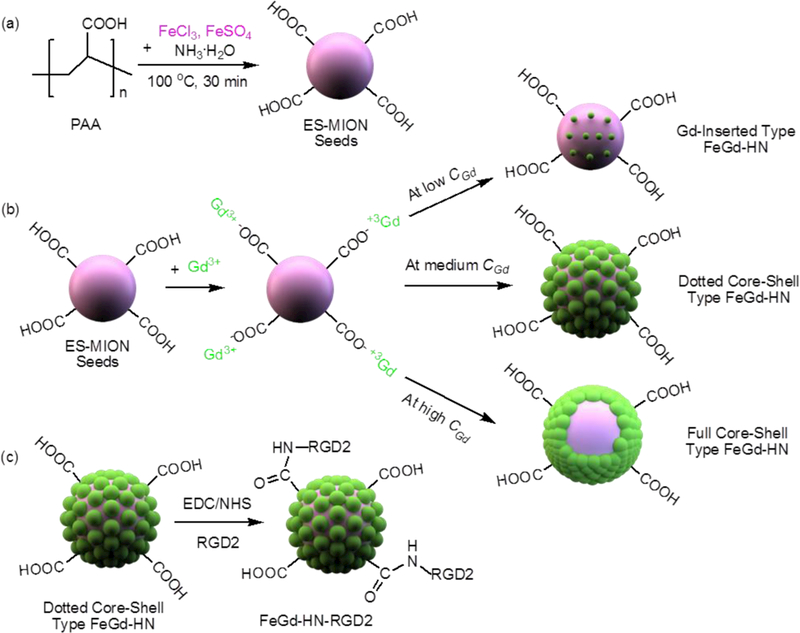
Scheme 1.
Schematic illustration of synthesis steps of our RGD2-conjugated dotted core-shell type ES-MION/GdON hybrid nanoparticles (FeGd-HN) with superhigh r 1 and very low r 2 / r 1 for high contrast T 1 -wei...
Figure images are served from the NIH/NLM PubMed Central Open Access Subset or Europe PMC; copyright remains with the publishers and authors.
💬 Discussion
0 commentsNo comments yet. Be the first to start a discussion!
Leave a Comment