Abstract
The optical and biomechanical properties of the cornea are largely governed by the collagen-rich stroma, a layer that represents approximately 90% of the total thickness. Within the stroma, the specific arrangement of superimposed lamellae provides the tissue with tensile strength, whilst the spatial arrangement of individual collagen fibrils within the lamellae confers transparency. In keratoconus, this precise stromal arrangement is lost, resulting in ectasia and visual impairment. In the normal cornea, we previously characterised the three-dimensional arrangement of an elastic fiber network spanning the posterior stroma from limbus-to-limbus. In the peripheral cornea/limbus there are elastin-containing sheets or broad fibers, most of which become microfibril bundles (MBs) with little or no elastin component when reaching the central cornea. The purpose of the current study was to compare this network with the elastic fiber distribution in post-surgical keratoconic corneal buttons, using serial block face scanning electron microscopy and transmission electron microscopy. We have demonstrated that the MB distribution is very different in keratoconus. MBs are absent from a region of stroma anterior to Descemet's membrane, an area that is densely populated in normal cornea, whilst being concentrated below the epithelium, an area in which they are absent in normal cornea. We contend that these latter microfibrils are produced as a biomechanical response to provide additional strength to the anterior stroma in order to prevent tissue rupture at the apex of the cone. A lack of MBs anterior to Descemet's membrane in keratoconus would alter the biomechanical properties of the tissue, potentially contributing to the pathogenesis of the disease.
🔬 Techniques
🧪 Sample Preparation
🏭 Microscope Brands
🧪 Reagent Suppliers
💻 Software Details
📋 Protocols
🏛️ Research Organizations (ROR)
Affiliated research institutions:
📋 Methods
Tissue specimens
Four human keratoconus buttons (7 mm diameter) were obtained from the Department of Ophthalmology, Kyoto Prefectural University, Japan, following penetrating keratoplasty. Button 1, from the right eye of a 47-year-old female, had a minor apical scar inferior to the pupil ( Fig. 1 A). Button 2, obtained from the right eye of a 57-year-old male was more severely scarred in the same region ( Fig. 1 B). Button 3, from the right eye of a 68-year-old female, was scarred superiorly (no image). Button 4, from the left eye of a 31-year-old male, had a small infero-nasal scar ( Fig. 1 C). All thinned cone regions were located para-centrally. Following surgery, buttons were immediately fixed in 4% paraformaldehyde before being transported to Cardiff on dry ice. Normal human corneas with scleral rim were obtained from Bristol Eye bank and stored in 4% paraformaldehyde until use. Local ethics committee approval was obtained for this study and the research was carried out in accordance with the tenets of the Declaration of Helsinki. Fig. 1 Corneal images in keratoconus before penetrating keratoplasty. Cornea 1 (A) has mild scarring inferior to the pupil, whilst cornea 2 (B) is at a more advanced stage of the disease, hence more severe scarring. Cornea 4 (C) has a small infero-nasal scar. Red circles indicate the regions where 7 mm buttons were taken. (For interpretation of the references to colour in this figure legend, the reader is referred to the web version of this article.) Fig. 1 2.2 Serial block face scanning electron microscopy Thin segments were dissected from each sample and fixed in modified Karnovsky's fixative (2.5% glutaraldehyde and 2% paraformaldehyde in 0.1M cacodylate buffer at pH 7.2) at room temperature before being stored in buffer at 4 °C overnight. Button 1 was processed en bloc using a tannic-acid based staining protocol, whereas buttons 2 and 3 were processed with a novel en bloc orcein staining protocol as below. Normal corneas were processed using both protocols. Sclera control was processed with the orcein protocol only. Tannic acid based staining protocol: Initially, tissue was processed according to methods described previously ( Lewis et al., 2016 ). Briefly, this consisted of post fixation with 1% osmium tetroxide, incubation in 0.5% tannic acid, staining with 2% aqueous uranyl acetate, dehydration in an ethanol series, further staining with 2% ethanoic uranyl acetate and a saturated solution of lead acetate ( Kushida, 1966 ) before embedding in epoxy resin. This method resulted in dark staining throughout the tissue with little contrast, therefore, additional tissue was processed using an orcein based protocol. Orcein based staining protocol: Orcein staining has been used to visualise elastic tissue in the cornea using TEM ( Lewis et al., 2016 ), using a protocol based on earlier work ( Nakamura et al., 1977 ). A novel en bloc orcein staining protocol was developed in an attempt to enhance contrast of keratoconic samples using SBF SEM. Samples were fixed with 1% osmium tetroxide before being washed in dH 2 O for 20 min and transferred to 70% ethanol for 10 min. Samples were stained with 0.3% orcein in 70% ethanol for 2 h. After a 30 min wash with 70% ethanol, specimens were dehydrated in 90% ethanol for 20 min, followed by 100% ethanol × 2 for 20 min. Following dehydration, the tissue was subjected to the same remaining steps described in the tannic acid based protocol. Specimens were examined using a Zeiss Sigma VP FEG SEM equipped with a Gatan 3View2 system, where data sets of up to 1000 images were acquired every 50 nm at 4k × 4k pixel resolution. Three-dimensional reconstructions of data sets were created using Amira 6 software (FEI, Mérignac, France).
Show full methods section
Tissue specimens
Four human keratoconus buttons (7 mm diameter) were obtained from the Department of Ophthalmology, Kyoto Prefectural University, Japan, following penetrating keratoplasty. Button 1, from the right eye of a 47-year-old female, had a minor apical scar inferior to the pupil ( Fig. 1 A). Button 2, obtained from the right eye of a 57-year-old male was more severely scarred in the same region ( Fig. 1 B). Button 3, from the right eye of a 68-year-old female, was scarred superiorly (no image). Button 4, from the left eye of a 31-year-old male, had a small infero-nasal scar ( Fig. 1 C). All thinned cone regions were located para-centrally. Following surgery, buttons were immediately fixed in 4% paraformaldehyde before being transported to Cardiff on dry ice. Normal human corneas with scleral rim were obtained from Bristol Eye bank and stored in 4% paraformaldehyde until use. Local ethics committee approval was obtained for this study and the research was carried out in accordance with the tenets of the Declaration of Helsinki. Fig. 1 Corneal images in keratoconus before penetrating keratoplasty. Cornea 1 (A) has mild scarring inferior to the pupil, whilst cornea 2 (B) is at a more advanced stage of the disease, hence more severe scarring. Cornea 4 (C) has a small infero-nasal scar. Red circles indicate the regions where 7 mm buttons were taken. (For interpretation of the references to colour in this figure legend, the reader is referred to the web version of this article.) Fig. 1 2.2 Serial block face scanning electron microscopy Thin segments were dissected from each sample and fixed in modified Karnovsky's fixative (2.5% glutaraldehyde and 2% paraformaldehyde in 0.1M cacodylate buffer at pH 7.2) at room temperature before being stored in buffer at 4 °C overnight. Button 1 was processed en bloc using a tannic-acid based staining protocol, whereas buttons 2 and 3 were processed with a novel en bloc orcein staining protocol as below. Normal corneas were processed using both protocols. Sclera control was processed with the orcein protocol only. Tannic acid based staining protocol: Initially, tissue was processed according to methods described previously ( Lewis et al., 2016 ). Briefly, this consisted of post fixation with 1% osmium tetroxide, incubation in 0.5% tannic acid, staining with 2% aqueous uranyl acetate, dehydration in an ethanol series, further staining with 2% ethanoic uranyl acetate and a saturated solution of lead acetate ( Kushida, 1966 ) before embedding in epoxy resin. This method resulted in dark staining throughout the tissue with little contrast, therefore, additional tissue was processed using an orcein based protocol. Orcein based staining protocol: Orcein staining has been used to visualise elastic tissue in the cornea using TEM ( Lewis et al., 2016 ), using a protocol based on earlier work ( Nakamura et al., 1977 ). A novel en bloc orcein staining protocol was developed in an attempt to enhance contrast of keratoconic samples using SBF SEM. Samples were fixed with 1% osmium tetroxide before being washed in dH 2 O for 20 min and transferred to 70% ethanol for 10 min. Samples were stained with 0.3% orcein in 70% ethanol for 2 h. After a 30 min wash with 70% ethanol, specimens were dehydrated in 90% ethanol for 20 min, followed by 100% ethanol × 2 for 20 min. Following dehydration, the tissue was subjected to the same remaining steps described in the tannic acid based protocol. Specimens were examined using a Zeiss Sigma VP FEG SEM equipped with a Gatan 3View2 system, where data sets of up to 1000 images were acquired every 50 nm at 4k × 4k pixel resolution. Three-dimensional reconstructions of data sets were created using Amira 6 software (FEI, Mérignac, France).
Transmission electron microscopy
All corneal samples that were en bloc stained for SBF SEM analysis were also used for TEM. 90 nm gold sections were cut using a Leica UC6 ultra-microtome, floated on distilled water, and mounted on copper grids. All section were visualised using a JEOL 1010 TEM.
Appendix A Supplementary data
The following is the supplementary data related to this article: Supplementary Fig. S1 High magnification TEM images of elastic material in human cornea. When viewed in cross-section, elastic material in central keratoconic (A) and normal (B) cornea appears as electron-dense MBs, whereas at the limbus of normal cornea (C), elastic material consists of a central amorphous component surrounded by electron-dense microfibrils. Scale bars = 200 nm. Supplementary Fig. S1
📊 Figures
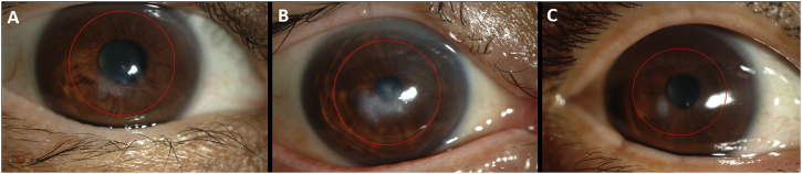
Fig.u00a01
Corneal images in keratoconus before penetrating keratoplasty. Cornea 1 (A) has mild scarring inferior to the pupil, whilst cornea 2 (B) is at a more advanced stage of the disease, hence more severe s...
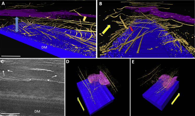
Fig.u00a02
Distribution of MBs anterior to Descemet's membrane (DM) in normal (Au2013B) and keratoconic button 2 (Cu2013E) corneas, using tannic acid staining. Blueu00a0=u00a0DM, Goldu00a0=u00a0MBs, Pink/Purpleu...
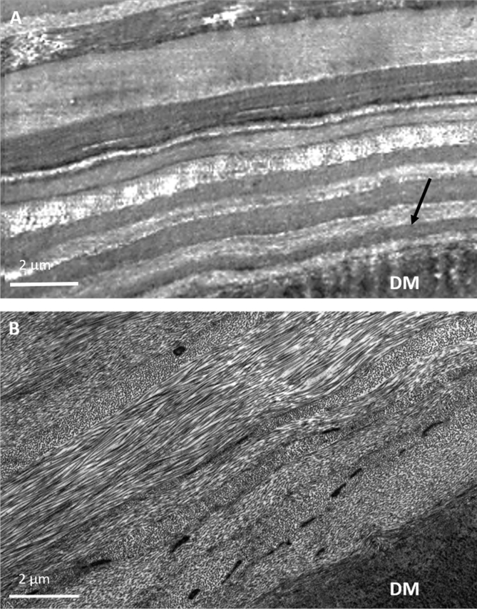
Fig.u00a03
The posterior stroma and Descemet's membrane (DM) stained with tannic acid. In keratoconic button 1 cornea (A), the TEM image shows the first u223c10u00a0u03bcm of stroma and reveals no MBs in the str...
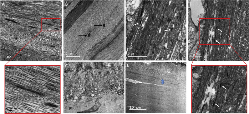
Fig.u00a04
TEM micrographs of normal (A) and keratoconic button 1 (Bu2013F) cornea using tannic acid staining. Normal cornea contains a concentration of MBs anterior to DM (A), with longitudinally sectioned stru...
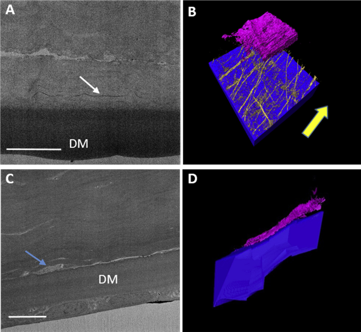
Fig.u00a05
Distribution of MBs anterior to Descemet's membrane (DM) in normal (Au2013B) and keratoconic button 2 (Cu2013D) cornea using orcein staining. Blueu00a0=u00a0DM, Goldu00a0=u00a0MBs, Pink/Purpleu00a0=u0...
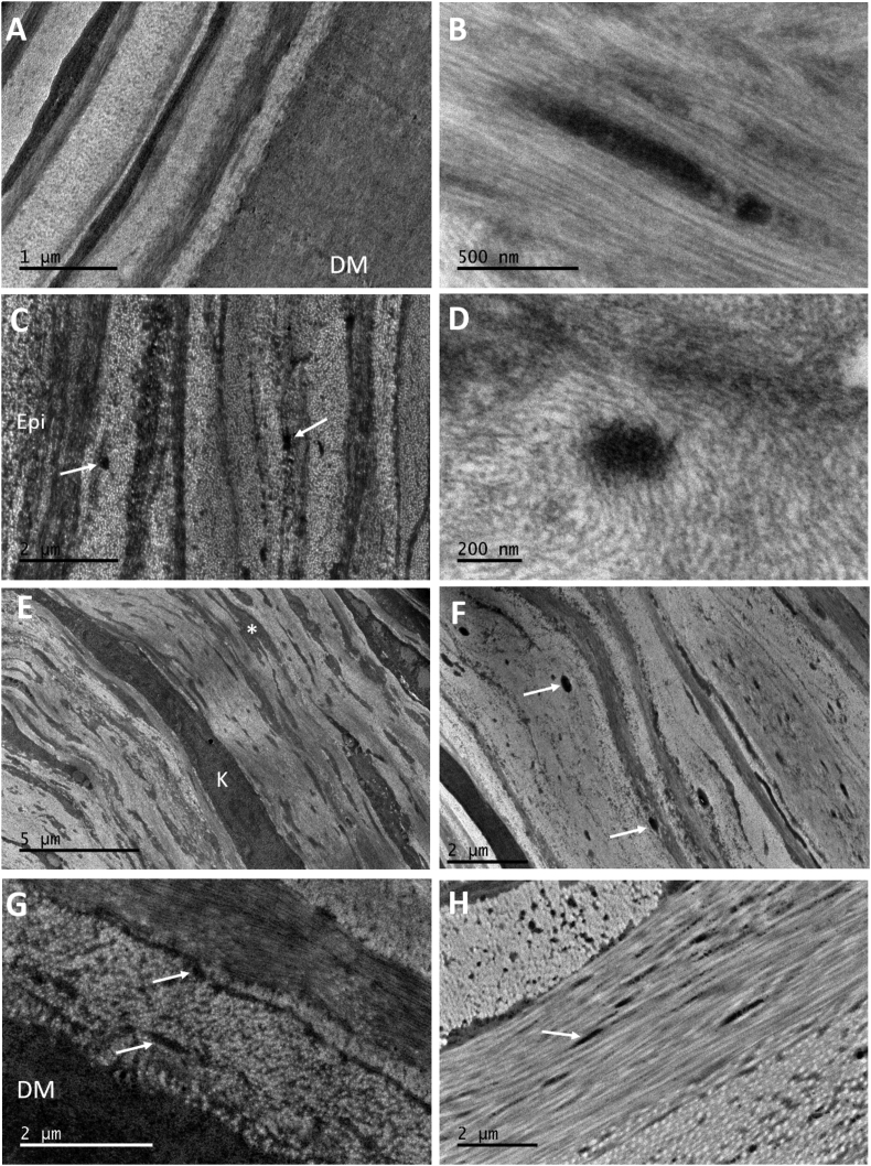
Fig.u00a06
TEM micrographs of orcein stained keratoconic cornea buttons 2 and 3 (Au2013F) and normal controls (Gu2013H), showing the presence of MBs (white arrows). No MBs are seen anterior to DM (A). MBs were o...
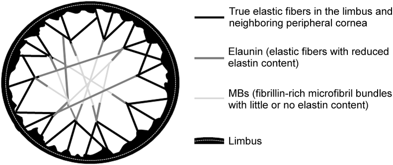
Fig.u00a07
Schematic diagram of the proposed elastic fiber network in the corneal stroma. Elastic fibers consisting of predominantly fibrillin microfibrils sheathing an amorphous elastin core occur pseudo-circum...
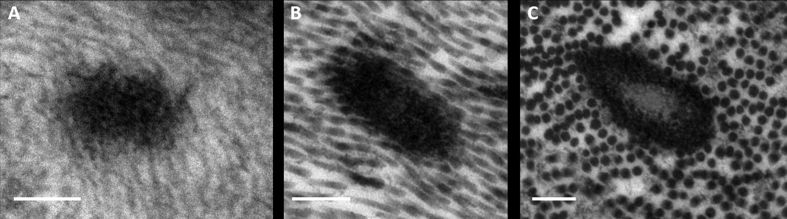
Supplementary Fig.u00a0S1
High magnification TEM images of elastic material in human cornea. When viewed in cross-section, elastic material in central keratoconic (A) and normal (B) cornea appears as electron-dense MBs, wherea...
Figure images are served from the NIH/NLM PubMed Central Open Access Subset or Europe PMC; copyright remains with the publishers and authors.
💬 Discussion
0 commentsNo comments yet. Be the first to start a discussion!
Leave a Comment