Abstract
BACKGROUND: Microscopic techniques enable real-space imaging of complex biological events and processes. They have become an essential tool to confirm and complement hypotheses made by biomedical scientists and also allow the re-examination of existing models, hence influencing future investigations. Particularly imaging live cells is crucial for an improved understanding of dynamic biological processes, however hitherto live cell imaging has been limited by the necessity to introduce probes within a cell without altering its physiological and structural integrity. We demonstrate herein that this hurdle can be overcome by effective cytosolic delivery. PRINCIPAL FINDINGS: We show the delivery within several types of mammalian cells using nanometre-sized biomimetic polymer vesicles (a.k.a. polymersomes) that offer both highly efficient cellular uptake and endolysomal escape capability without any effect on the cellular metabolic activity. Such biocompatible polymersomes can encapsulate various types of probes including cell membrane probes and nucleic acid probes as well as labelled nucleic acids, antibodies and quantum dots. SIGNIFICANCE: We show the delivery of sufficient quantities of probes to the cytosol, allowing sustained functional imaging of live cells over time periods of days to weeks. Finally the combination of such effective staining with three-dimensional imaging by confocal laser scanning microscopy allows cell imaging in complex three-dimensional environments under both mono-culture and co-culture conditions. Thus cell migration and proliferation can be studied in models that are much closer to the in vivo situation.
🔬 Techniques
🔭 Microscopes
💻 Software
✨ Fluorophores
🧪 Sample Preparation
🔬 Cell Lines
🏭 Microscope Brands
🧪 Reagent Suppliers
💻 Software Details
🏛️ Research Organizations (ROR)
Affiliated research institutions:
📋 Methods
Materials 2-(Methacryloyloxy)ethyl phosphorylcholine (MPC; >99%) was provided by Biocompatibles UK Ltd. 2-(Diisopropylamino)ethyl methacrylate (DPA; Scientific Polymer Products, USA) was passed through the column supplied by the manufacturer to remove inhibitor. Copper(I) chloride (CuCl; 99.995%), 2,2′-bipyridine (bpy, 99%), 2-bromoisobutyl bromide, triethyl-amine, methanol and isopropanol were purchased from Sigma-Aldrich UK (Poole, Dorset, UK) and were used as received. Column chromatography grade silica gel 60 (0.063–0.200 mm) used for removal of the atomic transfer radical polymerisation (ATRP) copper catalyst was purchased from E. Merck (Darmstadt, Germany). Regenerated Cellulose (RC) dialysis tubing (Spectra Por® 6, molecular weight cut-off 3.5 kDa) was purchased from Spectrum Labs (Rancho Dominguez, CA, USA). 2-(N-Morpholino)ethyl 2-bromo-2-methylpropanoate (ME-Br) was synthesized according to a previously reported procedure [38] . Chloroform was purchased from Fisher Scientific, phosphate buffer saline (PBS) tablets from Oxoid Ltd, sepharose 4B and rhodamine octadecyl ester perchlorate from Sigma-Aldrich. DMEM media and foetal calf serum were bought from Biosera (UK) and L-glutamine, penicillin streptomycin and Amphotericin B were bought from Sigma (UK). Collagenase A was purchased from Boehringer-Mannheim (Lewes, UK). For the MTT-ESTA assay, 3-(4,5-dimethylthiazol-2-yl)-2,5 diphenyl tetrazolium bromide (MTT) was purchased from Sigma-Aldrich (UK) and hydrochloric acid from BDH AnalaR. Cell tracker® red, propidium iodide, BODIPY TR ceramide, were purchased by Invitrogen (UK).Quantum dots (Lumidot™ CdSe/ZnS 69346 nanopowder) were purchased from Sigma-Aldrich UK (Poole, Dorset, UK). PMPC25-PDPA70 diblock copolymer was synthesized by ATRP as reported else where [39] . Briefly, a round bottom flask with a magnetic stirrer bar and rubber suba seal was charged with ME-Br initiator (100 mg, 0.36 mmol), bpy ligand (111 mg, 0.71 mmol), MPC monomer (2.67 g, 8.93 mmol) and dissolved in methanol (4 ml). This solution was deoxygenated by bubbling N2 for 30 min, before the addition of Cu(I)Cl (35 mg, 0.36 mmol). The relative molar ratios of [MPC]∶[ME-Br]∶[CuCl]∶[bpy] were 25∶1∶1∶2. After 65 min, a mixture of deoxygenated DPA (5.33 g, 25 mmol) and methanol (6 ml) was injected into the flask. After 48 h the reaction solution was diluted through the addition of isopropanol and then passed through a silica column to remove the residual Cu catalyst. To remove any bpy from reaction product, the copolymer was dissolved in the minimal amount of isopropanol and dialyzed against water for 7 days, with daily changes of water. The resulting dispersed copolymer was freeze-dried and a colour-less copolymer was obtained.
Show full methods section
Materials 2-(Methacryloyloxy)ethyl phosphorylcholine (MPC; >99%) was provided by Biocompatibles UK Ltd. 2-(Diisopropylamino)ethyl methacrylate (DPA; Scientific Polymer Products, USA) was passed through the column supplied by the manufacturer to remove inhibitor. Copper(I) chloride (CuCl; 99.995%), 2,2′-bipyridine (bpy, 99%), 2-bromoisobutyl bromide, triethyl-amine, methanol and isopropanol were purchased from Sigma-Aldrich UK (Poole, Dorset, UK) and were used as received. Column chromatography grade silica gel 60 (0.063–0.200 mm) used for removal of the atomic transfer radical polymerisation (ATRP) copper catalyst was purchased from E. Merck (Darmstadt, Germany). Regenerated Cellulose (RC) dialysis tubing (Spectra Por® 6, molecular weight cut-off 3.5 kDa) was purchased from Spectrum Labs (Rancho Dominguez, CA, USA). 2-(N-Morpholino)ethyl 2-bromo-2-methylpropanoate (ME-Br) was synthesized according to a previously reported procedure [38] . Chloroform was purchased from Fisher Scientific, phosphate buffer saline (PBS) tablets from Oxoid Ltd, sepharose 4B and rhodamine octadecyl ester perchlorate from Sigma-Aldrich. DMEM media and foetal calf serum were bought from Biosera (UK) and L-glutamine, penicillin streptomycin and Amphotericin B were bought from Sigma (UK). Collagenase A was purchased from Boehringer-Mannheim (Lewes, UK). For the MTT-ESTA assay, 3-(4,5-dimethylthiazol-2-yl)-2,5 diphenyl tetrazolium bromide (MTT) was purchased from Sigma-Aldrich (UK) and hydrochloric acid from BDH AnalaR. Cell tracker® red, propidium iodide, BODIPY TR ceramide, were purchased by Invitrogen (UK).Quantum dots (Lumidot™ CdSe/ZnS 69346 nanopowder) were purchased from Sigma-Aldrich UK (Poole, Dorset, UK). PMPC25-PDPA70 diblock copolymer was synthesized by ATRP as reported else where [39] . Briefly, a round bottom flask with a magnetic stirrer bar and rubber suba seal was charged with ME-Br initiator (100 mg, 0.36 mmol), bpy ligand (111 mg, 0.71 mmol), MPC monomer (2.67 g, 8.93 mmol) and dissolved in methanol (4 ml). This solution was deoxygenated by bubbling N2 for 30 min, before the addition of Cu(I)Cl (35 mg, 0.36 mmol). The relative molar ratios of [MPC]∶[ME-Br]∶[CuCl]∶[bpy] were 25∶1∶1∶2. After 65 min, a mixture of deoxygenated DPA (5.33 g, 25 mmol) and methanol (6 ml) was injected into the flask. After 48 h the reaction solution was diluted through the addition of isopropanol and then passed through a silica column to remove the residual Cu catalyst. To remove any bpy from reaction product, the copolymer was dissolved in the minimal amount of isopropanol and dialyzed against water for 7 days, with daily changes of water. The resulting dispersed copolymer was freeze-dried and a colour-less copolymer was obtained.
Methods
Production of polymersomes Polymersomes encapsulating amphiphiles/hydrophobic molecules: 1×10⁻³ moles of PMPC25-PDPA70 copolymer were dissolved in a glass vial in a 2∶1 chloroform: methanol solution. For samples encapsulating rhodamine 0.05×10⁻³ moles of rhodamine B octadecyl ester perchlorate were added (Sigma) to the co-polymer solution. For polymersomes encapsulating 1,1′-Dioctadecyl-3,3,3′,3′-tetramethylindodicarbocyanine perchlorate (Sigma) 10 µg of this compound were added to the polymer solution. For polymersomes encapsulating BODIPY TR ceramide 0.05×10⁻³ moles of phospholipids was added to the polymer solution. For polymersomes encapsulating fluorescein 1,2-dihexadecylphosphatidylethanolamine 0.05×10⁻³ moles of phospholipids were added to the polymer solution. For polymersomes encapsulating NBD Cholesterol 0.05×10⁻³ moles of that molecule were added to the polymer solution. Afterwards a copolymer film was formed by evaporating the solvent for 2 hours in a vacuum oven at 37°C. The film was then rehydrated using 2 ml pH 2 PBS (100 mM). Once the film dissolved the pH was gradually increased to 7.3 in order to obtain a 0.05 mM polymer solution. The polymersomes solution was then purified by gel permeation chromatography using a sepharose 4B size exclusion column to extract the fraction containing polymersomes and removing any un-encapsulated. Polymersomes encapsulating hydrophilic molecules: 1×10⁻³ moles of PMPC25-PDPA70 copolymer were dissolved in a glass vial in a 2∶1 chloroform: methanol solution. Afterward a copolymer film was formed by evaporating the solvent for 2 hours in a vacuum oven at 37°C. The film was then rehydrated using 2 ml pH 2 PBS (100 mM). Once the film dissolved the pH was gradually increased. At pH 6 payloads were added (0.01 mg of propidium iodide, 0.01 mg of quantum dots (Lumidot™ CdSe/ZnS 69346 nanopowder), 0.08 mg/ml of antibodies (mouse monoclonal anti-human-a-tubulin FITC-labelled IgG), 10 ng of RNA. The pH of the resulting solution was risen to 7.3 before sonication for 15 mins using a sonicator (Sonicor Instruments Corporation). The polymersomes solution was then purified by gel permeation chromatography using a sepharose 4B size exclusion column to extract the fraction containing polymersomes and removing any un-encapsulated. In all cases the polymersomes size distribution determined by Dynamic light scattering (DLS) was 200 nm±50 nm.
Cell culture
Primary human dermal fibroblasts: (HDF) were isolated from skin obtained from abdominoplasty or breast reduction operations (according to local ethically approved guidelines, NHS Trust, Sheffield, UK). Primary cultures of fibroblasts were established as previously described [40] . Briefly, the epidermal layer of the skin was removed by trypsinisation and the remaining dermal layer was washed in PBS. The dermis was then minced using surgical blades and incubated in 0.5% (w/v) collagenase A at 37°C overnight in a humidified CO2 incubator. A cellular pellet was collected from the digest and cultured in DMEM (Sigma, UK) supplemented with 10% (v/v) foetal calf serum, 2 mM L-glutamine, 100 IU/ml penicillin, 100 mg/ml streptomycin and 625 ng/ml amphotericin B. Cells were sub-cultured routinely using 0.02% (w/v) EDTA and used for experimentation between passages 4 and 8. Primary human oral fibroblasts (HOF) and normal human oral keratinocytes (HOK) were isolated from oral mucosal biopsies obtained from consenting patients during dental surgeries. The biopsies were incubated overnight at 4°C in 0.1% w/v Difco trypsin solution supplemented with 100 IU/ml penicillin, 100 mg/ml streptomycin and 0.625 µg/ml amphotericin B. The epithelium was then peeled from the connective tissue component and HOKs were gently scraped from the underside of the epithelium and the top side of the connective layer using a scalpel. HOKs were cultured according to the method by Rheinwald and Green. Briefly, cells are cultured on an irradiated mouse fibroblast (i3T3) feeder layer in Green's media. Green's media is composed of Dulbecco's modified Eagle's medium (DMEM) and Hams F12 medium in a 3∶1 (v/v) ratio supplemented with 10% (v/v) FCS, 100 IU/ml penicillin, 100 mg/ml streptomycin and 625 ng/ml amphotericin B, 100 nM cholera toxin, 10 ng/ml epidermal growth factor (EGF), 0.4 µgml-1 hydrocortisone, 0.18 mM adenine, 0.005 mg/ml insulin, 0.005 mg/ml transferrin, 2 mM glutamine and 200 nM triiodothyronine. HOKs were cultured until 80% confluent and the media was replenished every 3–4 days. i3T3 cells were detached using 0.02% EDTA solution before trypsinisation of the HOKs. HOFs were isolated from the connective tissue component of the oral biopsy. The connective tissue was finely minced and incubated at 37°C in a 5% CO2 humidified incubator overnight in 10 ml of 0.5% collagenase A. The isolated HOFs were cultured in DMEM supplemented with 10% (v/v) foetal calf serum (FCS), 100 IU ml-1 penicillin, 100 mg/ml streptomycin and 625 ng/ml amphotericin B. HOKs were used up to passage 3 and HOFs between passage 3 and 8. Human squamous cell carcinoma: (FADU) whrere cultured in RPMI supplemented with 10% FCS, 2 mM L-glutamine and 50 U/ml penicillin & streptomycin.
Wild type Chinese hamster ovary cells
(CHO) were cultured in Ham's F12 medium containing 10% FCS, 2 mM L-glutamine, 50 U/ml penicillin, and 0.05 mg/ml streptomycin and 300 ng/ml amphotericin B. Cells were sub-cultured routinely using 0.02% (w/v) EDTA. Macrophage and monocytes culture and extractions: Fresh isolated blood was collected into 50 ml Falcon tube containing 3.7% w/v sodium citrate. Blood solution was spun down for 20 minutes at 400 g/1400 rpm. Serum was collected to make platelet rich plasma derived serum (PRPDS). 0.026 ml of CACl2 1 mM was added to every ml of the serum and the solution was incubated at 37°C for 1 hour. Blood cells were then mixed 1∶1 with HBSS (Cambrex/BioWhittaker, Cat no. BE10-547F). Subsequentially 30 ml of blood/HBSS was overlaid onto 20 ml Ficoll-Hypaque Lymphocyte Separation Media (LSM1077 – PAA Labs, Cat no. J15-004). Blood fractions were spun for 40 minutes at the same rpm as before to be separated. At this point the Monocyte layer (approx 50 ml) was collected and dispersed in HBSS media. Then the tube was spun down for 15 minutes, and the supernatant was discarded. Finally the cell number was counted. Cells has been plate at 20×10 6 in a 6-well plate in IMDM (Sigma, Cat no. BE12-76F) +2% human AB serum (Sigma, H4522-100 ml)+2 mM L-Glutamine. To allow mononuclear cells adhesion cells were left for 2 hours at 37°C, 5% CO 2 . Then the medium was removed and the cells were washed 4 times with HBSS. Finally cells were left in 10 ml IMDM +2% AB serum +2 mM L-Glutamine and incubated at 37°C, 5% CO 2 . Cells treatment with polymersomes Cells were seeded in standard 6 well plates at a density of 5×10 4 cells per well and grown for two days in culture medium. Medium was fresh replenished added with 0.005 mM polymersomes encapsulating molecules. Cells were incubated for 24 hours. Treated samples were then washed 5 times with PBS and immediately visualized by live confocal fluorescent scanning microscopy. Results were compared with untreated samples. MTT-ESTA assay: All cells types were seeded in standard 24 well plates at a density of 3×10 4 cells per well and grown for two days in culture medium. Blood cells (macrophages, monocites and red blood cells) were left and treated in suspension. Cell monolayers/suspension was treated for 24 h with fresh medium containing 0.005 mM of polymersomes. Viable cell density was then assessed using the MTT (3-(4,5-dimethylthiazol-2-yl)-2,5 diphenyl tetrazolium bromide) assay. Briefly, treated/untreated cells were washed thoroughly twice with PBS and then incubated in MTT solution (0.5 mg/ml MTT in PBS, 1 ml per well of 24 well plate or per cm2 of cultured tissue) for 45 minutes at 37°C and 95% air/5% CO 2 . Intracellular metabolic activity reduces MTT to a purple formazan salt. Subsequently, the solution was aspirated and the insoluble formazan product was solubilised by adding isopropanol (0.5 ml per well of 24 well plate or 1 ml/cm 2 cultured tissue) and incubating for 10 minutes. The optical density at 540 nm was then measured using a plate reading spectrophotometer (Dynex Technologies, MRX II). The statistical analysis performed on the data was a paired Student's t-test. Cell treatment with cell tracker red The cell tracker red lyophilized product was dissolved DMSO to a final concentration of 10 mM. The stock solution was then diluted down to a final working concentration of 0.005 mM in serum-free medium and applied to cells at 37°C for 45 minutes. Cells were then washed three times with PBS and the applied solution was replenished with full medium. The statistical analysis performed on the data was a paired Student's t-test.
Culture of 3D tissue engineered oral mucosa
Skin obtained from consenting donors was de-cellularised in 1 mol/L sodium chloride for at least 8 hours and washed thoroughly in PBS and cell culture medium to create de-epithelialised dermis (DED). When HOFs and HOKs were 70–80% confluent, dye containing polymersomes were added to the cell culture medium giving a final concentration of 1 mg polymer/ml cell culture media. HOFs and HOKs were incubated for 24 hours with Rhodamine B octadecyl ester perchlorate and 1,1'-Dioctadecyl-3,3,3',3'-tetramethylindodicarbocyanine perchlorate containing polymersomes respectively. Cells were then trypsinized before being added to the 3D culture as a cell suspension. DED was cut into 2 cm×2 cm squares and placed into 6 well plates submerged in Green's media. 1 mL of cell suspension containing 5×10⁵ polymersome labelled HOKs and 5×10 5 polymersome labelled HOFs in Green's media was added into stainless steel rings pushed onto the DED. Green's media was also added outside the ring to stop the cell solution leaking out. After 2 days half the media inside the ring was changed. On day 3 the DED with cells attached was brought to an air liquid interface using a stainless steel grid and the first image was obtained. The underside of the model was in contact with Green's media while the top was exposed to the air to encourage epithelial stratification. Models were cultured for 10 days at the air-liquid interface (ALI) and imaged using an upright Zeiss LSM 510 Meta confocal microscope. Models were washed 3X with PBS before imaging and submerged in PBS during imaging.
Cell seeding using fibrin clot
Fibrin clot was used to seed cells at specific areas for some of the 2D and all the 3D culture experiments. Briefly, cell suspension was prepared by mixing fibrinogen (3.5 mg/ml), thrombin (10 units/ml) and HDFs (1×10 5 cells per ml of fibrin fibrinogen) together. Tiny drops (10–20 µm) of the cell suspension were quickly transferred to specific areas of either 2D or 3D culture systems. After incubated at 37°C for 20–40 minutes, the fibrinogen polymerised and the cells were embedded in the fibrin clot. Cell culture was subsequently carried out by submerging the cells and or the 3D scaffolds in DMEM medium. In this research, whether the HDFs were stained (with either vesicles or Cell TrackerTM) or not, the cell densities in all the cell suspensions were constant (1×10 5 cells per ml of fibrin fibrinogen). Slight modification was made to the previously developed 3D cell culture system using fibrin clot [41] . Briefly, plasminogen-free bovine fibrinogen (3.5 mg/ml) dissolved in serum free DMEM medium was mixed thoroughly with bovine thrombin (10 IU/ml), and then quickly transferred into a silicon mould attached to 6-well tissue culture plate to cast a thin fibrin gel with the thickness of 0.2–0.5 mm. After the fibrin matrix gelled within 20–40 minutes, HDFs were seeded specifically on one end of the pre-cast gel using the cell seeding method described earlier. Cell culture was then carried out by submerging the gel and the cells in DMEM medium at 37°C, 95% air/5% CO2. During cell culture in the 3D fibrin clot scaffold, the media were changed 2–3 times a week. To monitor the performance of the culture systems and or investigate cell-cell cell-scaffold interactions at different culture time points, the 6 well plates were taken out of the incubator and image analysis of the cells was performed from the bottom of the plate using inverted phase-contrast or epifluorescent microscopy as described in the following sections. For confocal laser scanning microscopy, immersion objective lenses were used to monitor the cell cultured in 2D or 3D environments directly.
Microscopy
Both epifluorescence microscopy and Confocal laser scanning microscopy were used to monitor and analyse cells cultured on either tissue culture surface or 3D scaffolds after the cells were stained with CellTracker™ or polymersomes encapsuling Rhodamine B octadecyl ester perchlorate. Epifluorescent or confocal images were taken using ImageXpress™ (AXON, USA), fluorescent microcope (Leica, Germany) with a 10×magnification lens and Confocal Laser Scanning Microcope LSM 510 META (Carl Zeiss, Germany) at λ ex = 543 nm, λ em = 600 nm (for vesicle rhodamine/Cell TrackerTM visualization) with a 40×magnification lens. In order to locate cells in fibrin clot for optical sectioning of the cells in the scaffold, cell nuclei were also stained with 1 µM of SYTO 9 dye (Invitrogen, UK) for 10–15 minutes and washing thoroughly with either PBS or cell culture media for laser scanning confocal microcopy at λ ex = 488 nm, λ em = 543 nm. In situ analysis of cell density, integrity and distribution in 2D or 3D culture was conducted using image analysis software (Openlab 4.0.2, Volocity 3.0.2 and LSM510 META). Reconstructed 3D images were also produced using LSM510 META software by taking separated z-stacked image planes through different layers of the tissues, followed by volume reconstruction.
Materials 2-(Methacryloyloxy)ethyl phosphorylcholine (MPC; >99%) was provided by Biocompatibles UK Ltd. 2-(Diisopropylamino)ethyl methacrylate (DPA; Scientific Polymer Products, USA) was passed through the column supplied by the manufacturer to remove inhibitor. Copper(I) chloride (CuCl; 99.995%), 2,2′-bipyridine (bpy, 99%), 2-bromoisobutyl bromide, triethyl-amine, methanol and isopropanol were purchased from Sigma-Aldrich UK (Poole, Dorset, UK) and were used as received. Column chromatography grade silica gel 60 (0.063–0.200 mm) used for removal of the atomic transfer radical polymerisation (ATRP) copper catalyst was purchased from E. Merck (Darmstadt, Germany). Regenerated Cellulose (RC) dialysis tubing (Spectra Por® 6, molecular weight cut-off 3.5 kDa) was purchased from Spectrum Labs (Rancho Dominguez, CA, USA). 2-(N-Morpholino)ethyl 2-bromo-2-methylpropanoate (ME-Br) was synthesized according to a previously reported procedure [38] . Chloroform was purchased from Fisher Scientific, phosphate buffer saline (PBS) tablets from Oxoid Ltd, sepharose 4B and rhodamine octadecyl ester perchlorate from Sigma-Aldrich. DMEM media and foetal calf serum were bought from Biosera (UK) and L-glutamine, penicillin streptomycin and Amphotericin B were bought from Sigma (UK). Collagenase A was purchased from Boehringer-Mannheim (Lewes, UK). For the MTT-ESTA assay, 3-(4,5-dimethylthiazol-2-yl)-2,5 diphenyl tetrazolium bromide (MTT) was purchased from Sigma-Aldrich (UK) and hydrochloric acid from BDH AnalaR. Cell tracker® red, propidium iodide, BODIPY TR ceramide, were purchased by Invitrogen (UK).Quantum dots (Lumidot™ CdSe/ZnS 69346 nanopowder) were purchased from Sigma-Aldrich UK (Poole, Dorset, UK). PMPC25-PDPA70 diblock copolymer was synthesized by ATRP as reported else where [39] . Briefly, a round bottom flask with a magnetic stirrer bar and rubber suba seal was charged with ME-Br initiator (100 mg, 0.36 mmol), bpy ligand (111 mg, 0.71 mmol), MPC monomer (2.67 g, 8.93 mmol) and dissolved in methanol (4 ml). This solution was deoxygenated by bubbling N2 for 30 min, before the addition of Cu(I)Cl (35 mg, 0.36 mmol). The relative molar ratios of [MPC]∶[ME-Br]∶[CuCl]∶[bpy] were 25∶1∶1∶2. After 65 min, a mixture of deoxygenated DPA (5.33 g, 25 mmol) and methanol (6 ml) was injected into the flask. After 48 h the reaction solution was diluted through the addition of isopropanol and then passed through a silica column to remove the residual Cu catalyst. To remove any bpy from reaction product, the copolymer was dissolved in the minimal amount of isopropanol and dialyzed against water for 7 days, with daily changes of water. The resulting dispersed copolymer was freeze-dried and a colour-less copolymer was obtained.
Methods
Production of polymersomes Polymersomes encapsulating amphiphiles/hydrophobic molecules: 1×10⁻³ moles of PMPC25-PDPA70 copolymer were dissolved in a glass vial in a 2∶1 chloroform: methanol solution. For samples encapsulating rhodamine 0.05×10⁻³ moles of rhodamine B octadecyl ester perchlorate were added (Sigma) to the co-polymer solution. For polymersomes encapsulating 1,1′-Dioctadecyl-3,3,3′,3′-tetramethylindodicarbocyanine perchlorate (Sigma) 10 µg of this compound were added to the polymer solution. For polymersomes encapsulating BODIPY TR ceramide 0.05×10⁻³ moles of phospholipids was added to the polymer solution. For polymersomes encapsulating fluorescein 1,2-dihexadecylphosphatidylethanolamine 0.05×10⁻³ moles of phospholipids were added to the polymer solution. For polymersomes encapsulating NBD Cholesterol 0.05×10⁻³ moles of that molecule were added to the polymer solution. Afterwards a copolymer film was formed by evaporating the solvent for 2 hours in a vacuum oven at 37°C. The film was then rehydrated using 2 ml pH 2 PBS (100 mM). Once the film dissolved the pH was gradually increased to 7.3 in order to obtain a 0.05 mM polymer solution. The polymersomes solution was then purified by gel permeation chromatography using a sepharose 4B size exclusion column to extract the fraction containing polymersomes and removing any un-encapsulated. Polymersomes encapsulating hydrophilic molecules: 1×10⁻³ moles of PMPC25-PDPA70 copolymer were dissolved in a glass vial in a 2∶1 chloroform: methanol solution. Afterward a copolymer film was formed by evaporating the solvent for 2 hours in a vacuum oven at 37°C. The film was then rehydrated using 2 ml pH 2 PBS (100 mM). Once the film dissolved the pH was gradually increased. At pH 6 payloads were added (0.01 mg of propidium iodide, 0.01 mg of quantum dots (Lumidot™ CdSe/ZnS 69346 nanopowder), 0.08 mg/ml of antibodies (mouse monoclonal anti-human-a-tubulin FITC-labelled IgG), 10 ng of RNA. The pH of the resulting solution was risen to 7.3 before sonication for 15 mins using a sonicator (Sonicor Instruments Corporation). The polymersomes solution was then purified by gel permeation chromatography using a sepharose 4B size exclusion column to extract the fraction containing polymersomes and removing any un-encapsulated. In all cases the polymersomes size distribution determined by Dynamic light scattering (DLS) was 200 nm±50 nm.
Cell culture
Primary human dermal fibroblasts: (HDF) were isolated from skin obtained from abdominoplasty or breast reduction operations (according to local ethically approved guidelines, NHS Trust, Sheffield, UK). Primary cultures of fibroblasts were established as previously described [40] . Briefly, the epidermal layer of the skin was removed by trypsinisation and the remaining dermal layer was washed in PBS. The dermis was then minced using surgical blades and incubated in 0.5% (w/v) collagenase A at 37°C overnight in a humidified CO2 incubator. A cellular pellet was collected from the digest and cultured in DMEM (Sigma, UK) supplemented with 10% (v/v) foetal calf serum, 2 mM L-glutamine, 100 IU/ml penicillin, 100 mg/ml streptomycin and 625 ng/ml amphotericin B. Cells were sub-cultured routinely using 0.02% (w/v) EDTA and used for experimentation between passages 4 and 8. Primary human oral fibroblasts (HOF) and normal human oral keratinocytes (HOK) were isolated from oral mucosal biopsies obtained from consenting patients during dental surgeries. The biopsies were incubated overnight at 4°C in 0.1% w/v Difco trypsin solution supplemented with 100 IU/ml penicillin, 100 mg/ml streptomycin and 0.625 µg/ml amphotericin B. The epithelium was then peeled from the connective tissue component and HOKs were gently scraped from the underside of the epithelium and the top side of the connective layer using a scalpel. HOKs were cultured according to the method by Rheinwald and Green. Briefly, cells are cultured on an irradiated mouse fibroblast (i3T3) feeder layer in Green's media. Green's media is composed of Dulbecco's modified Eagle's medium (DMEM) and Hams F12 medium in a 3∶1 (v/v) ratio supplemented with 10% (v/v) FCS, 100 IU/ml penicillin, 100 mg/ml streptomycin and 625 ng/ml amphotericin B, 100 nM cholera toxin, 10 ng/ml epidermal growth factor (EGF), 0.4 µgml-1 hydrocortisone, 0.18 mM adenine, 0.005 mg/ml insulin, 0.005 mg/ml transferrin, 2 mM glutamine and 200 nM triiodothyronine. HOKs were cultured until 80% confluent and the media was replenished every 3–4 days. i3T3 cells were detached using 0.02% EDTA solution before trypsinisation of the HOKs. HOFs were isolated from the connective tissue component of the oral biopsy. The connective tissue was finely minced and incubated at 37°C in a 5% CO2 humidified incubator overnight in 10 ml of 0.5% collagenase A. The isolated HOFs were cultured in DMEM supplemented with 10% (v/v) foetal calf serum (FCS), 100 IU ml-1 penicillin, 100 mg/ml streptomycin and 625 ng/ml amphotericin B. HOKs were used up to passage 3 and HOFs between passage 3 and 8. Human squamous cell carcinoma: (FADU) whrere cultured in RPMI supplemented with 10% FCS, 2 mM L-glutamine and 50 U/ml penicillin & streptomycin.
Wild type Chinese hamster ovary cells
(CHO) were cultured in Ham's F12 medium containing 10% FCS, 2 mM L-glutamine, 50 U/ml penicillin, and 0.05 mg/ml streptomycin and 300 ng/ml amphotericin B. Cells were sub-cultured routinely using 0.02% (w/v) EDTA. Macrophage and monocytes culture and extractions: Fresh isolated blood was collected into 50 ml Falcon tube containing 3.7% w/v sodium citrate. Blood solution was spun down for 20 minutes at 400 g/1400 rpm. Serum was collected to make platelet rich plasma derived serum (PRPDS). 0.026 ml of CACl2 1 mM was added to every ml of the serum and the solution was incubated at 37°C for 1 hour. Blood cells were then mixed 1∶1 with HBSS (Cambrex/BioWhittaker, Cat no. BE10-547F). Subsequentially 30 ml of blood/HBSS was overlaid onto 20 ml Ficoll-Hypaque Lymphocyte Separation Media (LSM1077 – PAA Labs, Cat no. J15-004). Blood fractions were spun for 40 minutes at the same rpm as before to be separated. At this point the Monocyte layer (approx 50 ml) was collected and dispersed in HBSS media. Then the tube was spun down for 15 minutes, and the supernatant was discarded. Finally the cell number was counted. Cells has been plate at 20×10 6 in a 6-well plate in IMDM (Sigma, Cat no. BE12-76F) +2% human AB serum (Sigma, H4522-100 ml)+2 mM L-Glutamine. To allow mononuclear cells adhesion cells were left for 2 hours at 37°C, 5% CO 2 . Then the medium was removed and the cells were washed 4 times with HBSS. Finally cells were left in 10 ml IMDM +2% AB serum +2 mM L-Glutamine and incubated at 37°C, 5% CO 2 . Cells treatment with polymersomes Cells were seeded in standard 6 well plates at a density of 5×10 4 cells per well and grown for two days in culture medium. Medium was fresh replenished added with 0.005 mM polymersomes encapsulating molecules. Cells were incubated for 24 hours. Treated samples were then washed 5 times with PBS and immediately visualized by live confocal fluorescent scanning microscopy. Results were compared with untreated samples. MTT-ESTA assay: All cells types were seeded in standard 24 well plates at a density of 3×10 4 cells per well and grown for two days in culture medium. Blood cells (macrophages, monocites and red blood cells) were left and treated in suspension. Cell monolayers/suspension was treated for 24 h with fresh medium containing 0.005 mM of polymersomes. Viable cell density was then assessed using the MTT (3-(4,5-dimethylthiazol-2-yl)-2,5 diphenyl tetrazolium bromide) assay. Briefly, treated/untreated cells were washed thoroughly twice with PBS and then incubated in MTT solution (0.5 mg/ml MTT in PBS, 1 ml per well of 24 well plate or per cm2 of cultured tissue) for 45 minutes at 37°C and 95% air/5% CO 2 . Intracellular metabolic activity reduces MTT to a purple formazan salt. Subsequently, the solution was aspirated and the insoluble formazan product was solubilised by adding isopropanol (0.5 ml per well of 24 well plate or 1 ml/cm 2 cultured tissue) and incubating for 10 minutes. The optical density at 540 nm was then measured using a plate reading spectrophotometer (Dynex Technologies, MRX II). The statistical analysis performed on the data was a paired Student's t-test. Cell treatment with cell tracker red The cell tracker red lyophilized product was dissolved DMSO to a final concentration of 10 mM. The stock solution was then diluted down to a final working concentration of 0.005 mM in serum-free medium and applied to cells at 37°C for 45 minutes. Cells were then washed three times with PBS and the applied solution was replenished with full medium. The statistical analysis performed on the data was a paired Student's t-test.
Culture of 3D tissue engineered oral mucosa
Skin obtained from consenting donors was de-cellularised in 1 mol/L sodium chloride for at least 8 hours and washed thoroughly in PBS and cell culture medium to create de-epithelialised dermis (DED). When HOFs and HOKs were 70–80% confluent, dye containing polymersomes were added to the cell culture medium giving a final concentration of 1 mg polymer/ml cell culture media. HOFs and HOKs were incubated for 24 hours with Rhodamine B octadecyl ester perchlorate and 1,1'-Dioctadecyl-3,3,3',3'-tetramethylindodicarbocyanine perchlorate containing polymersomes respectively. Cells were then trypsinized before being added to the 3D culture as a cell suspension. DED was cut into 2 cm×2 cm squares and placed into 6 well plates submerged in Green's media. 1 mL of cell suspension containing 5×10⁵ polymersome labelled HOKs and 5×10 5 polymersome labelled HOFs in Green's media was added into stainless steel rings pushed onto the DED. Green's media was also added outside the ring to stop the cell solution leaking out. After 2 days half the media inside the ring was changed. On day 3 the DED with cells attached was brought to an air liquid interface using a stainless steel grid and the first image was obtained. The underside of the model was in contact with Green's media while the top was exposed to the air to encourage epithelial stratification. Models were cultured for 10 days at the air-liquid interface (ALI) and imaged using an upright Zeiss LSM 510 Meta confocal microscope. Models were washed 3X with PBS before imaging and submerged in PBS during imaging.
Cell seeding using fibrin clot
Fibrin clot was used to seed cells at specific areas for some of the 2D and all the 3D culture experiments. Briefly, cell suspension was prepared by mixing fibrinogen (3.5 mg/ml), thrombin (10 units/ml) and HDFs (1×10 5 cells per ml of fibrin fibrinogen) together. Tiny drops (10–20 µm) of the cell suspension were quickly transferred to specific areas of either 2D or 3D culture systems. After incubated at 37°C for 20–40 minutes, the fibrinogen polymerised and the cells were embedded in the fibrin clot. Cell culture was subsequently carried out by submerging the cells and or the 3D scaffolds in DMEM medium. In this research, whether the HDFs were stained (with either vesicles or Cell TrackerTM) or not, the cell densities in all the cell suspensions were constant (1×10 5 cells per ml of fibrin fibrinogen). Slight modification was made to the previously developed 3D cell culture system using fibrin clot [41] . Briefly, plasminogen-free bovine fibrinogen (3.5 mg/ml) dissolved in serum free DMEM medium was mixed thoroughly with bovine thrombin (10 IU/ml), and then quickly transferred into a silicon mould attached to 6-well tissue culture plate to cast a thin fibrin gel with the thickness of 0.2–0.5 mm. After the fibrin matrix gelled within 20–40 minutes, HDFs were seeded specifically on one end of the pre-cast gel using the cell seeding method described earlier. Cell culture was then carried out by submerging the gel and the cells in DMEM medium at 37°C, 95% air/5% CO2. During cell culture in the 3D fibrin clot scaffold, the media were changed 2–3 times a week. To monitor the performance of the culture systems and or investigate cell-cell cell-scaffold interactions at different culture time points, the 6 well plates were taken out of the incubator and image analysis of the cells was performed from the bottom of the plate using inverted phase-contrast or epifluorescent microscopy as described in the following sections. For confocal laser scanning microscopy, immersion objective lenses were used to monitor the cell cultured in 2D or 3D environments directly.
Microscopy
Both epifluorescence microscopy and Confocal laser scanning microscopy were used to monitor and analyse cells cultured on either tissue culture surface or 3D scaffolds after the cells were stained with CellTracker™ or polymersomes encapsuling Rhodamine B octadecyl ester perchlorate. Epifluorescent or confocal images were taken using ImageXpress™ (AXON, USA), fluorescent microcope (Leica, Germany) with a 10×magnification lens and Confocal Laser Scanning Microcope LSM 510 META (Carl Zeiss, Germany) at λ ex = 543 nm, λ em = 600 nm (for vesicle rhodamine/Cell TrackerTM visualization) with a 40×magnification lens. In order to locate cells in fibrin clot for optical sectioning of the cells in the scaffold, cell nuclei were also stained with 1 µM of SYTO 9 dye (Invitrogen, UK) for 10–15 minutes and washing thoroughly with either PBS or cell culture media for laser scanning confocal microcopy at λ ex = 488 nm, λ em = 543 nm. In situ analysis of cell density, integrity and distribution in 2D or 3D culture was conducted using image analysis software (Openlab 4.0.2, Volocity 3.0.2 and LSM510 META). Reconstructed 3D images were also produced using LSM510 META software by taking separated z-stacked image planes through different layers of the tissues, followed by volume reconstruction.
📊 Figures
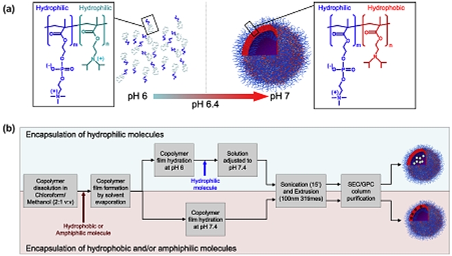
Figure 1
Polymersomes preparation.
(a) Chemical structure and solution behaviour of the PMPC-PDPA copolymer in water at different pHs. (b) Process of encapsulations for both hydrophilic, hydrophobic and amphiphilic molecules.
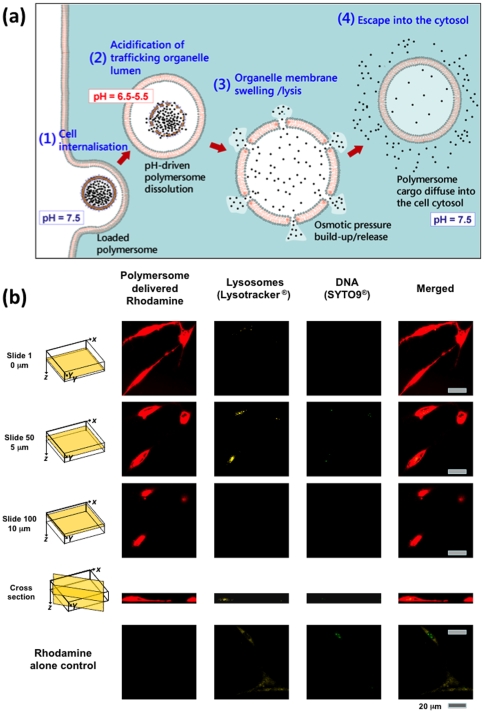
Figure 2
Polymersomes intracellular delivery.
(a) Mechanism of polymersome-mediated cytosolic delivery. (b) Primary human dermal fibroblast (HDF) exposed to Rhodamine-loaded (red) polymersomes imaged at different focal levels (0 u00b5m, 5 u00b5m,...
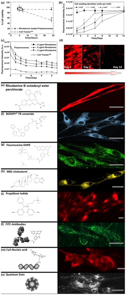
Figure 3
Tracking period and cytotoxicity induced by polymersome-mediated staining compared with commercial method.
( a ) Cell viability determined by MTT assay at different times for HDF cells exposed to either Rhodamine-loaded polymersomes or CellTracker (nu200a=u200a3, error baru200a=u200aSEM; *p<0.05). ( b )...
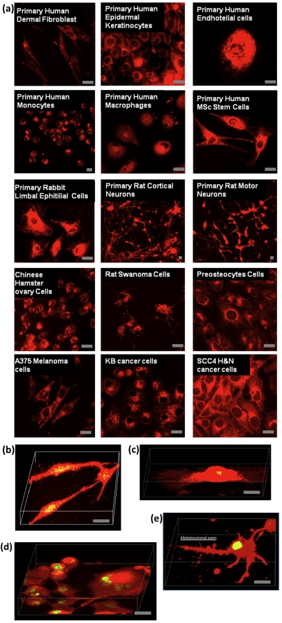
Figure 4
Selected examples of cells treated with Rhodamine-loaded polymersomes.
( a ) Live primary human dermal fibroblast (HDF), primary human epidermal keratinocytes (HEK), primary human endothelial cells (HE), primary human monocytes (HMC), primary human macrophages (HMP), pri...
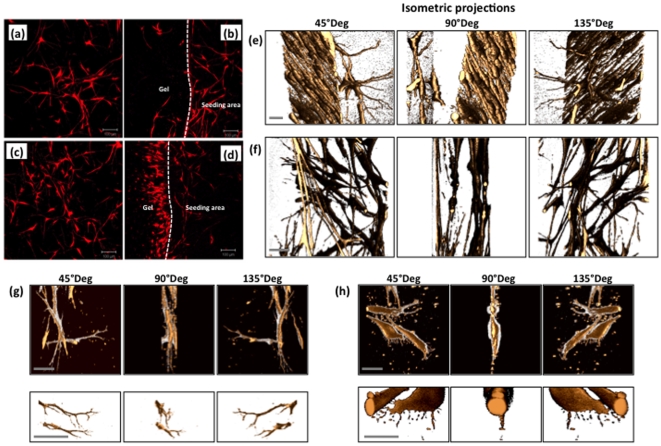
Figure 5
HDF cells seeded in a pre-cast fibrin clot gel stained using Rhodamine B octadecyl ester-loaded polymersomes and subsequently monitored using fluorescent microscopy.
( a ) Imaged after 7 days at the initial cell seeding area. ( b ) Imaged after 7 days at the interface between the initial cell seeding area and the pre-cast gel. ( c ) Imaged after 14 days at the ini...
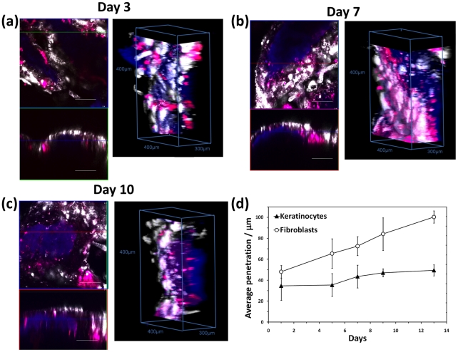
Figure 6
Tracking different cell types in 3D models.
( a ) Tissue-engineered human oral mucosa imaged by confocal laser scanning microscopy after 3 days, ( b ) 7 days and ( c ) 10 days. The keratinocytes (HOKs) and the fibroblasts (HOFs) that comprise t...
Figure images are served from the NIH/NLM PubMed Central Open Access Subset or Europe PMC; copyright remains with the publishers and authors.
💬 Discussion
0 commentsNo comments yet. Be the first to start a discussion!
Leave a Comment