Abstract
Expression of multiple reporter or effector transgenes in the same cell from a single construct is increasingly necessary in various experimental paradigms. The discovery of short, virus-derived peptide sequences that mediate a ribosome-skipping event enables generation of multiple separate peptide products from one mRNA. Here we describe methods and vectors to facilitate easy production of polycistronic-like sequences utilizing these 2A peptides tailored for expression in Drosophila both in vitro and in vivo. We tested the separation efficiency of different viral 2A peptides in cultured Drosophila cells and in vivo and found that the 2A peptides from porcine teschovirus-1 (P2A) and Thosea asigna virus (T2A) worked best. To demonstrate the utility of this approach, we used the P2A peptide to co-express the red fluorescent protein tdTomato and the genetically-encoded calcium indicator GCaMP5G in larval motorneurons. This technique enabled ratiometric calcium imaging with motion correction allowing us to record synaptic activity at the neuromuscular junction in an intact larval preparation through the cuticle. The tools presented here should greatly facilitate the generation of 2A peptide-mediated expression of multiple transgenes in Drosophila.
🔬 Techniques
🔭 Microscopes
🧬 Organisms
💻 Software
✨ Fluorophores
🧪 Sample Preparation
🏭 Microscope Brands
🧪 Reagent Suppliers
📷 Detectors
🔎 Objectives
🎨 Filters
💻 Software Details
🏛️ Research Organizations (ROR)
Affiliated research institutions:
📋 Methods
DNA Cloning
DNA sequences coding for codon-optimized 2A peptides were generated from the oligonucleotides in Table 1 , adding 3 amino acids to the N-terminus and 4 amino acids to the C-terminus of the peptide ( [7] ) (BamHI and BglII sites underlined). 2A peptide sequence variants used are shown in Table 2 . 2A sequences were generated by annealing oligos, cutting with BamHI and BglII and cloning into the BglII site of pC5-Kan ( [8] ) to produce pC5-Kan 2A. This vector contains 18 commonly used restriction enzyme sites N-terminal to the 2A sequence and 9 additional C-terminal sites that facilitate cloning genes of interest on both sides of the 2A sequence, as well as rare 8-cutter sites for shuttling the gene-2A-gene cassette into the pUAS-C5 ( [8] ) expression vector. 10.1371/journal.pone.0100637.t001 Table 1 Oligonucleotides used in this paper (added restriction enzyme sites underlined). Name Sequence F-E2A aaa ggatcc GGCCAGTGCACCAACTACGCCCTGCTGAAGCTGGCCGGCGACGTGG R-E2A ttt agatct GGGGGCGGGGCCGGGGTTGGACTCCACGTCGCCGGCCAGCTTCAGC F-F2A aaa ggatcc GGCGTGAAGCAGACCCTGAACTTCGACCTGCTGAAGCTGGCCGGCG R-F2A ttt agatct GGGGGCGGGGCCGGGGTTGGACTCCACGTCGCCGGCCAGCTTCAGCAGGTCG F-P2A aaa ggatcc GGCGCCACCAACTTCTCCCTGCTGAAGCAGGCCGGCGACGTGGAGG R-P2A ttt agatct GGGGGCGGGGCCGGGGTTCTCCTCCACGTCGCCGGCCTGC F-T2A aaa ggatcc GGCGAGGGCCGCGGCTCCCTGCTGACCTGCGGCGACGTGGAGG R-T2A ttt agatct GGGGGCGGGGCCGGGGTTCTCCTCCACGTCGCCGCAGGTCAGC F-attB NdeI GGAACC CATATG GTCGACGATGTAGG R-attB NdeI GGCTAGA CATATG CTCGACATGCCCGCCG F-myrGFP BclI aaa tgatca ATCAAAATGGGCAACAAATGCTG R-GFPo StuI aaa aggcct CTTGTAGAGCTCATCCATGCCGTG F-dsRed BamHI Aa ggatcc atggcctcctccgagg R-dsRed XbaI Ga tctaga gtcgcggccggc F-tdTomato BamHI aaa ggatcc ATGGTGAGCAAGGGCGAGG R-tdTomato HpaI aaa gttaac CTTGTACAGCTCGTCCATGCCG F-GCaMP5G BamHI aaa ggatcc ATGGGTTCTCATCATCATCATCATCATGG R-GCaMP5G XbaI aaa tctaga TCACTTCGCTGTCATCATTTGTACAAACTC F-act5c EcoRI caggaaacagctatgaccatgattac gaattc R-act5c NotI aaa gcggccgc ggtctctggattagacgactgctgg 10.1371/journal.pone.0100637.t002 Table 2 2A peptide sequences. E2A QCTNYALLKLAGDVESNPGP F2A VKQTLNFDLLKLAGDVESNPGP P2A ATNFSLLKQAGDVEENPGP T2A EGRGSLLTCGDVEENPGP MyrGFP was created by PCR using F-myrGFP BclI and R-GFPo StuI and pJFRC19 template ( [9] ) which contains a fly codon-optimized myristylation sequence derived from the first 85 amino acids of the Drosophila Src homolog (Src64B) followed by a codon-optimized EGFP (F64L, S65T). This PCR product was cut with BclI and StuI and ligated with pC5-Kan 2A cut with BamHI/StuI. DsRed-nls is also called RedStinger ( [10] ), which is a DsRed variant containing a nuclear localization signal from the Drosophila transformer gene. It was amplified by PCR with F-dsRed BamHI and R-dsRed XbaI, and cloned into pCR8GW TOPO, then excised with BamHI/XbaI and cloned into pC5-Kan myrGFP-2A cut with BglII/AvrII to make pC5-Kan myrGFP-2A–RedStinger . pPac-C5 was generated by modifying the MCS from pPac-PL (C.S. Thummel, unpublished). A BamHI/AvrII fragment from pC5-Kan was inserted into BamHI/XbaI sites of pPacPL to make pPac-C5 with a MCS including the following unique restriction sites: BamHI, XbaI, XhoI, StuI, AgeI, MluI, NheI, KpnI, NotI. A SpeI/AscI fragment from pC5-Kan myrGFP-2A–RedStinger was then cloned into XbaI/MluI sites of pPac-C5 to generate pPac-C5 myrGFP-2A–RedStinger . To make pPac-C5 myrGFP-dsRed-nls (no 2A peptide), myrGFP was subcloned from pC5-Kan myrGFP P2A into pBS-C5 ( [8] ) using EcoRI/StuI. Then dsRed-nls PCR cut with BamHI/XbaI (as described above) was added into BglII/NheI sites of pBS-C5 myrGFP. myrGFP-dsRed-nls was then cut with SpeI/AscI and ligated with pPac-C5 cut with XbaI/MluI to generate pPac-C5 myrGFP dsRed-nls (no 2A peptide control). pUAS-C5 attB was made by inserting a 285 bp attB site created from PCR with F-attB NdeI and R-attB NdeI using pattB as template ( [11] ), then inserted into NdeI site of pUAS-C5 ( [8] ). A SpeI/AscI fragment from pC5-Kan myrGFP-2A–RedStinger was subcloned into pUAS-C5 attB cut with SpeI/AscI and injected into a fly harboring the p{CaryIP}Su(hw)attP1 site at 87B13 on the third chromosome using standard techniques to generate UAS- myrGFP-2A–RedStinger flies. To make pUAS-C5 attB tdTomato-P2A–GCamP5G , tdTomato was made by PCR with F-tdTomato BamHI and R-tdTomato HpaI using pRSET-B tdTomato template (R. Tsien; [12] ), cut with BamHI/HpaI and inserted into the BamHI/StuI sites of pC5-Kan P2A . GCamP5G was amplified from Addgene plasmid #31788 with F-GCaMP5G BamHI and R-GCaMP5G XbaI, then cut with BamHI/XbaI and inserted into BglII/AvrII sites of pC5-Kan tdTomato P2A . The tdTomato-P2A–GCaMP5G fragment was then subcloned into pUAS-C5 attB with PacI/AscI and injected into p{CaryIP}Su(hw)attP1 site at 87B13 on the third chromosome. Act5c-LexAGADfl flies were generated by amplifying the act5c promoter using F-act5c and R-act5c primers, cloning into pCR8-GW-TOPO (Invitrogen), and performing an L/R reaction with pBPnlsLexAGADflUw ( [9] ). The resulting construct was integrated into the p{CaryP}attP40 site at 25C6 on the second chromosome. pC5-Kan P2A has been deposited with Addgene (plasmid #70838) and GenBank ( KJ470630 ). Other plasmids, sequences, and flies are available upon request.
Show full methods section
DNA Cloning
DNA sequences coding for codon-optimized 2A peptides were generated from the oligonucleotides in Table 1 , adding 3 amino acids to the N-terminus and 4 amino acids to the C-terminus of the peptide ( [7] ) (BamHI and BglII sites underlined). 2A peptide sequence variants used are shown in Table 2 . 2A sequences were generated by annealing oligos, cutting with BamHI and BglII and cloning into the BglII site of pC5-Kan ( [8] ) to produce pC5-Kan 2A. This vector contains 18 commonly used restriction enzyme sites N-terminal to the 2A sequence and 9 additional C-terminal sites that facilitate cloning genes of interest on both sides of the 2A sequence, as well as rare 8-cutter sites for shuttling the gene-2A-gene cassette into the pUAS-C5 ( [8] ) expression vector. 10.1371/journal.pone.0100637.t001 Table 1 Oligonucleotides used in this paper (added restriction enzyme sites underlined). Name Sequence F-E2A aaa ggatcc GGCCAGTGCACCAACTACGCCCTGCTGAAGCTGGCCGGCGACGTGG R-E2A ttt agatct GGGGGCGGGGCCGGGGTTGGACTCCACGTCGCCGGCCAGCTTCAGC F-F2A aaa ggatcc GGCGTGAAGCAGACCCTGAACTTCGACCTGCTGAAGCTGGCCGGCG R-F2A ttt agatct GGGGGCGGGGCCGGGGTTGGACTCCACGTCGCCGGCCAGCTTCAGCAGGTCG F-P2A aaa ggatcc GGCGCCACCAACTTCTCCCTGCTGAAGCAGGCCGGCGACGTGGAGG R-P2A ttt agatct GGGGGCGGGGCCGGGGTTCTCCTCCACGTCGCCGGCCTGC F-T2A aaa ggatcc GGCGAGGGCCGCGGCTCCCTGCTGACCTGCGGCGACGTGGAGG R-T2A ttt agatct GGGGGCGGGGCCGGGGTTCTCCTCCACGTCGCCGCAGGTCAGC F-attB NdeI GGAACC CATATG GTCGACGATGTAGG R-attB NdeI GGCTAGA CATATG CTCGACATGCCCGCCG F-myrGFP BclI aaa tgatca ATCAAAATGGGCAACAAATGCTG R-GFPo StuI aaa aggcct CTTGTAGAGCTCATCCATGCCGTG F-dsRed BamHI Aa ggatcc atggcctcctccgagg R-dsRed XbaI Ga tctaga gtcgcggccggc F-tdTomato BamHI aaa ggatcc ATGGTGAGCAAGGGCGAGG R-tdTomato HpaI aaa gttaac CTTGTACAGCTCGTCCATGCCG F-GCaMP5G BamHI aaa ggatcc ATGGGTTCTCATCATCATCATCATCATGG R-GCaMP5G XbaI aaa tctaga TCACTTCGCTGTCATCATTTGTACAAACTC F-act5c EcoRI caggaaacagctatgaccatgattac gaattc R-act5c NotI aaa gcggccgc ggtctctggattagacgactgctgg 10.1371/journal.pone.0100637.t002 Table 2 2A peptide sequences. E2A QCTNYALLKLAGDVESNPGP F2A VKQTLNFDLLKLAGDVESNPGP P2A ATNFSLLKQAGDVEENPGP T2A EGRGSLLTCGDVEENPGP MyrGFP was created by PCR using F-myrGFP BclI and R-GFPo StuI and pJFRC19 template ( [9] ) which contains a fly codon-optimized myristylation sequence derived from the first 85 amino acids of the Drosophila Src homolog (Src64B) followed by a codon-optimized EGFP (F64L, S65T). This PCR product was cut with BclI and StuI and ligated with pC5-Kan 2A cut with BamHI/StuI. DsRed-nls is also called RedStinger ( [10] ), which is a DsRed variant containing a nuclear localization signal from the Drosophila transformer gene. It was amplified by PCR with F-dsRed BamHI and R-dsRed XbaI, and cloned into pCR8GW TOPO, then excised with BamHI/XbaI and cloned into pC5-Kan myrGFP-2A cut with BglII/AvrII to make pC5-Kan myrGFP-2A–RedStinger . pPac-C5 was generated by modifying the MCS from pPac-PL (C.S. Thummel, unpublished). A BamHI/AvrII fragment from pC5-Kan was inserted into BamHI/XbaI sites of pPacPL to make pPac-C5 with a MCS including the following unique restriction sites: BamHI, XbaI, XhoI, StuI, AgeI, MluI, NheI, KpnI, NotI. A SpeI/AscI fragment from pC5-Kan myrGFP-2A–RedStinger was then cloned into XbaI/MluI sites of pPac-C5 to generate pPac-C5 myrGFP-2A–RedStinger . To make pPac-C5 myrGFP-dsRed-nls (no 2A peptide), myrGFP was subcloned from pC5-Kan myrGFP P2A into pBS-C5 ( [8] ) using EcoRI/StuI. Then dsRed-nls PCR cut with BamHI/XbaI (as described above) was added into BglII/NheI sites of pBS-C5 myrGFP. myrGFP-dsRed-nls was then cut with SpeI/AscI and ligated with pPac-C5 cut with XbaI/MluI to generate pPac-C5 myrGFP dsRed-nls (no 2A peptide control). pUAS-C5 attB was made by inserting a 285 bp attB site created from PCR with F-attB NdeI and R-attB NdeI using pattB as template ( [11] ), then inserted into NdeI site of pUAS-C5 ( [8] ). A SpeI/AscI fragment from pC5-Kan myrGFP-2A–RedStinger was subcloned into pUAS-C5 attB cut with SpeI/AscI and injected into a fly harboring the p{CaryIP}Su(hw)attP1 site at 87B13 on the third chromosome using standard techniques to generate UAS- myrGFP-2A–RedStinger flies. To make pUAS-C5 attB tdTomato-P2A–GCamP5G , tdTomato was made by PCR with F-tdTomato BamHI and R-tdTomato HpaI using pRSET-B tdTomato template (R. Tsien; [12] ), cut with BamHI/HpaI and inserted into the BamHI/StuI sites of pC5-Kan P2A . GCamP5G was amplified from Addgene plasmid #31788 with F-GCaMP5G BamHI and R-GCaMP5G XbaI, then cut with BamHI/XbaI and inserted into BglII/AvrII sites of pC5-Kan tdTomato P2A . The tdTomato-P2A–GCaMP5G fragment was then subcloned into pUAS-C5 attB with PacI/AscI and injected into p{CaryIP}Su(hw)attP1 site at 87B13 on the third chromosome. Act5c-LexAGADfl flies were generated by amplifying the act5c promoter using F-act5c and R-act5c primers, cloning into pCR8-GW-TOPO (Invitrogen), and performing an L/R reaction with pBPnlsLexAGADflUw ( [9] ). The resulting construct was integrated into the p{CaryP}attP40 site at 25C6 on the second chromosome. pC5-Kan P2A has been deposited with Addgene (plasmid #70838) and GenBank ( KJ470630 ). Other plasmids, sequences, and flies are available upon request.
Fly Stocks
The following genotypes were used: act5c -Gal4 (stock #3954, deposited by Dr. Yashushi Hiromi, unpublished), tubulin-Gal4 (stock #5138, deposited by Dr. Liqun Luo ( [13] ) and n-syb -Gal4 (stock #51635, generated by Dr. Julie Simpson, unpublished) (from Bloomington stock center). UAS- myrGFP-2A–RedStinger and UAS- tdTomato-P2A–GCaMP5G flies were generated as described above. Act5c-LexA (this paper) and LexAop:myrGFP (stock #32212) were used as negative controls for the western blot from adult flies.
S2 Cell Culture
S2 cells (selected for adhesion; gift of Dr. Cedric Wesley, University of Wisconsin-Madison) were grown in DMEM (Invitrogen) supplemented with 10% FBS and 1% penicillin-streptomycin and transfected using Effectene (QIAgen) according to manufacturer’s protocol.
Western Blotting
S2 cells were harvested, washed with PBS and resuspended in 2x SDS sample buffer, then boiled and centrifuged. Supernatant was then loaded on a 14% SDS-PAGE gel. 15 adult flies were anesthetized, transferred to tubes and homogenized in 2x SDS sample buffer, boiled and centrifuged. Approximately 1 fly was loaded per lane. Protein was transferred to a PVDF membrane, blocked with Odyssey blocking buffer (Licor) and probed with rabbit anti-GFP (A6455, Invitrogen) at 1∶1000, followed by donkey anti-rabbit 800 IRDye (Licor) at 1∶10,000. Quantitative imaging was performed using an Odyssey infrared imaging system (Licor). Integrated intensity measurements were performed with Odyssey software. Separation efficiency was calculated as the lower band intensity divided by the sum of lower and upper band intensities after subtracting the background.
Immunohistochemistry and Imaging
S2 cells were harvested 48 hours post-transfection and resuspended in fresh media and then plated on a poly-lysine coated coverslip for 10 minutes. The solution was removed and replaced with PBS and then 4% parafomaldehyde in 0.1 M phosphate buffer, pH 7.20 for 5 minutes. Fixative was removed and the coverslip washed with PBS before placing on a droplet of Vectashield (Vector Laboratories) on a slide and sealed with nail polish. 3 rd instar larval salivary glands were fixed 20 minutes in 4% PFA freshly diluted from 16% stock (EM Sciences) in 0.1 M phosphate buffer pH 7.20 with 0.2% Triton-X 100, then washed with PBS containing 0.1% Triton-X 100 and To-Pro3 (Invitrogen) before equilibrating in 70% glycerol and mounting in Vectashield. Samples were imaged on a Zeiss LSM 510 confocal microscope with an Imager.M1 module using 20X/0.8 NA Plan-APOCHROMAT, 40X/1.3NA Plan-NEOFLUAR or 63X/1.4NA Plan-APOCHROMAT objectives. Fluorescence was excited using an Argon laser at 488 nm, a DPSS 561 laser at 561 nm, and a HeNe laser at 643 nm.
Image Analysis The 3D Object Counter plugin in ImageJ software
(FIJI package,NIH) was used to determine the size and abundance of bright GFP/RFP-positive aggregates from confocal stacks of larval muscles.
Live Imaging
Live imaging was performed ex vivo in modified BES-buffered hemolymph-like solution 6 (0mM NaHCO 3 − , 19mM BES, pH 7.3; [14] ) supplemented with 2mM Ca 2+ and 7mM L-glutamic acid (LGA; Cat. no. G1626; Sigma-Aldrich) to prevent muscle contraction during nerve stimulation. Wide-field microscopy was performed on an Olympus BX51WI microscope fitted with a 60× (0.9 NA) water-immersion objective. A Lumen Dynamics (Mississauga, Ontario, Canada) X-Cite XLED1 LED excitation unit with GYX (540–600 nm) and BDX (450–495 nm) modules was used to supply excitation light which was then further filtered with a dual-band pass 470/556 nm filter. Images were captured with an Andor Technology (Belfast, Northern Ireland) EMCCD camera (DV887). A Cairn Instruments (Kent, UK) Optosplit II emission beam-splitter was placed before the camera for dual-emission wavelength imaging. Simultaneous imaging of tdTomato (ex./em. 556/640 nm, [12] ) and GCaMP5 (ex./em 480/530 nm, [15] ) was achieved using a primary 500/530 nm bandpass dichroic mirror in the microscope housing, a secondary 600 nm dichroic mirror mounted in the emission beam-splitter, and 512/25 nm and 660/20 nm emission filters in the beam-splitter. Filters and dichroic mirrors were obtained from Chroma Technology (Bellows Falls, VT, USA) or Semrock (Lake Forest, IL, USA). The imaging system was controlled through a Dell PC running Andor iQ software (version 2.7.1). All imaging was performed at room temperature (∼24°C) on MN termini of axons innervating muscle 13 of abdominal segment 4 with type-Ib ‘big’ boutons (MN13-Ib), with regions of interest drawn around at least three boutons. The image acquisition rate was 5 Hz in all cases.
Preparation of Specimens
For ex vivo imaging, female wandering 3 rd instar Drosophila larvae were collected and fillet-dissected in chilled HL6 to expose the longitudinal muscles of the body wall and the segmental nerves that project from the ventral ganglion. The posterior ventral ganglion was cut from the cerebral ganglia to stop endogenous nerve activity ( [17] ) and a loop of a segmental nerve was drawn into the lumen of a glass micropipette with the ventral ganglion still attached ( [14] ). Nerves were stimulated as described previously ( [18] ). For in vivo imaging, intact larvae were restrained between a glass slide and coverslip.
Statistical Analysis and Figure Preparation
SigmaPlot (version 12.3; Systat Software) was used for statistical analysis. One-way ANOVA (with a Bonferroni post hoc test) was used for comparisons. Differences were considered to be statistically significant at α-values of P ≤0.05.
📊 Figures
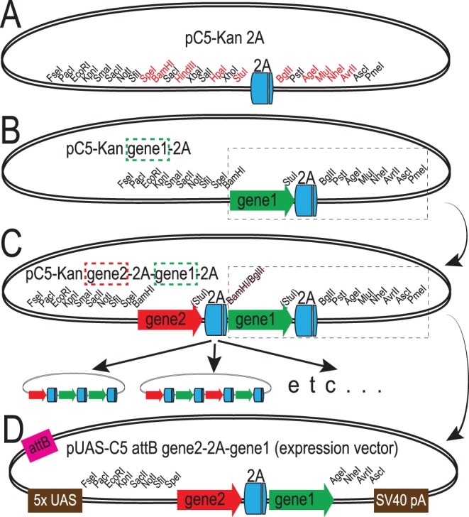
Figure 1
Outline of vector design for cloning 2A peptide-linked transgenes.
A. Diagram of pC5-Kan 2A, a vector containing a codon-optimized 2A peptide coding sequence flanked on each side by multiple cloning sites that facilitates cloning. Restriction sites in frame with the ...
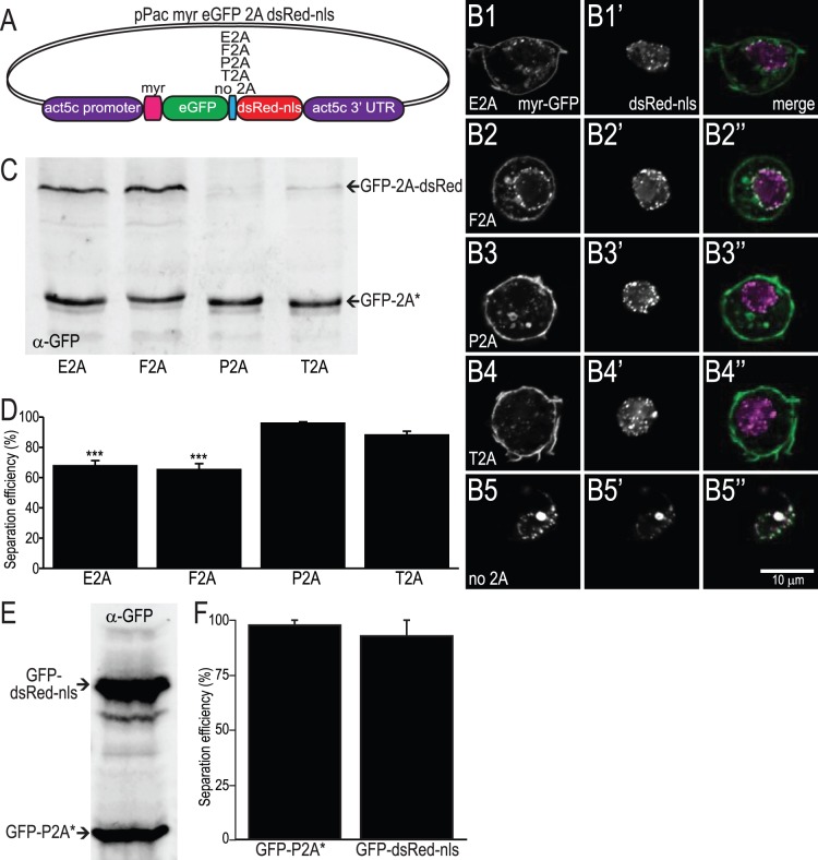
Figure 2
Demonstration of separation of proteins linked by different 2A peptides in cultured cells.
A. Diagram of vector used to transform S2 cells to express myristylated GFP and nuclear-localized dsRed. B. Single optical sections from confocal micrographs of fixed S2 cells transfected with vector ...
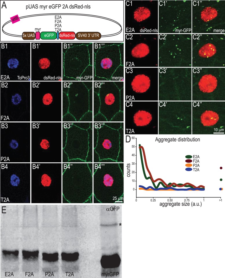
Figure 3
Testing the separation efficiency of 2A peptides in vivo .
A. Diagram of the DNA construct injected into flies placing expression of myrGFP-2A-dsRed-nls under the control of Gal4. B. Single confocal sections showing separation of myrGFP and dsRed-nls in saliv...
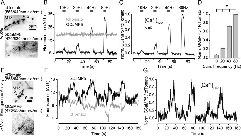
Figure 4
Application of the P2A peptide system for ratiometric calcium imaging at the larval NMJ.
A. Dual-channel imaging of tdTomato (top) and GCaMP5G (bottom) at the neuromuscular junctions on muscle 13 of a dissected third instar larva expressing UAS-tdTomato-P2Au2013GCaMP5G driven by n-syb-Gal...
Figure images are served from the NIH/NLM PubMed Central Open Access Subset or Europe PMC; copyright remains with the publishers and authors.
💬 Discussion
0 commentsNo comments yet. Be the first to start a discussion!
Leave a Comment