Abstract
As a part of our programme to develop nanobioconjugates for the treatment of cancer, we first synthesized extracellular, protein-capped, highly stable and well-dispersed gadolinium oxide (Gd2O3) nanoparticles by using thermophilic fungus Humicola sp. The biodistribution of the nanoparticles in rats was checked by radiolabelling with Tc-99m. Finally, these nanoparticles were bioconjugated with the chemically modified anticancer drug taxol with the aim of characterizing the role of this bioconjugate in the treatment of cancer. The biosynthesized Gd2O3 nanoparticles were characterized by UV-vis spectroscopy, transmission electron microscopy (TEM), X-ray diffraction (XRD) and X-ray photoemission spectroscopy (XPS). The Gd2O3-taxol bioconjugate was confirmed by UV-vis spectroscopy and fluorescence microscopy and was purified by using high performance liquid chromatography (HPLC).
🔬 Techniques
🔬 Cell Lines
🏭 Microscope Brands
🧪 Reagent Suppliers
🏛️ Research Organizations (ROR)
Affiliated research institutions:
📋 Methods
Experimental Materials Gadolinium chloride (GdCl 3 ) and sodium carbonate were obtained from Sigma Aldrich. Malt extract, yeast extract, glucose and peptone were obtained from HiMedia and used as received.
Methods
The thermophilic fungus Humicola sp. was cultured and maintained by us as described previously [ 21 ]. Biosynthesis of gadolinium oxide nanoparticles The harvested mycelial mass weighing 20 g [ 21 ] was suspended in 100 mL of 10 −3 M aqueous gadolinium chloride solution in a 250 mL Erlenmeyer flask at pH 9. The whole mixture was put onto a shaker at 50 °C (200 rpm) and maintained in the dark.
Characterization of gadolinium oxide nanoparticles UV–vis spectroscopy
To check the synthesis of gadolinium oxide nanoparticles, the mixture was monitored by periodic sampling of aliquots (2 mL) of the aqueous component. The measurement was carried out on a Shimadzu dual-beam spectrophotometer (model UV-1601 PC) operated at a resolution of 1 nm. Transmission electron microscopy (TEM) TEM analyses of gadolinium oxide nanoparticles were carried out on a JEOL model 1200 EX operated at 80 kV. Samples were prepared by drop-casting the particles (suspended in water) on carbon coated copper grids. High resolution (HR)-TEM HR-TEM analysis was carried out on a TECHNAI G2 F30 S-TWIN instrument operated at an acceleration voltage of 300 kV with a lattice resolution of 0.14 nm and a point image resolution of 0.20 nm. A sample was prepared by drop-casting the particles (suspended in water) on carbon coated copper grid. The selected area electron diffraction (SAED) pattern analysis was carried out on the same grid. X-ray diffraction (XRD) X-ray diffraction (XRD) measurements of biosynthesized Gd 2 O 3 nanoparticles were carried out by coating the Gd 2 O 3 powder on a glass substrate on a Philips X’PERT PRO instrument equipped X’celerator. Iron-filtered Cu Kα radiation (λ = 1.5406 Å) was used and the sample was scanned by using X’celerator with 121 active channels. XRD patterns were recorded in the 2θ range of 10–80° with a step size of 0.02° and a time of 5 seconds per step at 40 kV voltage and a current of 30 mA. X-ray photoemission spectroscopy (XPS) XPS of Gd 2 O 3 nanoparticles powder was carried out on a VG microtech ESCA (XPS) 3000 spectrometer. The base pressure during XPS analysis was 1 × 10 −9 Torr and Mg Kα X-ray radiation (1253.6 eV) at a power of 200 watts was used. The binding energy of Au (4f7/2) at 84.0 ± 0.1 eV was used to calibrate the binding energy scale of the spectrometer. Any charging shift produced in the spectrum was corrected by referencing to the C (1s) position (284.6 eV) Background correction of core level spectra was performed by using the Shirley algorithm. The chemically distinct species were resolved by a nonlinear least square fitting procedure. Radiolabelling and biodistribution studies Radiolabelling of gadolinium oxide (Gd 2 O 3 ) nanoparticles with Tc-99m To fabricate Tc-99m–Gd 2 O 3 nanoparticles, 10 mg of Gd 2 O 3 nanoparticles were dissolved in 1 mL of distilled water, to which 100 μg of SnCl 2 ·2H 2 O was added, and the pH was brought to 6.5. A 0.22 μm membrane filter was employed to filter the contents into a sterile vial to which approximately 2 mCi of Tc-99m was added and the mixture was incubated for 10 min. The instant thin layer chromatography (ITLC) method was used to determine the percentage of radiolabeling [ 22 ]. Radiochemical purity (RCP) ITLC with silica gel coated fiber sheets was used to estimate the radiochemical purity of Tc-99m with Gd 2 O 3 nanoparticles employing 100% acetone and 0.9% saline as the mobile phase. To the ITLC-SG strip, 2–3 μL of the radiolabeled complex was applied at a point 1 cm from the end and allowed to run for approximately 10 cm. ITLC as the stationary phase and pyridine/acetic acid/water (3:5:1.5 v/v) as the mobile phase were used in determining the amount of reduced/hydrolyzed Tc-99m. A radioactivity well counter (ECIL) was employed in determining the radioactivity distribution over the strip. The fraction of radioactivity remaining at the origin determined the radiochemical purity (RCP), which was designated as % RCP. Biodistribution of radiolabelled nanoparticles A male Sprauge Dawley rat weighing 180–220 g was chosen to evaluate the localization of the labeled complex. The Tc-99m–Gd 2 O 3 nanoparticles of 14.8 MBq were administered into the rat through its penile vein. The biodistribution studies of these nanoparticles were conducted 45 min post-injection. Bioconjugation of taxol with Gd 2 O 3 nanoparticles Materials Glutaric anhydride, pyridine, 1,1’-carbonyldiimidazole (CDI), tert -butyldimethylsilyl chloride, imidazole, dimethylformamide (DMF), succinic anhydride, 4-dimethylaminopyridine, 1-ethyl-3-(3-dimethylaminopropyl)carbodiimide (EDC), 3-nitro-L-tyrosine ethyl ester hydrochloride (NTEE), 1-hydroxybenzotriazol (HBT), 2-( N -morpholino)ethanesulfonic acid (MES) and 2-[4-(2-hydroxyethyl)piperazin-1-yl]ethanesulfonic acid (HEPES) were purchased from Sigma, and HPLC grade solvents (acetonitrile, chloroform, etc) were purchased from Merck. Methods/modification of taxol Synthesis of 2’-glutaryltaxol: 2’-Glutaryltaxol was prepared by reacting 10 mg of taxol, dissolved in 1.2 mL of pyridine, with 140 mg of glutaric anhydride [ 23 ]. The reaction was carried out at room temperature for about 2 h and was monitored on TLC by using a mobile phase of chloroform/acetonitrile (7:3). After the incubation period, the solvent was evaporated under high vacuum, and the residue was washed twice with water. The obtained product was precipitated by using acetone and further purified by preparative TLC by using the mobile phase chloroform/acetonitrile (7:3). Synthesis of 2’-glutarylhexanediamine taxol: The recovered 2’-glutaryltaxol from the preparative TLC was solvent dried and dissolved in 100 µL of dry acetonitrile, 5 µmol of 1,1’-carbonyldiimidazole (CDI) was added, and it was heated at 45 °C for about 15 min. After the reaction mixture was at room temperature, 5 µmol of 1,6-hexanediamine·2HCl was added, and it was incubated at room temperature for 1 h. The reaction was monitored on TLC and purified as described above. Estimation of free carboxyl groups on Gd 2 O 3 nanoparticles and bioconjugation with taxol Biologically synthesized Gd 2 O 3 nanoparticles have a natural protein coat. The carboxyl groups present on this protein capped nanoparticles were targeted to couple with the free amino group present in 2’-glutarylhexanediamine taxol and estimated by the following procedure: The total reaction mixture of 3 mL containing 100 μg of Gd 2 O 3 nanoparticles in 50 mM MES/HEPES buffer (75:25 v/v) pH 6.0, 50 mM EDC and 30 mM NTEE was incubated at 30 °C for 45 min. Subsequently, the reaction was terminated with the addition of 1 mL of 10% TCA, and the precipitated Gd-peptide complex was collected by centrifugation, washed extensively with chilled acetone, air-dried and dissolved in 1 mL of 100 mM NaOH. The number of nitrotyrosyl groups was determined spectrophotometrically at 430 nm by using a molar absorption coefficient of 4600 M −1 cm −1 . 2’-Glutarylhexanediamine taxol (400 µg) was dissolved in anhydrous DMF (300 µL), and EDC (1.2 µmol, 1.1 equiv) along with 1-hydroxybenzotriazol (HBT) (4 µmol, 2.2 equiv). The reaction mixture was stirred at room temperature for about 1 h, and a solution of Gd 2 O 3 nanoparticles in phosphate buffer of pH 7.2 was added. After stirring for 12 h at room temperature, the reaction mixture was concentrated under a high vacuum. Further purification of the 2’-glutarylhexanediamine-taxol–Gd 2 O 3 bioconjugate was carried out by HPLC. Characterization of Gd 2 O 3 –taxol bioconjugate UV–vis spectroscopy The UV–vis spectroscopic analysis of Gd 2 O 3 –taxol bioconjugate was carried out on a Shimadzu dual-beam spectrophotometer (model UV-1601 PC) operated at a resolution of 1 nm.
Show full methods section
Experimental Materials Gadolinium chloride (GdCl 3 ) and sodium carbonate were obtained from Sigma Aldrich. Malt extract, yeast extract, glucose and peptone were obtained from HiMedia and used as received.
Methods
The thermophilic fungus Humicola sp. was cultured and maintained by us as described previously [ 21 ]. Biosynthesis of gadolinium oxide nanoparticles The harvested mycelial mass weighing 20 g [ 21 ] was suspended in 100 mL of 10 −3 M aqueous gadolinium chloride solution in a 250 mL Erlenmeyer flask at pH 9. The whole mixture was put onto a shaker at 50 °C (200 rpm) and maintained in the dark.
Characterization of gadolinium oxide nanoparticles UV–vis spectroscopy
To check the synthesis of gadolinium oxide nanoparticles, the mixture was monitored by periodic sampling of aliquots (2 mL) of the aqueous component. The measurement was carried out on a Shimadzu dual-beam spectrophotometer (model UV-1601 PC) operated at a resolution of 1 nm. Transmission electron microscopy (TEM) TEM analyses of gadolinium oxide nanoparticles were carried out on a JEOL model 1200 EX operated at 80 kV. Samples were prepared by drop-casting the particles (suspended in water) on carbon coated copper grids. High resolution (HR)-TEM HR-TEM analysis was carried out on a TECHNAI G2 F30 S-TWIN instrument operated at an acceleration voltage of 300 kV with a lattice resolution of 0.14 nm and a point image resolution of 0.20 nm. A sample was prepared by drop-casting the particles (suspended in water) on carbon coated copper grid. The selected area electron diffraction (SAED) pattern analysis was carried out on the same grid. X-ray diffraction (XRD) X-ray diffraction (XRD) measurements of biosynthesized Gd 2 O 3 nanoparticles were carried out by coating the Gd 2 O 3 powder on a glass substrate on a Philips X’PERT PRO instrument equipped X’celerator. Iron-filtered Cu Kα radiation (λ = 1.5406 Å) was used and the sample was scanned by using X’celerator with 121 active channels. XRD patterns were recorded in the 2θ range of 10–80° with a step size of 0.02° and a time of 5 seconds per step at 40 kV voltage and a current of 30 mA. X-ray photoemission spectroscopy (XPS) XPS of Gd 2 O 3 nanoparticles powder was carried out on a VG microtech ESCA (XPS) 3000 spectrometer. The base pressure during XPS analysis was 1 × 10 −9 Torr and Mg Kα X-ray radiation (1253.6 eV) at a power of 200 watts was used. The binding energy of Au (4f7/2) at 84.0 ± 0.1 eV was used to calibrate the binding energy scale of the spectrometer. Any charging shift produced in the spectrum was corrected by referencing to the C (1s) position (284.6 eV) Background correction of core level spectra was performed by using the Shirley algorithm. The chemically distinct species were resolved by a nonlinear least square fitting procedure. Radiolabelling and biodistribution studies Radiolabelling of gadolinium oxide (Gd 2 O 3 ) nanoparticles with Tc-99m To fabricate Tc-99m–Gd 2 O 3 nanoparticles, 10 mg of Gd 2 O 3 nanoparticles were dissolved in 1 mL of distilled water, to which 100 μg of SnCl 2 ·2H 2 O was added, and the pH was brought to 6.5. A 0.22 μm membrane filter was employed to filter the contents into a sterile vial to which approximately 2 mCi of Tc-99m was added and the mixture was incubated for 10 min. The instant thin layer chromatography (ITLC) method was used to determine the percentage of radiolabeling [ 22 ]. Radiochemical purity (RCP) ITLC with silica gel coated fiber sheets was used to estimate the radiochemical purity of Tc-99m with Gd 2 O 3 nanoparticles employing 100% acetone and 0.9% saline as the mobile phase. To the ITLC-SG strip, 2–3 μL of the radiolabeled complex was applied at a point 1 cm from the end and allowed to run for approximately 10 cm. ITLC as the stationary phase and pyridine/acetic acid/water (3:5:1.5 v/v) as the mobile phase were used in determining the amount of reduced/hydrolyzed Tc-99m. A radioactivity well counter (ECIL) was employed in determining the radioactivity distribution over the strip. The fraction of radioactivity remaining at the origin determined the radiochemical purity (RCP), which was designated as % RCP. Biodistribution of radiolabelled nanoparticles A male Sprauge Dawley rat weighing 180–220 g was chosen to evaluate the localization of the labeled complex. The Tc-99m–Gd 2 O 3 nanoparticles of 14.8 MBq were administered into the rat through its penile vein. The biodistribution studies of these nanoparticles were conducted 45 min post-injection. Bioconjugation of taxol with Gd 2 O 3 nanoparticles Materials Glutaric anhydride, pyridine, 1,1’-carbonyldiimidazole (CDI), tert -butyldimethylsilyl chloride, imidazole, dimethylformamide (DMF), succinic anhydride, 4-dimethylaminopyridine, 1-ethyl-3-(3-dimethylaminopropyl)carbodiimide (EDC), 3-nitro-L-tyrosine ethyl ester hydrochloride (NTEE), 1-hydroxybenzotriazol (HBT), 2-( N -morpholino)ethanesulfonic acid (MES) and 2-[4-(2-hydroxyethyl)piperazin-1-yl]ethanesulfonic acid (HEPES) were purchased from Sigma, and HPLC grade solvents (acetonitrile, chloroform, etc) were purchased from Merck. Methods/modification of taxol Synthesis of 2’-glutaryltaxol: 2’-Glutaryltaxol was prepared by reacting 10 mg of taxol, dissolved in 1.2 mL of pyridine, with 140 mg of glutaric anhydride [ 23 ]. The reaction was carried out at room temperature for about 2 h and was monitored on TLC by using a mobile phase of chloroform/acetonitrile (7:3). After the incubation period, the solvent was evaporated under high vacuum, and the residue was washed twice with water. The obtained product was precipitated by using acetone and further purified by preparative TLC by using the mobile phase chloroform/acetonitrile (7:3). Synthesis of 2’-glutarylhexanediamine taxol: The recovered 2’-glutaryltaxol from the preparative TLC was solvent dried and dissolved in 100 µL of dry acetonitrile, 5 µmol of 1,1’-carbonyldiimidazole (CDI) was added, and it was heated at 45 °C for about 15 min. After the reaction mixture was at room temperature, 5 µmol of 1,6-hexanediamine·2HCl was added, and it was incubated at room temperature for 1 h. The reaction was monitored on TLC and purified as described above. Estimation of free carboxyl groups on Gd 2 O 3 nanoparticles and bioconjugation with taxol Biologically synthesized Gd 2 O 3 nanoparticles have a natural protein coat. The carboxyl groups present on this protein capped nanoparticles were targeted to couple with the free amino group present in 2’-glutarylhexanediamine taxol and estimated by the following procedure: The total reaction mixture of 3 mL containing 100 μg of Gd 2 O 3 nanoparticles in 50 mM MES/HEPES buffer (75:25 v/v) pH 6.0, 50 mM EDC and 30 mM NTEE was incubated at 30 °C for 45 min. Subsequently, the reaction was terminated with the addition of 1 mL of 10% TCA, and the precipitated Gd-peptide complex was collected by centrifugation, washed extensively with chilled acetone, air-dried and dissolved in 1 mL of 100 mM NaOH. The number of nitrotyrosyl groups was determined spectrophotometrically at 430 nm by using a molar absorption coefficient of 4600 M −1 cm −1 . 2’-Glutarylhexanediamine taxol (400 µg) was dissolved in anhydrous DMF (300 µL), and EDC (1.2 µmol, 1.1 equiv) along with 1-hydroxybenzotriazol (HBT) (4 µmol, 2.2 equiv). The reaction mixture was stirred at room temperature for about 1 h, and a solution of Gd 2 O 3 nanoparticles in phosphate buffer of pH 7.2 was added. After stirring for 12 h at room temperature, the reaction mixture was concentrated under a high vacuum. Further purification of the 2’-glutarylhexanediamine-taxol–Gd 2 O 3 bioconjugate was carried out by HPLC. Characterization of Gd 2 O 3 –taxol bioconjugate UV–vis spectroscopy The UV–vis spectroscopic analysis of Gd 2 O 3 –taxol bioconjugate was carried out on a Shimadzu dual-beam spectrophotometer (model UV-1601 PC) operated at a resolution of 1 nm.
Fluorescence microscopy
Fluorescence measurements of Gd 2 O 3 –taxol bioconjugate were carried out by using a Perkin Elmer LS-50B spectrofluorimeter with a slit width of 7 nm for both monochromators and a scan speed of 100 nm/min. Purification of Gd 2 O 3 –taxol bioconjugate by HPLC The bioconjugate from other chemical contaminants was purified by HPLC (Waters model 2489 with UV–vis detector) by using Acetonitrile 5–95% on a C 18 symmetry column. The compounds eluted from the columns were detected at 227 nm and 325 nm by using a dual wavelength detector.
Materials Gadolinium chloride (GdCl 3 ) and sodium carbonate were obtained from Sigma Aldrich. Malt extract, yeast extract, glucose and peptone were obtained from HiMedia and used as received.
Methods
The thermophilic fungus Humicola sp. was cultured and maintained by us as described previously [ 21 ].
Materials Glutaric anhydride, pyridine, 1,1’-carbonyldiimidazole (CDI), tert -butyldimethylsilyl chloride, imidazole, dimethylformamide (DMF), succinic anhydride, 4-dimethylaminopyridine, 1-ethyl-3-(3-dimethylaminopropyl)carbodiimide (EDC), 3-nitro-L-tyrosine ethyl ester hydrochloride (NTEE), 1-hydroxybenzotriazol (HBT), 2-( N -morpholino)ethanesulfonic acid (MES) and 2-[4-(2-hydroxyethyl)piperazin-1-yl]ethanesulfonic acid (HEPES) were purchased from Sigma, and HPLC grade solvents (acetonitrile, chloroform, etc) were purchased from Merck.
Methods/modification of taxol Synthesis of 2’-glutaryltaxol: 2’-Glutaryltaxol was prepared by reacting 10 mg of taxol, dissolved in 1.2 mL of pyridine, with 140 mg of glutaric anhydride [ 23 ]. The reaction was carried out at room temperature for about 2 h and was monitored on TLC by using a mobile phase of chloroform/acetonitrile (7:3). After the incubation period, the solvent was evaporated under high vacuum, and the residue was washed twice with water. The obtained product was precipitated by using acetone and further purified by preparative TLC by using the mobile phase chloroform/acetonitrile (7:3). Synthesis of 2’-glutarylhexanediamine taxol: The recovered 2’-glutaryltaxol from the preparative TLC was solvent dried and dissolved in 100 µL of dry acetonitrile, 5 µmol of 1,1’-carbonyldiimidazole (CDI) was added, and it was heated at 45 °C for about 15 min. After the reaction mixture was at room temperature, 5 µmol of 1,6-hexanediamine·2HCl was added, and it was incubated at room temperature for 1 h. The reaction was monitored on TLC and purified as described above.
📊 Figures
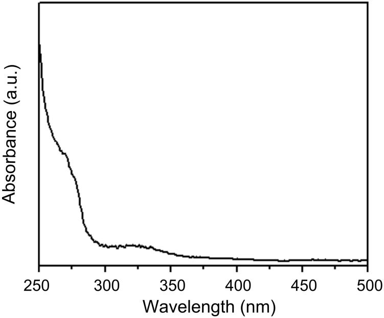
Figure 1
UVu2013vis spectrum of biosynthesized gadolinium oxide nanoparticles solution after 96 h of reaction with the fungal biomass.
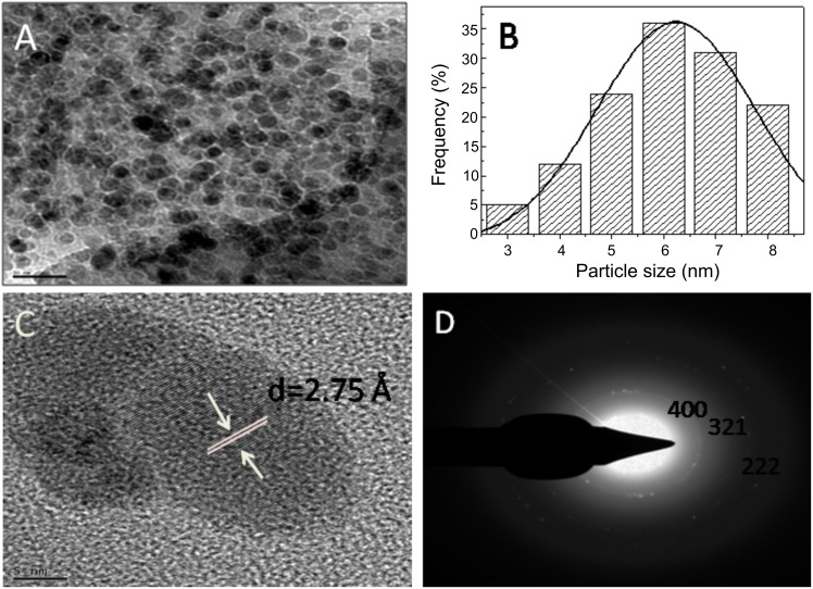
Figure 2
(A) TEM micrograph recorded from drop-cast films of Gd 2 O 3 nanoparticle solution formed by the reaction of GdCl 3 with the fungal biomass of Humicola sp. for 96 h. (B) Particle size distribution det...
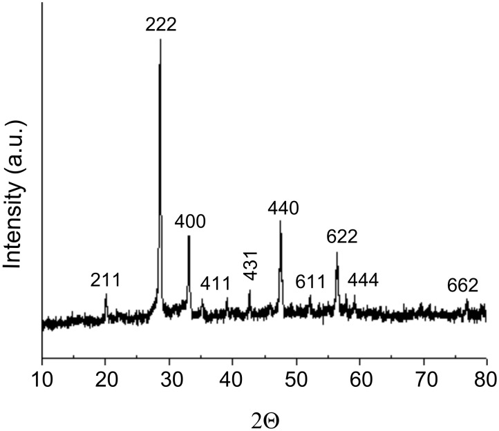
Figure 3
XRD measurements of biosynthesized Gd 2 O 3 nanoparticles.
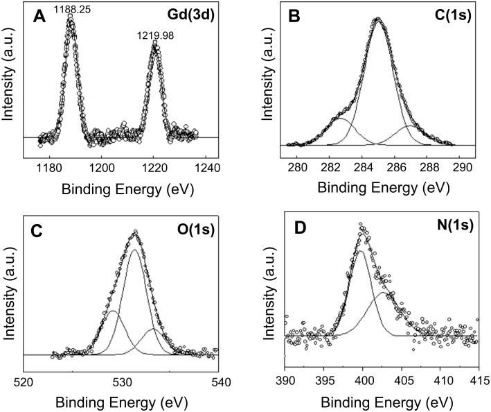
Figure 4
XPS data showing the (A) Gd(3d), (B) C(1s), (C) O(1s) and (D) N(1s) core level spectra recorded from biosynthesized Gd 2 O 3 nanoparticles film cast onto a Si substrate. The raw data are shown in the ...
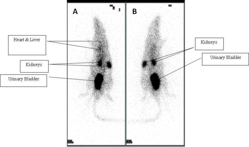
Figure 5
Gamma scintigraphic image of the biodistribution of Tc-99mu2013Gd 2 O 3 nanoparticles in a rat showing a dorsal (A) and a ventral (B) view.
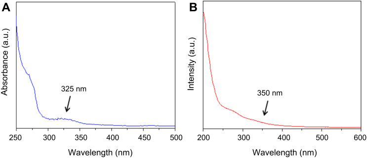
Figure 6
UVu2013vis spectroscopy of (A) Gd 2 O 3 nanoparticles showing a peak at 325 nm and (B) Gd 2 O 3 u2013taxol bioconjugate showing a shoulder at 350 nm.
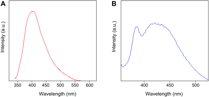
Figure 7
(A) Fluorescence spectra of Gd 2 O 3 nanoparticles excited at 320 nm giving emission at 400 nm and (B) Gd 2 O 3 u2013taxol bioconjugate excited at 320 nm giving emission at 440 nm.
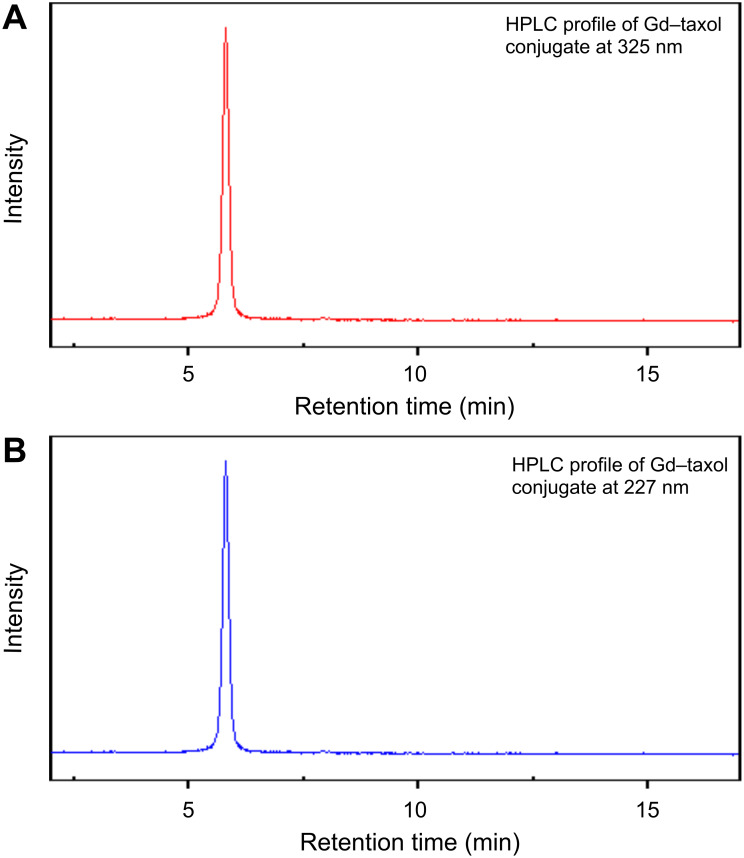
Figure 8
HPLC profile of Gd 2 O 3 u2013taxol bioconjugate showing absorbance at (A) 325 nm and (B) 227 nm.
Figure images are served from the NIH/NLM PubMed Central Open Access Subset or Europe PMC; copyright remains with the publishers and authors.
💬 Discussion
0 commentsNo comments yet. Be the first to start a discussion!
Leave a Comment