Abstract
AbstractTelocytes have been reported to play an important role in long‐distance heterocellular communication in normal and diseased heart, both through direct contact (atypical junctions), as well as by releasing extracellular vesicles (EVs) which may act as paracrine mediators. Exosomes and ectosomes are the two main types of EVs, as classified by size and the mechanism of biogenesis. Using electron microscopy (EM) and electron tomography (ET) we have found that telocytes in culture release at least three types of EVs: exosomes (released from endosomes; 45 ± 8 nm), ectosomes (which bud directly from the plasma membrane; 128 ± 28 nm) and multivesicular cargos (MVC; 1 ± 0.4 μm), the latter containing tightly packaged endomembrane‐bound vesicles (145 ± 35 nm). Electron tomography revealed that endomembrane vesicles are released into the extracellular space as a cargo enclosed by plasma membranes (estimated area of up to 3 μm2). This new type of EV, also released by telocytes in tissue, likely represents an essential component in the paracrine secretion of telocytes and may consequently be directly involved in heart physiology and regeneration.
🔬 Techniques
🔭 Microscopes
💻 Software
✨ Fluorophores
🧪 Sample Preparation
🏭 Microscope Brands
🧪 Reagent Suppliers
📷 Detectors
💻 Software Details
🏛️ Research Organizations (ROR)
Affiliated research institutions:
📋 Methods
Interstitial cell culture
The study was approved by the Bioethics Committee of the ‘Victor Babes’ National Institute of Pathology, Bucharest. Three month old Wistar rats were treated with 1000 U/kg heparin, then killed by cervical dislocation. The hearts were removed and placed in Hank's Balanced Salt Solution (HBSS) supplemented with 1% PSF (Penicilin, Streptomycin and Fungizone) and HEPES 0.01 mM, then dissected under the stereomicroscope and mechanically minced into ∼1 mm 3 fragments. Enzymatic dissociation was done using 300 U/ml collagenase I in DMEM/F12 supplemented with 0.01 mM HEPES. The resulting solution was then filtered using a 40 μm strainer, then cells washed and resuspended in DMEM/F12 culture medium supplemented with 10% fetal calf serum, 100 U/ml penicillin and 100 mg/ml streptomycin. Cells were grown to confluence (80%) in 25 cm 2 flasks, and then dislodged from the culture vessel with 2 mM EDTA at 37°C for 5 min. Finally, cells were re-plated on 35 mm Petri dishes for glutaraldehyde fixation. Unless otherwise specified, buffers and chemical reagents were supplied by Sigma-Aldrich Company Ltd., USA.
Transmission electron microscopy
Cells were fixed with 2.5% glutaraldehyde in 0.1 M cacodylate buffer with 1.4% sucrose at pH 7.4 and 37°C, for 5 min. Cells were scraped, resuspended in the same fixative for 4 hr at 4°C and then post-fixed for 1 hr in buffered 1% OsO 4 with 1.5% K 4 Fe(CN) 6 (potassium ferrocyanide-reduced osmium). Fixed cells were spun at 850 × g, embedded in 1% agar gel, and further processed for final epoxy resin embedding (Agar 100). The epoxy resin was polymerized at 37°C for 48 hr. Thick sections (1 μm) were obtained using an MTXL RMC ultramicrotome (Boeckeler Instruments Inc., Arizona, USA) and then stained with 1% toluidine blue for visualization using light microscopy. The ultra-thin sections were cut using a diamond knife at 60 and 300 nm, double stained with 1% uranyl acetate and Reynolds lead citrate. The 60 nm thin sections were visualized using a Morgagni 268 TEM (FEI Company, Eindhoven, The Netherlands) at 80 kV. Digital electron micrographs were recorded with a MegaView III CCD and images processed using iTEM-SIS software (Olympus, Münster, Germany). All measurements were performed within the same software package.
Show full methods section
Interstitial cell culture
The study was approved by the Bioethics Committee of the ‘Victor Babes’ National Institute of Pathology, Bucharest. Three month old Wistar rats were treated with 1000 U/kg heparin, then killed by cervical dislocation. The hearts were removed and placed in Hank's Balanced Salt Solution (HBSS) supplemented with 1% PSF (Penicilin, Streptomycin and Fungizone) and HEPES 0.01 mM, then dissected under the stereomicroscope and mechanically minced into ∼1 mm 3 fragments. Enzymatic dissociation was done using 300 U/ml collagenase I in DMEM/F12 supplemented with 0.01 mM HEPES. The resulting solution was then filtered using a 40 μm strainer, then cells washed and resuspended in DMEM/F12 culture medium supplemented with 10% fetal calf serum, 100 U/ml penicillin and 100 mg/ml streptomycin. Cells were grown to confluence (80%) in 25 cm 2 flasks, and then dislodged from the culture vessel with 2 mM EDTA at 37°C for 5 min. Finally, cells were re-plated on 35 mm Petri dishes for glutaraldehyde fixation. Unless otherwise specified, buffers and chemical reagents were supplied by Sigma-Aldrich Company Ltd., USA.
Transmission electron microscopy
Cells were fixed with 2.5% glutaraldehyde in 0.1 M cacodylate buffer with 1.4% sucrose at pH 7.4 and 37°C, for 5 min. Cells were scraped, resuspended in the same fixative for 4 hr at 4°C and then post-fixed for 1 hr in buffered 1% OsO 4 with 1.5% K 4 Fe(CN) 6 (potassium ferrocyanide-reduced osmium). Fixed cells were spun at 850 × g, embedded in 1% agar gel, and further processed for final epoxy resin embedding (Agar 100). The epoxy resin was polymerized at 37°C for 48 hr. Thick sections (1 μm) were obtained using an MTXL RMC ultramicrotome (Boeckeler Instruments Inc., Arizona, USA) and then stained with 1% toluidine blue for visualization using light microscopy. The ultra-thin sections were cut using a diamond knife at 60 and 300 nm, double stained with 1% uranyl acetate and Reynolds lead citrate. The 60 nm thin sections were visualized using a Morgagni 268 TEM (FEI Company, Eindhoven, The Netherlands) at 80 kV. Digital electron micrographs were recorded with a MegaView III CCD and images processed using iTEM-SIS software (Olympus, Münster, Germany). All measurements were performed within the same software package.
Electron tomography and 3D reconstruction
In the case of the 300 nm sections, goat anti-rabbit conjugated gold particles with a diameter of 10 nm (Aurion, Wageningen, The Netherlands) were used as fiducial markers. These were attached to both sides of the section by floating the grids on top of the gold-containing droplets, placed on a sheet of parafilm. Grids were then washed with ultrapure (Type 1) water to allow detachment of unbound particles and contaminants. Electron tomography was performed at room temperature using a FEI Tecnai G2 Spirit TEM, equipped with a LaB 6 cathode, at an acceleration voltage of 100 kV. Grid areas of interest were recorded by means of MegaView G2 CCD camera (Olympus) at nominal magnifications ranging from 21,000× to 60,000×. Single axis tilt series were collected using the Xplore3D (FEI) acquisition program, with final object sampling ranging from 2.6 to 4 nm/pixel. The angular tilt range was typically set from −60° to +60°, with increments of 1°. The tilt series images were aligned using the 10 nm gold fiducial markers visible on each section. Then, tomograms were generated from the aligned series using the eTOMO package. Finally 3D reconstructions were done by manually drawing coloured contours on each of the stacked images, using IMOD 4.5.3 software 27 . When required, short animations were generated using the freely available video-editing software VideoMach ( http://gromada.com/videomach/ ).
Supporting Information Additional Supporting Information may be found in the online version of this article: Video S1 Electron tomography of a Multivesicular Cargo .avi .
📊 Figures
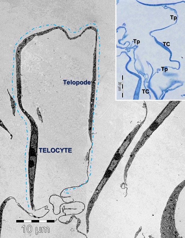
Figure 1
Electron microscopy image of interstitial cardiac cell culture shows a telocyte with about 100u00a0u00b5m long telopode. The inset shows a light microscopy image on blue-sections of telocytes (TC) in ...
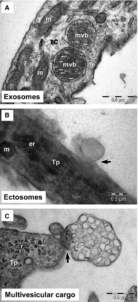
Figure 2
Electron microscopy of telocytes (TC) in culture demonstrates: ( A ) the presence of numerous intraluminal vesicles (small arrows) in two multivesicular bodies (mvb); ( B ) the ectosome budding (arrow...
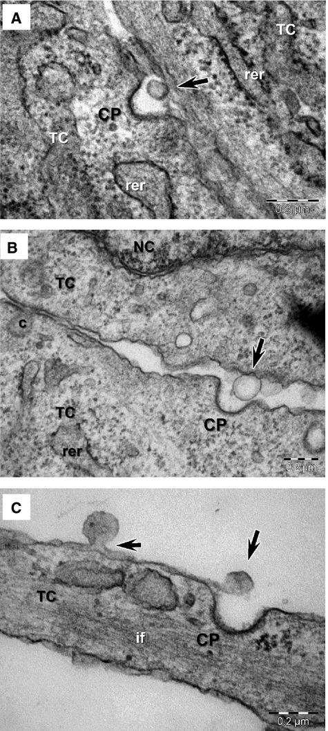
Figure 3
( A u2013 C ) Electron microscopy images of ectosomes released by telocytes (TC) in culture (arrows) frequently show the presence of clathrin coated pits (CP) in the vicinity. rer, rough endoplasmic r...
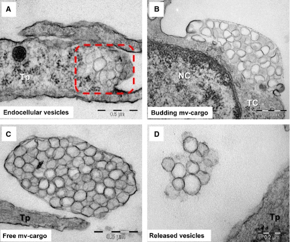
Figure 4
Possible mechanism of multivesicular cargo biogenesis based on electron microscopy images of telocytes (TC) in culture : ( A ) vesicles aggregation in the distinct space of cortical cytoplasm (square ...
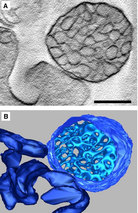
Figure 5
Electron tomography of multivesicular cargos (MVC): ( A ) Central tomogram section of a MVC, in direct contact with the cell membrane, revealing clustered vesicles enclosed by a membrane; ( B ) 3D mod...
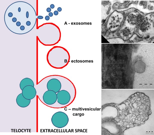
Figure 6
Schematic representation of the 3 types of extracellular vesicles released by telocytes and correspondent electron microscopy images of exosomes, ectosomes and multivesicular cargos.
Figure images are served from the NIH/NLM PubMed Central Open Access Subset or Europe PMC; copyright remains with the publishers and authors.
💬 Discussion
0 commentsNo comments yet. Be the first to start a discussion!
Leave a Comment