Abstract
AbstractInorganic dispersion is of great importance for actual implementation of advanced properties of organic–inorganic composites. Currently, electron microscopy is the most conventional approach for observing dispersion of inorganic fillers from ultrathin sections of organic–inorganic composites at the nanoscale by professional technicians. However, direct visualization of macrodispersion of inorganic fillers in organic–inorganic composites using high-contrast fluorescent imaging method is hampered. Here we design and synthesize a unique fluorescent surfactant, which combines the properties of the aggregation-induced emission (AIE) and amphiphilicity, to image macrodispersion of montmorillonite and layered double hydroxide fillers in polymer matrix. The proposed fluorescence imaging provides a number of important advantages over electron microscope imaging, and opens a new avenue in the development of direct three-dimensional observation of inorganic filler macrodispersion in organic–inorganic composites.
🔬 Techniques
🔭 Microscopes
🧪 Sample Preparation
🏭 Microscope Brands
🧪 Reagent Suppliers
📋 Protocols
🏛️ Research Organizations (ROR)
Affiliated research institutions:
📋 Methods
Materials Zinc dust and 4-hydroxyl benzophenone were purchased from Sigma-Aldrich Chemical Co. Sodium hydride (NaH), TiCl 4 , poly(vinyl chloride) with Mn=67,750 (polydispersity index (PDI)=2.32), dioctyl phthalate (DOP), CTAB, calcium stearate, zinc stearate, NaCl and K 2 CO 3 were purchased from J&K Chemical Ltd. Anhydrous dimethyl formamide (DMF), tetrahydrofuran (THF) and sodium sulfate were purchased from Alfa Aesar. 1-Bromooctane, 1,4-dibromobutane and trimethylamine in THF (100 ml, 1.0 M) were purchased from Tokyo Chemical Industry. Ethanol, PE, acetone, ethyl acetate (EA) and dichloromethane were purchased from Beijing Chemical Reagent Company. Sodium montmorillonite (Na + -MMT) with cation exchange capacity values of 145 meq per 100 g (from Nanocor, PGW grades) was used without further purification. Carbonate intercalated Mg-Al LDHs (with a molar ratio of 2:1 between Mg 2+ and Al 3+ ) were synthesized and characterized according to the literature 54 . TPE-SDS was synthesized and characterized according to our previous work 36 . All reagents were of analytical grade and used without further purification. Water was purified with a Milli-Q purification system (Milli-Q). Synthesis of compound 1 In a 250-ml, two-necked, round-bottom flask equipped with a condenser, zinc dust (2.9 g, 44 mmol) and 4-hydroxybenzophenone (2.0 g, 10 mmol) were dissolved in 100 ml dry THF under nitrogen. The mixture was cooled to −78 °C and TiCl 4 (2.5 ml, 22 mmol) was added dropwise. After the addition, the mixture was allowed to warm to room temperature in 0.5 h, and then was heated to reflux for overnight. The reaction was quenched with 10% aqueous K 2 CO 3 solution. The mixture was extracted with diethyl ether for three times and the combined organic layer was washed with brine twice and dried over sodium sulfate. After solvent evaporation, the crude product was separated through silica-gel chromatography flushed with PE/EA (v/v 1:1). 1.56 g product was obtained as a white solid with a yield of 86%. 1 H NMR (600 MHz, CDCl 3 , δ ): 6.94−7.05 (m, 10H), 6.76−6.81 (m, 4H), 6.47−6.52 (m, 4H). Synthesis of compound 2 Under N 2 atmosphere, sodium hydride (0.022 g, 0.55 mmol) was added to the solution of 1 (0.182 g, 0.50 mmol) in dry DMF (10 ml) and the mixture was stirred for extra 30 min at room temperature. Then 1-bromooctane (0.145 g, 0.75 mmol) was added and the reaction mixture was stirred at 60 °C for 8.0 h. When the reaction completed, the solvent DMF was removed and the residue was redissolved with EA, and the resulting solution was washed with water for three times and dried over sodium sulfate. The solution was concentrated and the residue was purified through silica gel chromatography flushed with PE/EA (7:1 v/v), compound 2 (0.121 g, 0.25 mmol) as a yellow liquid was obtained with a yield of 51%. 1 H NMR (600 MHz, CDCl 3 , δ ): 6.98−7.11 (m, 10H), 6.83−6.92 (m, 4H), 6.52−6.63 (m, 4H), 3.82−3.88 (m, 2H), 1.67−1.76 (m, 2H), 1.37−1.41 (m, 2H), 1.24−1.29 (m, 8H), 0.85−0.88 (t, 3H). Synthesis of compound 3 The compound 2 (0.174 g, 0.50 mmol) and K 2 CO 3 (0.076 g, 0.55 mmol) were mixed in 20 ml acetone. After stirred for 1 h, 1,4-dibromobutane (0.118 g, 0.55 mmol) was added and the resulting mixture was stirred for 24 h at 60 °C. After evaporation of acetone, the obtained solids were first redispersed in EA and then filtered to remove the insoluble K 2 CO 3 . The purified product was obtained in 74% yield after purification separation by silica gel chromatography using PE/EA (10:1 (v/v)). 1 H NMR (600 MHz, CDCl 3 , δ ): 7.00−7.12 (m, 10H), 6.88−6.95 (m, 4H), 6.59−6.65 (m, 4H), 4.20−4.25 (m, 2H), 3.84−3.93 (m, 4H), 2.43−2.46 (m, 2H), 1.81−1.86 (m, 4H), 1.70−1.76 (m, 2H), 1.38−1.43 (m, 2H), 1.26−1.32 (m, 8H), 0.87−0.89 (t, 3H). Synthesis of TPE-DTAB (compound 4 ) A 100-ml flask with a magnetic spin bar was charged with 3 (0.3 g, 0.50 mmol) dissolved in 20 ml of THF. To this solution, trimethylamine (1.0 M, 5 ml) was added. The mixture was heated to reflux and stirred for 3 days. During this period, 5 ml of trimethylamine in THF was added at several intervals. After THF and extra trimethylamine were evaporated, the residue was washed with chloroform and acetone and then dried overnight in vacuo at 60 °C. A yellowish product was obtained in 88% yield. 1 H NMR (600 MHz, DMSO-d 6 , δ ): 7.07−7.15 (m, 6H), 6.92−6.98 (m, 4H), 6.80−6.88 (m, 4H), 6.64−6.73 (m, 4H), 3.91−3.95 (m, 2H), 3.82−3.87 (m, 2H), 3.34−3.39 (m, 2H), 3.07−3.09 (t, 9H), 1.79−1.85 (m, 2H), 1.61−1.72 (m, 4H), 1.34−1.36 (m, 2H), 1.24−1.27 (m, 8H), 0.84−0.86 (t, 3H). 13 C NMR (600 MHz, DMSO-d 6 , δ ): 157.54, 157.29, 144.27, 144.20, 139.82, 139.64, 136.22, 132.41, 132.36, 131.19, 128.28, 128.17, 126.79, 126.76, 114.23, 114.19, 114.14, 114.09, 67.67, 66.97, 65.40, 52.60, 31.69, 29.20, 29.16, 29.12, 26.11, 26.02, 25.99, 22.55, 19.72, 14.43. MS: m / z : 590.3996 ([M-Br] + , calculated for C 41 H 52 NO 2 , 590.3993).
Show full methods section
Materials Zinc dust and 4-hydroxyl benzophenone were purchased from Sigma-Aldrich Chemical Co. Sodium hydride (NaH), TiCl 4 , poly(vinyl chloride) with Mn=67,750 (polydispersity index (PDI)=2.32), dioctyl phthalate (DOP), CTAB, calcium stearate, zinc stearate, NaCl and K 2 CO 3 were purchased from J&K Chemical Ltd. Anhydrous dimethyl formamide (DMF), tetrahydrofuran (THF) and sodium sulfate were purchased from Alfa Aesar. 1-Bromooctane, 1,4-dibromobutane and trimethylamine in THF (100 ml, 1.0 M) were purchased from Tokyo Chemical Industry. Ethanol, PE, acetone, ethyl acetate (EA) and dichloromethane were purchased from Beijing Chemical Reagent Company. Sodium montmorillonite (Na + -MMT) with cation exchange capacity values of 145 meq per 100 g (from Nanocor, PGW grades) was used without further purification. Carbonate intercalated Mg-Al LDHs (with a molar ratio of 2:1 between Mg 2+ and Al 3+ ) were synthesized and characterized according to the literature 54 . TPE-SDS was synthesized and characterized according to our previous work 36 . All reagents were of analytical grade and used without further purification. Water was purified with a Milli-Q purification system (Milli-Q). Synthesis of compound 1 In a 250-ml, two-necked, round-bottom flask equipped with a condenser, zinc dust (2.9 g, 44 mmol) and 4-hydroxybenzophenone (2.0 g, 10 mmol) were dissolved in 100 ml dry THF under nitrogen. The mixture was cooled to −78 °C and TiCl 4 (2.5 ml, 22 mmol) was added dropwise. After the addition, the mixture was allowed to warm to room temperature in 0.5 h, and then was heated to reflux for overnight. The reaction was quenched with 10% aqueous K 2 CO 3 solution. The mixture was extracted with diethyl ether for three times and the combined organic layer was washed with brine twice and dried over sodium sulfate. After solvent evaporation, the crude product was separated through silica-gel chromatography flushed with PE/EA (v/v 1:1). 1.56 g product was obtained as a white solid with a yield of 86%. 1 H NMR (600 MHz, CDCl 3 , δ ): 6.94−7.05 (m, 10H), 6.76−6.81 (m, 4H), 6.47−6.52 (m, 4H). Synthesis of compound 2 Under N 2 atmosphere, sodium hydride (0.022 g, 0.55 mmol) was added to the solution of 1 (0.182 g, 0.50 mmol) in dry DMF (10 ml) and the mixture was stirred for extra 30 min at room temperature. Then 1-bromooctane (0.145 g, 0.75 mmol) was added and the reaction mixture was stirred at 60 °C for 8.0 h. When the reaction completed, the solvent DMF was removed and the residue was redissolved with EA, and the resulting solution was washed with water for three times and dried over sodium sulfate. The solution was concentrated and the residue was purified through silica gel chromatography flushed with PE/EA (7:1 v/v), compound 2 (0.121 g, 0.25 mmol) as a yellow liquid was obtained with a yield of 51%. 1 H NMR (600 MHz, CDCl 3 , δ ): 6.98−7.11 (m, 10H), 6.83−6.92 (m, 4H), 6.52−6.63 (m, 4H), 3.82−3.88 (m, 2H), 1.67−1.76 (m, 2H), 1.37−1.41 (m, 2H), 1.24−1.29 (m, 8H), 0.85−0.88 (t, 3H). Synthesis of compound 3 The compound 2 (0.174 g, 0.50 mmol) and K 2 CO 3 (0.076 g, 0.55 mmol) were mixed in 20 ml acetone. After stirred for 1 h, 1,4-dibromobutane (0.118 g, 0.55 mmol) was added and the resulting mixture was stirred for 24 h at 60 °C. After evaporation of acetone, the obtained solids were first redispersed in EA and then filtered to remove the insoluble K 2 CO 3 . The purified product was obtained in 74% yield after purification separation by silica gel chromatography using PE/EA (10:1 (v/v)). 1 H NMR (600 MHz, CDCl 3 , δ ): 7.00−7.12 (m, 10H), 6.88−6.95 (m, 4H), 6.59−6.65 (m, 4H), 4.20−4.25 (m, 2H), 3.84−3.93 (m, 4H), 2.43−2.46 (m, 2H), 1.81−1.86 (m, 4H), 1.70−1.76 (m, 2H), 1.38−1.43 (m, 2H), 1.26−1.32 (m, 8H), 0.87−0.89 (t, 3H). Synthesis of TPE-DTAB (compound 4 ) A 100-ml flask with a magnetic spin bar was charged with 3 (0.3 g, 0.50 mmol) dissolved in 20 ml of THF. To this solution, trimethylamine (1.0 M, 5 ml) was added. The mixture was heated to reflux and stirred for 3 days. During this period, 5 ml of trimethylamine in THF was added at several intervals. After THF and extra trimethylamine were evaporated, the residue was washed with chloroform and acetone and then dried overnight in vacuo at 60 °C. A yellowish product was obtained in 88% yield. 1 H NMR (600 MHz, DMSO-d 6 , δ ): 7.07−7.15 (m, 6H), 6.92−6.98 (m, 4H), 6.80−6.88 (m, 4H), 6.64−6.73 (m, 4H), 3.91−3.95 (m, 2H), 3.82−3.87 (m, 2H), 3.34−3.39 (m, 2H), 3.07−3.09 (t, 9H), 1.79−1.85 (m, 2H), 1.61−1.72 (m, 4H), 1.34−1.36 (m, 2H), 1.24−1.27 (m, 8H), 0.84−0.86 (t, 3H). 13 C NMR (600 MHz, DMSO-d 6 , δ ): 157.54, 157.29, 144.27, 144.20, 139.82, 139.64, 136.22, 132.41, 132.36, 131.19, 128.28, 128.17, 126.79, 126.76, 114.23, 114.19, 114.14, 114.09, 67.67, 66.97, 65.40, 52.60, 31.69, 29.20, 29.16, 29.12, 26.11, 26.02, 25.99, 22.55, 19.72, 14.43. MS: m / z : 590.3996 ([M-Br] + , calculated for C 41 H 52 NO 2 , 590.3993).
Synthesis of TPE-DTAB-modified MMT and TPE-SDS-modified LDH TPE-DTAB-modified
MMT was prepared from Na + -MMT by ion exchange method. Typically, a 0.5 g portion of Na + -MMT was mixed with 50 ml of deionized water. Then, 0.5 g of TPE-DTAB was added in the MMT solution. The ion exchange was carried out under stirring for 1 h at 60 °C. Then, the reaction solution was centrifuged at 5,000 r.p.m. for 5 min, and the precipitate was washed with distilled water to remove the physically adsorbed TPE-DTAB. The obtained TPE-DTAB-modified MMT was dried under vacuum at 60 °C, and then finely powdered in an agate mortar for further use. TPE-SDS-modified LDH powder was prepared according to the same procedure. Synthesis of CTAB/TPE-DTAB-modified MMT Na + -MMT (0.5 g) was dispersed in 50 ml of deionized water. Then, a mixture of TPE-DTAB (0.05 g) and CTAB (0.25 g) was added to the MMT dispersion and stirred for 1 h at 60 °C. After centrifugation and washing, the obtained CTAB/TPE-DTAB-modified MMT solid was dried under vacuum at 60 °C and ground into powder. Preparation of PVC/MMT composite The PVC/TPE-DTAB-modified MMT composite, containing 10.0 g PVC powder, 5.0 g DOP and 0.5 g TPE-DTAB-modified MMT powder, was prepared by blending in a heated double-roller mixer for 5 min at 140 °C. The resulting composites were molded at 120 °C and then cooled at room temperature to give thin films with a thickness of 1 mm. The PVC/CTAB/TPE-DTAB-modified MMT composite was prepared according to the same procedure. Preparation of PVC/LDH composite The PVC/TPE-SDS-modified LDH composite, containing 10.0 g PVC powder, 5.0 g DOP, 0.2 g TPE-SDS-modified LDH powder, 0.23 g calcium stearate and 0.1 g zinc stearate, was prepared by blending in a heated double-roller mixer for 5 min at 140 °C. The resulting composite was molded at 120 °C and then cooled at room temperature to give thin films with a thickness of 1 mm.
TEM sample preparation
The composite films were ultrathin-sectioned with a diamond knife at −120 °C using a Leica EM UC6 ultramicrotome. The obtained ultrathin sections were then collected in a trough filled with deionized water and placed on 200-mesh copper grids.
Characterization
Proton and carbon-13 nuclear magnetic resonance ( 1 H NMR and 13 C NMR) spectra were recorded at room temperature with a 600-MHz Bruker spectrometer (Bruker). MS was carried out with Quattro microtriple quadrupole mass spectrometer (Waters). Electrical conductivity measurements were performed using a EC 215 conductivity meter (Shanghai Jingmi Instrumental Co.). TEM photographs were performed on a Tecnai G220 TEM (FEI Company) at an accelerating voltage of 200 kV. Ultraviolet–visible spectra were measured on a USB 4000 miniature fibre optic spectrometer in absorbance mode with a DH–2000 deuterium and tungsten halogen light source (Ocean Optics). Fluorescence spectra were obtained using a F–7000 fluorescence spectrophotometer at a slit of 5.0 nm with a scanning rate of 1,200 nm min −1 . X-ray diffraction measurements of MMT and TPE-DTAB-modified MMT were performed with a Brucker D8 ADVANCE X-ray diffractometer (Bruker) equipped with graphite–monochromatized Cu/Kα radiation ( λ =0.1541, nm). The samples as unoriented powders were step-scanned in steps of 0.02° (2 θ ) in the range of 2–10°. Fourier transform infrared spectroscopy experiments were carried out on Nicolet 380 system (Thermo) containing a controlled environment chamber equipped with CaF 2 windows. Zeta potential was determined using a Malvern Zetasizer 3000HS nano-granularity analyzer. The quantum yield values were obtained from the reconvolution fit analysis (Edinburgh F980 analysis software) equipped with an integrating sphere. Fluorescence microscope images were recorded on a confocal laser scanning microscope (Leica, TCS SP8).
Data availability
The data that support the findings of this study are available from the corresponding author upon request.
Materials Zinc dust and 4-hydroxyl benzophenone were purchased from Sigma-Aldrich Chemical Co. Sodium hydride (NaH), TiCl 4 , poly(vinyl chloride) with Mn=67,750 (polydispersity index (PDI)=2.32), dioctyl phthalate (DOP), CTAB, calcium stearate, zinc stearate, NaCl and K 2 CO 3 were purchased from J&K Chemical Ltd. Anhydrous dimethyl formamide (DMF), tetrahydrofuran (THF) and sodium sulfate were purchased from Alfa Aesar. 1-Bromooctane, 1,4-dibromobutane and trimethylamine in THF (100 ml, 1.0 M) were purchased from Tokyo Chemical Industry. Ethanol, PE, acetone, ethyl acetate (EA) and dichloromethane were purchased from Beijing Chemical Reagent Company. Sodium montmorillonite (Na + -MMT) with cation exchange capacity values of 145 meq per 100 g (from Nanocor, PGW grades) was used without further purification. Carbonate intercalated Mg-Al LDHs (with a molar ratio of 2:1 between Mg 2+ and Al 3+ ) were synthesized and characterized according to the literature 54 . TPE-SDS was synthesized and characterized according to our previous work 36 . All reagents were of analytical grade and used without further purification. Water was purified with a Milli-Q purification system (Milli-Q).
Supplementary Material Supplementary Information Supplementary Figures 1-12
📊 Figures
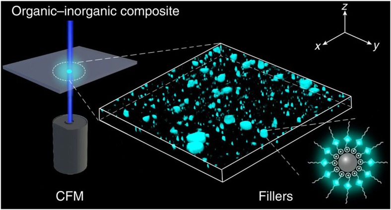
Figure 1
Schematic representation of visualization of 3D macrodispersion of fillers in organicu2013inorganic composites.
The inorganic fillers modified and bound with AIE molecules are dispersed inside the organic matrix, and then directly visualized by CFM.
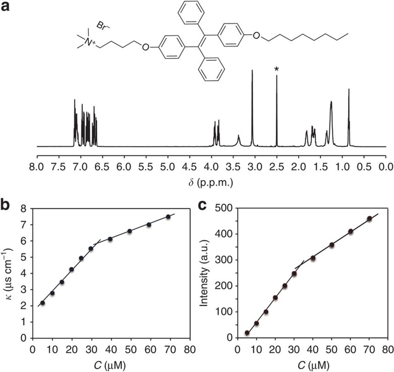
Figure 2
Molecular structure and characterization of TPE-DTAB.
( a ) 1 H NMR spectrum of TPE-DTAB in [D 6 ]dimethyl sulfoxide (the solvent peak is marked with asterisk). Plots of conductivity ( b ) and fluorescence intensity at 490u2009nm ( c ) versus the concent...
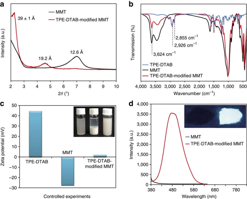
Figure 3
Characterizations of TPE-DTAB-modified MMT.
( a ) Powder X-ray diffraction patterns of Na + -MMT and TPE-DTAB-modified MMT. ( b ) Fourier transform infrared spectra of TPE-DTAB, Na + -MMT and TPE-DTAB-modified MMT. ( c ) u03b6 potential measure...
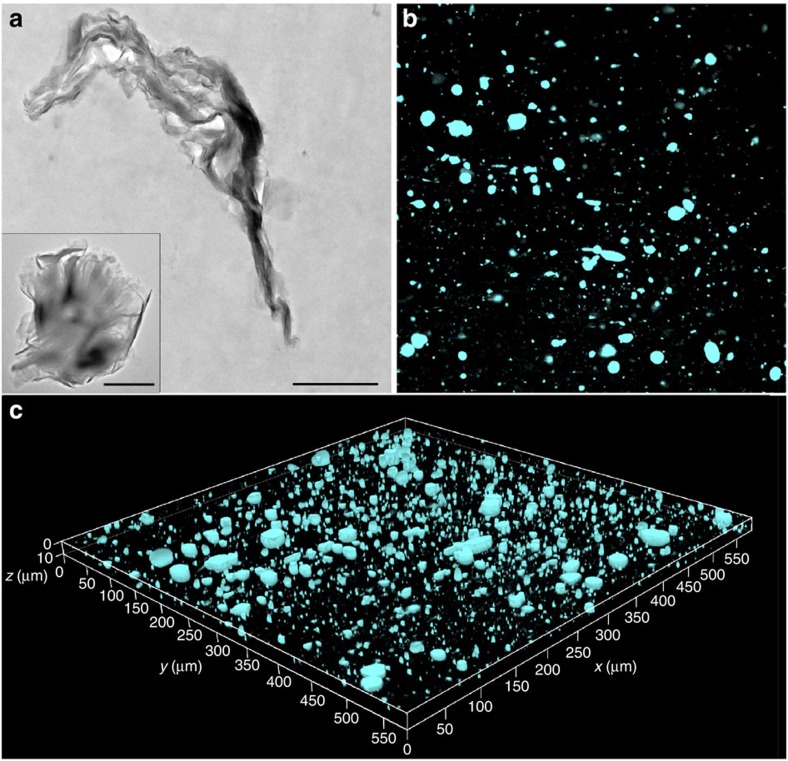
Figure 4
Macrodispersion of organo-modified MMT in PVC/MMT composite.
( a ) Cross-sectional TEM micrograph of PVC/TPE-DTAB-modified MMT (5u2009wt%) composite; the inset showed TEM image of TPE-DTAB-modified MMT. Scale bar, 1u2009u03bcm. ( b ) Fluorescence microscopy ima...
Figure images are served from the NIH/NLM PubMed Central Open Access Subset or Europe PMC; copyright remains with the publishers and authors.
💬 Discussion
0 commentsNo comments yet. Be the first to start a discussion!
Leave a Comment