Abstract
Heparanase is a heparan sulfate (HS) degrading endoglycosidase participating in extracellular matrix degradation and remodeling. Apart of its well characterized enzymatic activity, heparanase was noted to exert also enzymatic-independent functions. Non-enzymatic activities of heparanase include enhanced adhesion of tumor-derived cells and primary T-cells. Attempting to identify functional domains of heparanase that would serve as targets for drug development, we have identified heparin binding domains of heparanase. A corresponding peptide (residues Lys(158)-Asp(171), termed KKDC) was demonstrated to physically associate with heparin and HS, and to inhibit heparanase enzymatic activity. We hypothesized that the pro-adhesive properties of heparanase are mediated by its interaction with cell surface HS proteoglycans, and utilized the KKDC peptide to examine this possibility. We provide evidence that the KKDC peptide interacts with cell membrane HS, resulting in clustering of syndecan-1 and syndecan-4. We applied classical analysis of cell morphology, fluorescent and time-lapse microscopy and demonstrated that the KKDC peptide efficiently stimulates the adhesion and spreading of various cell types, mediated by PKC, Src, and the small GTPase Rac1. These results support, and further substantiate the notion that heparanase function is not limited to its enzymatic activity.
🔬 Techniques
✨ Fluorophores
🧪 Sample Preparation
🔬 Cell Lines
🏭 Microscope Brands
🧪 Reagent Suppliers
📷 Detectors
🏛️ Research Organizations (ROR)
Affiliated research institutions:
📋 Methods
Antibodies and reagents Anti syndecan-1 monoclonal antibody
(B-B4) was purchased from Serotec (Oxford, UK). Antibodies to syndecan-4 and paxillin were purchased from Santa Cruz Biotechnology (Sanata Cruz, CA). Anti-Rac 1 monoclonal antibody was purchased from Becton Dickinson Biosciences (San Diego, CA), and PAK PBD-agarose beads were from Cell Biolabs Inc (San Diego, CA). Anti-vinculin monoclonal antibody, phalloidin-TRITC, heparin, fibronectin, and the 110 kDa fibronectin-like genetically engineered protein, were purchased from Sigma (St. Louis, MO). Avidin-FITC was purchased from Vector (Burlingame, CA). The selective p38 (SB-203580), Src (PP2), Erk (PD-98059), PKC [Bisindolylmaleimide I (Bis)], Rock (Y27632) and Rac (NSC 23766) inhibitors were purchased from Calbiochem (San Diego, CA) and were dissolved in DMSO as stock solutions. DMSO was added to the cell culture as a control. The KKDC and its control, scrambled peptide were synthesized as described previously [29] . The introduced C-terminal cysteine residue mediates spontaneous peptide dimerization that can be further facilitated by enhanced oxygenation generated by stirring. For biotin labeling, an extra lysine residue was introduced at position 12 to create the sequence KKFKNSTYSRS K biotin SVDC. This approach was undertaken in order to preserve the N-terminal lysine residues critical for the peptide interaction with HS [29] . A peptide from the COOH-terminal heparin-binding domain of fibronectin (Hep II peptide, WQPPRARI) [38] was purchased from Sigma (St. Louis, MI). Cells and cell culture U87 MG human glioma and Colo 320 colon carcinoma cells were purchased from the American Type Culture Collection (ATCC). Rat C6 glioma cells were kindly provided by Dr. Eli Keshet (The Hebrew University Hadassah Medical School, Jerusalem) [39] , and human skin fibroblasts were kindly provided by Dr. H. Mendel (Rambam Health Care Campus, Haifa). ARH-77 leukemia-derived F cells were kindly provided by Dr. Ben-Zion Katz (Soraski Medical Center, Tel-Aviv, Israel). F cells are a sub-population of the ARH-77 cell line which poorly adhere and mainly floats (F) once plated on fibronectin-coated dishes [40] . Cells were grown in Dulbecco's Modified Eagle's Medium (DMEM) supplemented with glutamine, pyruvate, antibiotics and 10% fetal calf serum in a humidified atmosphere containing 5% CO 2 at 37°C. Wild type Chinese hamster ovary (CHO) K1 and mutant cells (pgs A-745) deficient in xylosyltransferase and unable to initiate glycosaminoglycan synthesis, were kindly provided by Dr. Jeffery Esko (University of California, San Diego), and grown in RPMI 1640 medium supplemented with 10% FCS and antibiotics [39] . Recombinant wild type and enzymatically-inactive heparanase mutated at glutamic acid residues 225 and 343 [double mutant (DM)] critical for enzymatic catalysis [41] were purified from the conditioned medium of HEK 293 transfected cells, essentially as described [42] .
Show full methods section
Antibodies and reagents Anti syndecan-1 monoclonal antibody
(B-B4) was purchased from Serotec (Oxford, UK). Antibodies to syndecan-4 and paxillin were purchased from Santa Cruz Biotechnology (Sanata Cruz, CA). Anti-Rac 1 monoclonal antibody was purchased from Becton Dickinson Biosciences (San Diego, CA), and PAK PBD-agarose beads were from Cell Biolabs Inc (San Diego, CA). Anti-vinculin monoclonal antibody, phalloidin-TRITC, heparin, fibronectin, and the 110 kDa fibronectin-like genetically engineered protein, were purchased from Sigma (St. Louis, MO). Avidin-FITC was purchased from Vector (Burlingame, CA). The selective p38 (SB-203580), Src (PP2), Erk (PD-98059), PKC [Bisindolylmaleimide I (Bis)], Rock (Y27632) and Rac (NSC 23766) inhibitors were purchased from Calbiochem (San Diego, CA) and were dissolved in DMSO as stock solutions. DMSO was added to the cell culture as a control. The KKDC and its control, scrambled peptide were synthesized as described previously [29] . The introduced C-terminal cysteine residue mediates spontaneous peptide dimerization that can be further facilitated by enhanced oxygenation generated by stirring. For biotin labeling, an extra lysine residue was introduced at position 12 to create the sequence KKFKNSTYSRS K biotin SVDC. This approach was undertaken in order to preserve the N-terminal lysine residues critical for the peptide interaction with HS [29] . A peptide from the COOH-terminal heparin-binding domain of fibronectin (Hep II peptide, WQPPRARI) [38] was purchased from Sigma (St. Louis, MI). Cells and cell culture U87 MG human glioma and Colo 320 colon carcinoma cells were purchased from the American Type Culture Collection (ATCC). Rat C6 glioma cells were kindly provided by Dr. Eli Keshet (The Hebrew University Hadassah Medical School, Jerusalem) [39] , and human skin fibroblasts were kindly provided by Dr. H. Mendel (Rambam Health Care Campus, Haifa). ARH-77 leukemia-derived F cells were kindly provided by Dr. Ben-Zion Katz (Soraski Medical Center, Tel-Aviv, Israel). F cells are a sub-population of the ARH-77 cell line which poorly adhere and mainly floats (F) once plated on fibronectin-coated dishes [40] . Cells were grown in Dulbecco's Modified Eagle's Medium (DMEM) supplemented with glutamine, pyruvate, antibiotics and 10% fetal calf serum in a humidified atmosphere containing 5% CO 2 at 37°C. Wild type Chinese hamster ovary (CHO) K1 and mutant cells (pgs A-745) deficient in xylosyltransferase and unable to initiate glycosaminoglycan synthesis, were kindly provided by Dr. Jeffery Esko (University of California, San Diego), and grown in RPMI 1640 medium supplemented with 10% FCS and antibiotics [39] . Recombinant wild type and enzymatically-inactive heparanase mutated at glutamic acid residues 225 and 343 [double mutant (DM)] critical for enzymatic catalysis [41] were purified from the conditioned medium of HEK 293 transfected cells, essentially as described [42] .
Cell morphology and immunocytochemistry
Cells were plated on glass cover slips uncoated or coated with fibronectin or the 110 kDa fibronectin-like protein for the time indicated in the absence (Con) or presence of the indicated peptides [scrambled (Scr), or KKDC; (50 µg/ml)], proteins [latent 65 kDa or mutated, enzymatically inactive heparanase (1 µg/ml)], or inhibitors. Cells were then fixed with 4% paraformaldehyde in PBS for 15 minutes, and visualized. For immunofluorescent staining, cells were fixed with cold methanol for 10 minutes, washed with PBS and subsequently incubated in PBS containing 10% normal goat serum for 1 hour at room temperature, followed by 2 hours incubation with the indicated primary antibody. Cells were then extensively washed with PBS and incubated with the relevant Cy2/Cy3-conjugated secondary antibody (Jackson ImmunoResearch, West Grove, PA) for 1 hour, washed and mounted (Vectashield, Vector, Burlingame, CA). Staining was observed under a fluorescent confocal microscope. For actin staining, cells were fixed with 4% paraformaldehyde, permeabilized with 0.5% triton X-100 for 2 minutes, washed and incubated with TRITC-phalloidin (Sigma) for 30 minutes and visualized by confocal microscopy, as described [43] , [44] .
Cell lysates and protein blotting
Cell cultures were incubated for 24 hours under serum-free conditions, pretreated with 1 mM orthovanadate for 10 minutes at 37°C, washed twice with ice cold PBS containing 1 mM orthovanadate and scraped into lysis buffer (50 mM Tris-HCl, pH 7.4, 150 mM NaCl, 1% Triton-X100, 1 mM orththovanadate, 1 mM PMSF) containing a cocktail of protease inhibitors (Roche, Mannheim, Germany). Total cellular protein concentration was determined by the BCA assay, according to the manufacturer's (Pierce, Rockford, IL) instructions. Thirty micrograms of cellular protein were subjected to SDS polyacrylamid gel (SDS-PAGE) and immunoblotting, as described [29] , [45] . For estimation of Rac1 activation, lysis buffer included 50 mM Tris-HCl, pH 7.4, 300 mM NaCl, 10 mM MgCl, 4% glycerol, and 1% Triton X-100. The amount of GTP-bound Rac1 was analyzed by incubating total cell lysates (200 µg) with 20 µg of the p21-binding domain (PBD) of PAK-agarose beads. Following 30 minutes incubation, the beads were washed and, after electrophoresis and blotting, membranes were probed with anti-Rac1 antibodies [26] .
Time Lapse microscopy
Cells were grown on Corning/Nunc 6 wells dishes in DMEM medium and viewed with inverted motorized microscope (DMIRE2 Leica, Germany). Images were captured using cooled B/W CCD camera (Retiga EXI, Qimaging, USA). Time-lapse acquisition was made with custom written software (Provided by Uri Alon, Weizmann Institute, Rehovot, Israel), inserted into Image-Pro software (MediaCybernetics, USA). The system is equipped with an on stage incubator (Cube & Box, LIS, Switzerland).
Supporting Information Movie S1 Time lapse microscopy. Rat C6 glioma cells (2×10⁴) were plated in a 6-well plate in complete growth medium, followed by 20 hours incubation in serum-free medium. Cells were then incubated with control scrambled peptide. Six fields in each well were randomly selected, and were examined every 10 min for 10 hours by the time lapse system. Representative time lapse movie is shown. (6.15 MB AVI) Click here for additional data file. Movie S2 Time lapse microscopy. Rat C6 glioma cells (2×10⁴) were plated in a 6-well plate in complete growth medium, followed by 20 hours incubation in serum-free medium. Cells were then incubated with heparanase (1 µg/ml); Six fields in each well were randomly selected, and were examined every 10 min for 10 hours by the time lapse system. Representative time lapse movie is shown. (7.14 MB AVI) Click here for additional data file. Movie S3 Time lapse microscopy. Rat C6 glioma cells (2×10⁴) were plated in a 6-well plate in complete growth medium, followed by 20 hours incubation in serum-free medium. Cells were then incubated with the KKDC peptide; Six fields in each well were randomly selected, and were examined every 10 min for 10 hours by the time lapse system. Representative time lapse movie is shown. (6.99 MB AVI) Click here for additional data file. Movie S4 Time lapse microscopy. Rat C6 glioma cells (2×10⁴) were plated in a 6-well plate in complete growth medium, followed by 20 hours incubation in serum-free medium. Cells were then incubated with the KKDC peptide with heparin (10µg/ml); Six fields in each well were randomly selected, and were examined every 10 min for 10 hours by the time lapse system. Representative time lapse movie is shown. (6.15 MB AVI) Click here for additional data file. Figure S1 KKDC peptide dimerization significantly improves heparin binding and anti-heparanase properties. A. Heparin binding. KKDC peptide was synthesized in the absence (KKDC) or presence of ethandithiol (EDT), which binds covalently to the sulfate group of cysteine and prevents di-sulfide bridge formation between cysteine residues. Peptides (50 µM) were incubated (2 hours, 4°C) with heparin-Sepharose beads in PBS, washed with PBS supplemented with NaCl to a final concentration of 0.35 M, followed by one wash with PBS. Dye-free sample buffer was added and the beads were boiled for 5 minuntes, centrifuged and the supernatants were loaded on Tris-Tricine gel. Subsequently, gels were stained with Coomassie blue to visualize bound peptides (arrow). B. Heparanase enzymatic activity. B16 melanoma cells (2×10⁶) were resuspended in RPMI medium and incubated (18 hours, 37°C) with 35S-labeled ECM in the absence (filled rectangle) or presence of control scrambled peptide dimer (green rectangle; 50 µM), KKDC peptide undergoing spontaneous dimerization (filled triangle; 50 µM), KKDC peptide following enhanced dimerization (blue rectangle; 50 µM), or heparin (filled circle; 15 µg/ml). The incubation medium (1 ml) containing sulfate labeled degradation fragments was subjected to gel filtration on a Sepharose CL-6B column. Fractions (0.2 ml) were eluted with PBS and their radioactivity counted in a beta-scintillation counter. Degradation fragments of HS side chains are eluted at 0.5
📊 Figures
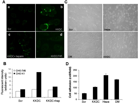
Figure 1
The KKDC peptide interacts with plasma membrane heparan sulfate.
A,B: peptide binding. Wild type (CHO K1; au2013c) and heparan sulfate-deficient (CHO-745; d) cells were incubated (2 hours) with biotinylated KKDC (bu2013d) or control peptide (Scr; a) in the absence ...
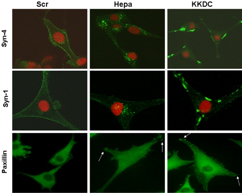
Figure 2
The KKDC peptide induces syndecan clustering and enhances cell spreading.
U87 cells were serum starved for 24 hours and were then incubated with the KKDC/Scr peptides (50 u00b5M) or heparanase (1 u00b5g/ml) for 1 hour. Cells were then gently washed, fixed with cold methanol...
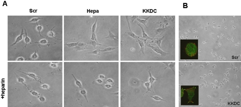
Figure 3
The KKDC peptide induces cell spreading.
A. C6 glioma cells were serum-starved for 24 hours, and were then incubated with heparanase (1 u00b5g/ml), KKDC or control (Scr) peptides (50 u00b5M) for 18 hours in the absence (upper panels) or pres...
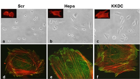
Figure 4
Heparanase and the KKDC peptide enhance cell spreading and formation of actin stress fibers.
CHO cells (au2013c) and human fibroblasts (du2013f) were plated on the 110 kDa fibronectin-like protein and incubated with heparanase (Hepa; 1 u00b5g/ml) or the KKDC/Scr peptides (50 u00b5M) for 18 ho...
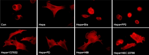
Figure 5
Cell spreading induced by heparanase is mediated by PKC, Rac, and Src.
CHO cells were plated on the 110 kDa fibronectin-like protein, without (Con) or with heparanase, in the absence (Hepa) or presence of selective PKC (Bis), Src (PP2), ROCK (Y27632), MAPK (PD), PKA (H89...
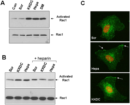
Figure 6
Heparanase and the KKDC peptide induce Rac1 activation.
A. F cells were washed with serum free medium and were then treated for 30 minutes with the KKDC/Scr peptides (50 u00b5M), wild type heparanase (Hepa) or inactive double mutant heparanase (DM) in seru...
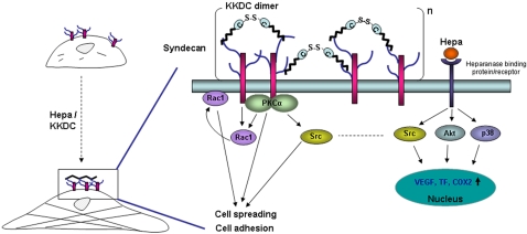
Figure 7
A schematic diagram of HSPG activation by heparanase/KKDC.
Clustering of syndecan family members, and possibly glypicans, by the KKDC dimer or the two heparin binding domains of heparanase facilitates cell-adhesion and cell-spreading (left). Enhanced cell adh...
Figure images are served from the NIH/NLM PubMed Central Open Access Subset or Europe PMC; copyright remains with the publishers and authors.
💬 Discussion
0 commentsNo comments yet. Be the first to start a discussion!
Leave a Comment