Abstract
Respiratory complex I is a redox-driven proton pump, accounting for a large part of the electrochemical gradient that powers mitochondrial adenosine triphosphate synthesis. Complex I dysfunction is associated with severe human diseases. Assembly of the one-megadalton complex I in the inner mitochondrial membrane requires assembly factors and chaperones. We have determined the structure of complex I from the aerobic yeast Yarrowia lipolytica by electron cryo-microscopy at 3.2-Å resolution. A ubiquinone molecule was identified in the access path to the active site. The electron cryo-microscopy structure indicated an unusual lipid-protein arrangement at the junction of membrane and matrix arms that was confirmed by molecular simulations. The structure of a complex I mutant and an assembly intermediate provide detailed molecular insights into the cause of a hereditary complex I-linked disease and complex I assembly in the inner mitochondrial membrane.
🔬 Techniques
🔭 Microscopes
💻 Software
✨ Fluorophores
🧪 Sample Preparation
🏭 Microscope Brands
🧪 Reagent Suppliers
📷 Detectors
💻 Software Details
💾 Data Repositories
🏛️ Research Organizations (ROR)
Affiliated research institutions:
📋 Methods
Purification of complex I variants from Y. lipolytica Complex
I was purified from Y. lipolytica strain GB30 carrying a deletion for the sulfur transferase (ST1) gene to enhance sample homogeneity ( 40 ). ST1 is a substoichiometric component of Y. lipolytica complex I, and the enzyme complex is fully functional in the absence of this polypeptide. Protein purification was carried out by His-tag affinity and size exclusion chromatography as described in ( 41 ), but on the Ni-NTA Sepharose column, the detergent was exchanged from 0.025% DDM to 0.025% LMNG. The complex I assembly intermediate accumulating in the Δ numm strain ( 34 ) (NUMM corresponds to NDUFS6; see table S2) was purified in the same way and polished by ion exchange chromatography on a MonoQ column. The complex I mutant from strain Δ nuym ( 12 ) (NUYM corresponds to NDUFS4; see table S2) was purified in the same way as the GB30 complex but without the detergent exchange. Activity measurements NADH:decylubiquinone (DBQ) oxidoreductase activity was measured in 20 mM Na + -Mops (pH 7.2), 50 mM NaCl, 2 mM KCN, and 0.025% LMNG using 150 μM DBQ and 100 μM NADH as substrates. The residual activity in the presence of 2 μM 2- n -decyl-quinazolin-4-yl-amine (DQA) was subtracted. For assignment of our preparation to the A or D form, we tested the sensitivity of complex I toward 5 mM MgCl 2 or 2 mM NEM. Activity measurements were carried out in 50 mM tris-HCl (pH 8.5), 80 mM KCl, 0.2 mM EDTA, 1.1 mM NaCN, and 0.025% LMNG with 150 μM DBQ and 100 μM NADH ( 9 ). Proton pumping assay with reconstituted complex I Asolectin (10 mg ml −1 ) from soybean was solubilized in 1.6% n -octyl glucopyranoside, 20 mM K + -Mops (pH 7.2), and 80 mM KCl. Purified complex I (200 μg) was reconstituted into proteoliposomes at a protein-to-lipid ratio of 1:50 (w/w) according to ( 42 ), but with 30-min intervals for the addition of BioBeads (Bio-Rad). Proteoliposomes were centrifuged at 100,000 g (1 hour, 4°C) and resuspended in 200 μl of 20 mM K + -Mops (pH 7.2) and 80 mM KCl. Proteoliposomes containing 5 μg of complex I were added to 2 ml of buffer [20 mM K + -Mops (pH 7.2), 50 mM KCl, and 0.5 μM valinomycin] in a stirred cuvette. Proton pumping was monitored by ACMA fluorescence quenching. The fluorophore was added to a final concentration of 0.5 μM. Measurements were performed in a Shimadzu RF-5001 fluorimeter at an excitation wavelength of 430 nm and an emission wavelength of 475 nm (bandpass, 5 nm each; integration time, 1 s) at 30°C. The reaction was started by the successive addition of 60 μM DBQ and 100 μM NADH. Last, 1 μM FCCP was added to collapse the pH gradient.
Show full methods section
Purification of complex I variants from Y. lipolytica Complex
I was purified from Y. lipolytica strain GB30 carrying a deletion for the sulfur transferase (ST1) gene to enhance sample homogeneity ( 40 ). ST1 is a substoichiometric component of Y. lipolytica complex I, and the enzyme complex is fully functional in the absence of this polypeptide. Protein purification was carried out by His-tag affinity and size exclusion chromatography as described in ( 41 ), but on the Ni-NTA Sepharose column, the detergent was exchanged from 0.025% DDM to 0.025% LMNG. The complex I assembly intermediate accumulating in the Δ numm strain ( 34 ) (NUMM corresponds to NDUFS6; see table S2) was purified in the same way and polished by ion exchange chromatography on a MonoQ column. The complex I mutant from strain Δ nuym ( 12 ) (NUYM corresponds to NDUFS4; see table S2) was purified in the same way as the GB30 complex but without the detergent exchange. Activity measurements NADH:decylubiquinone (DBQ) oxidoreductase activity was measured in 20 mM Na + -Mops (pH 7.2), 50 mM NaCl, 2 mM KCN, and 0.025% LMNG using 150 μM DBQ and 100 μM NADH as substrates. The residual activity in the presence of 2 μM 2- n -decyl-quinazolin-4-yl-amine (DQA) was subtracted. For assignment of our preparation to the A or D form, we tested the sensitivity of complex I toward 5 mM MgCl 2 or 2 mM NEM. Activity measurements were carried out in 50 mM tris-HCl (pH 8.5), 80 mM KCl, 0.2 mM EDTA, 1.1 mM NaCN, and 0.025% LMNG with 150 μM DBQ and 100 μM NADH ( 9 ). Proton pumping assay with reconstituted complex I Asolectin (10 mg ml −1 ) from soybean was solubilized in 1.6% n -octyl glucopyranoside, 20 mM K + -Mops (pH 7.2), and 80 mM KCl. Purified complex I (200 μg) was reconstituted into proteoliposomes at a protein-to-lipid ratio of 1:50 (w/w) according to ( 42 ), but with 30-min intervals for the addition of BioBeads (Bio-Rad). Proteoliposomes were centrifuged at 100,000 g (1 hour, 4°C) and resuspended in 200 μl of 20 mM K + -Mops (pH 7.2) and 80 mM KCl. Proteoliposomes containing 5 μg of complex I were added to 2 ml of buffer [20 mM K + -Mops (pH 7.2), 50 mM KCl, and 0.5 μM valinomycin] in a stirred cuvette. Proton pumping was monitored by ACMA fluorescence quenching. The fluorophore was added to a final concentration of 0.5 μM. Measurements were performed in a Shimadzu RF-5001 fluorimeter at an excitation wavelength of 430 nm and an emission wavelength of 475 nm (bandpass, 5 nm each; integration time, 1 s) at 30°C. The reaction was started by the successive addition of 60 μM DBQ and 100 μM NADH. Last, 1 μM FCCP was added to collapse the pH gradient.
Mass spectrometry Purified complex
I from Y. lipolytica was denatured in 2 M guanidine hydrochloride. Proteins were digested with trypsin (sequencing grade, Promega) and analyzed by mass spectrometry using Thermo Scientific Q Exactive Plus equipped with an ultra-high performance liquid chromatography (UHPLC) unit (Thermo Scientific Dionex UltiMate 3000). Peptides and proteins (false discovery ratio,
📊 Figures
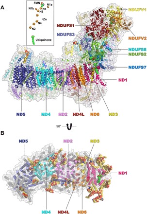
Fig. 1
Cryo-EM structure of respiratory complex I from Y. lipolytica .
( A ) Side view; inset shows FMN and FeS clusters [assignment of EPR signatures N1 to N5 according to ( 72 )] and a Q molecule in the access pathway. ( B ) Top view with the matrix arm omitted for cla...
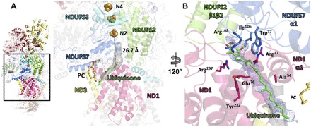
Fig. 2
Q molecule in the access pathway.
( A ) Slice of membrane arm/matrix arm interface viewed from the back of complex I, a cavity (gray) opens in ND1 and permits access of Q from the bilayer to the Q reduction site near cluster N2. ( B )...
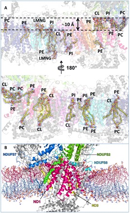
Fig. 3
Lipid binding sites and unique protein lipid arrangement at the junction of the membrane and matrix arms.
( A ) Lipid and detergent binding sites in the membrane arm. CL, cardiolipin; PC, phosphatidylcholine; PE, phosphatidylethanolamine; PI, phosphatidylinositol. ( B ) Cross section of a simulation snaps...
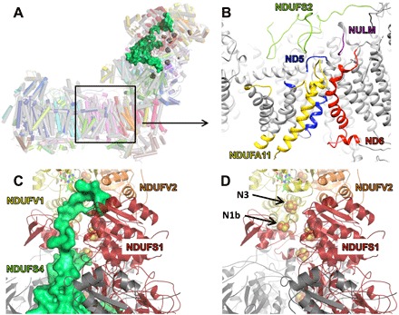
Fig. 4
Mutant lacking NDUFS4.
( A ) Overlay of wild type (gray, NDUFS4 green surface representation) and mutant (cartoon, color as in Fig. 1 ). ( B ) Zoom into membrane arm. Disordered protein in the mutant is highlighted by color...
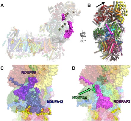
Fig. 5
Cryo-EM structure of a complex I assembly intermediate.
( A ) Overlay of wild type (gray) and assembly intermediate (color) with assembly factor NDUFAF2 (purple surface). ( B ) Overlay as in (A) viewed from the back. The arrow indicates the tilted matrix a...
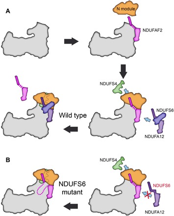
Fig. 6
Function of the assembly factor NDUFAF2.
( A ) Assembly factor NDUFAF2 associates with a complex I assembly intermediate that lacks the N module (NDUFS1, NDUFV1, and NDUFV2). NDUFAF2 provides a platform for N module attachment (dashed line) ...
Figure images are served from the NIH/NLM PubMed Central Open Access Subset or Europe PMC; copyright remains with the publishers and authors.
💬 Discussion
0 commentsNo comments yet. Be the first to start a discussion!
Leave a Comment