Abstract
Recognition of ligands by toll-like receptor (TLR) 2 requires interactions with other TLRs. TLRs form a combinatorial repertoire to discriminate between the diverse microbial ligands. Diversity results from extracellular and intracellular interactions of different TLRs. This paper demonstrates that TLR1 and TLR2 are required for ara-lipoarabinomannan- and tripalmitoyl cysteinyl lipopeptide-stimulated cytokine secretion from mononuclear cells. Confocal microscopy revealed that TLR1 and TLR2 cotranslationally form heterodimeric complexes on the cell surface and in the cytosol. Simultaneous cross-linking of both receptors resulted in ligand-independent signal transduction. Using chimeric TLRs, we found that expression of the extracellular domains along with simultaneous expression of the intracellular domains of both TLRs was necessary to achieve functional signaling. The domains from each receptor did not need to be contained within a single contiguous protein. Chimeric TLR analysis further defined the toll/IL-1R domains as the area of crucial intracellular TLR1-TLR2 interaction.
🔬 Techniques
💻 Software
✨ Fluorophores
🧪 Sample Preparation
🔬 Cell Lines
🏭 Microscope Brands
🧪 Reagent Suppliers
💻 Software Details
🏛️ Research Organizations (ROR)
Affiliated research institutions:
📋 Methods
DNA expression vectors Human
TLR2 and TLR4 cDNAs were obtained from Tularik. The TLR cDNAs encoded Flag-epitope–tagged proteins and had been cloned into the pFlag-CMV-1 vector. Chimeric TLR2–TLR1 (TLR [2–1]) and TLR1–TLR2 (TLR [1–2]) constructs were generated by PCR as follows: An XhoI restriction site was added just upstream of Cys 577 of TLR1 and Cys 585 of TLR2 and used for the domain swapping. Intracellular and extracellular domains were PCR amplified using Pfu Turbo DNA polymerase (Stratagene) and cross-assembled into pBlueScript ® II KS(+) (Stratagene). The transmembrane and cytoplasmic regions of TLR1 and TLR2 were PCR amplified using primers as follows: T2cyto-5, 5′-GCGCCTCGAGTGTCACAGGACAGCACTGGTGTCTG-3′; TLR2–3, 5′-CGCGGGTACCCTAGGACTTTATCGCAGCTCTCAG-3′; T1cyto-5, 5′-GCGCCTCGAGTGCAACATAACTCTGCTGATCGTCACC-3′; and TLR1-3, 5′-CGCGGGTAC-CCTATTTCTTTGCTTGCTCTGTCAGC-3′. The PCR products were digested and cloned into the KpnI and XhoI sites of pBlueScript ® II KS (+) (Stratagene). A portion of the extracellular domain of TLR1 was PCR amplified using primers as follows: TL1-Bam, 5′-CTTTCATTAGGATCCTCCAGCTGGTTTG-3′; and T1ex-3, 5′-GCGCCTCGAGTTCAGACATGTGAAAGTCCTTTAGTAGG-3′. The PCR product was digested with BamHI and XhoI and cloned into the BamHI and XhoI sites of the pBlueScript ® vector already containing the cytoplasmic region of TLR2. This vector was then digested with BamHI and KpnI (KpnI site was blunted with Klenow enzyme), and the fragment containing the regions of TLR1 and TLR2 was inserted into the BamHI and SmaI sites of pFlag-CMV-1 TLR1.II KS(+) (Stratagene). A portion of the extracellular domain of TLR2 was PCR amplified using primers as follows: TL2-RV, 5′-CTAACATTGATATCAGTAAGAATAGTTTTC-3′; and T2ex-3, 5′-GCGCCTCGAGCACCGAGAGGCGGACATCCTGAACC-3′. The PCR product was digested with EcoRV and XhoI and cloned into the EcoRV and XhoI sites of the pBlueScript ® vector already containing the cytoplasmic region of TLR1. This vector was then digested with EcoRV and KpnI (KpnI site was blunted with Klenow enzyme), and the fragment containing the cytoplasmic region of TLR1 was inserted into the EcoRV and SmaI sites of pFlag-CMV-1 TLR2. To construct the vectors TLR [1–2 TIR] and TLR [2–1 TIR], the domain swapping was done using a NsiI restriction site that is conserved in TLR1 and TLR2. pCMV-Flag TLR1 was cut with NotI and NsiI (NsiI partial digestion), and the fragment of 1,917 bp was cloned into the NotI and NsiI sites of the pBlueScript ® containing the cytoplasmic region of TLR2. The resulting plasmid was then cut with NotI and KpnI, and the fragment containing TLR [1–2 TIR] was inserted into the NotI and KpnI sites of pFlag-CMV-1. Similar strategy was used to construct the TLR [2–1 TIR] vector. Stimulants Yeast zymosan was purchased from Sigma-Aldrich. Mycobacterial araLAM, purified from rapidly growing avirulent mycobacteria, was provided by Dr. John Belisle (Colorado State University, Fort Collins, CO) under the National Institutes of Health, National Institute of Allergy and Infectious Diseases contract N01-AI-75320 entitled “Tuberculosis Research Materials and Vaccine Testing.” Pam 3 CSK 4 was obtained from EMC Microcollections. Rough LPS (Sigma-Aldrich) was phenol extracted and used at 10 ng/ml for stimulation assays. Recombinant human IL-1β was purchased from R&D Systems, and used as a positive control for NFκB activation. In vitro stimulation of human PBMCs: antibody-blocking experiments Human PBMCs were isolated from peripheral blood using Lymphocyte Separation Medium (Mediatech). PBMCs were cultured in RPMI 1640 medium supplemented with 10% FCS (Atlanta Biologicals) in 24-well plates at 10 6 cells/well. For blocking experiments, PBMCs were preincubated for 30 min at 37°C in 5% CO 2 with anti-TLR1 (clone GD2.F4; eBiosciences), or anti-TLR2 (clone 11G7) mAbs or isotype control antibody (eBiosciences) at 10 μg/ml concentration/well before the addition of stimulants. Culture supernatants were collected after an overnight incubation at 37°C in 5% CO 2 . Secreted IL-6 levels were determined by ELISA according to manufacturer's instructions (OptEIA; BD Biosciences). Data are representative of at least three independent experiments.
Show full methods section
DNA expression vectors Human
TLR2 and TLR4 cDNAs were obtained from Tularik. The TLR cDNAs encoded Flag-epitope–tagged proteins and had been cloned into the pFlag-CMV-1 vector. Chimeric TLR2–TLR1 (TLR [2–1]) and TLR1–TLR2 (TLR [1–2]) constructs were generated by PCR as follows: An XhoI restriction site was added just upstream of Cys 577 of TLR1 and Cys 585 of TLR2 and used for the domain swapping. Intracellular and extracellular domains were PCR amplified using Pfu Turbo DNA polymerase (Stratagene) and cross-assembled into pBlueScript ® II KS(+) (Stratagene). The transmembrane and cytoplasmic regions of TLR1 and TLR2 were PCR amplified using primers as follows: T2cyto-5, 5′-GCGCCTCGAGTGTCACAGGACAGCACTGGTGTCTG-3′; TLR2–3, 5′-CGCGGGTACCCTAGGACTTTATCGCAGCTCTCAG-3′; T1cyto-5, 5′-GCGCCTCGAGTGCAACATAACTCTGCTGATCGTCACC-3′; and TLR1-3, 5′-CGCGGGTAC-CCTATTTCTTTGCTTGCTCTGTCAGC-3′. The PCR products were digested and cloned into the KpnI and XhoI sites of pBlueScript ® II KS (+) (Stratagene). A portion of the extracellular domain of TLR1 was PCR amplified using primers as follows: TL1-Bam, 5′-CTTTCATTAGGATCCTCCAGCTGGTTTG-3′; and T1ex-3, 5′-GCGCCTCGAGTTCAGACATGTGAAAGTCCTTTAGTAGG-3′. The PCR product was digested with BamHI and XhoI and cloned into the BamHI and XhoI sites of the pBlueScript ® vector already containing the cytoplasmic region of TLR2. This vector was then digested with BamHI and KpnI (KpnI site was blunted with Klenow enzyme), and the fragment containing the regions of TLR1 and TLR2 was inserted into the BamHI and SmaI sites of pFlag-CMV-1 TLR1.II KS(+) (Stratagene). A portion of the extracellular domain of TLR2 was PCR amplified using primers as follows: TL2-RV, 5′-CTAACATTGATATCAGTAAGAATAGTTTTC-3′; and T2ex-3, 5′-GCGCCTCGAGCACCGAGAGGCGGACATCCTGAACC-3′. The PCR product was digested with EcoRV and XhoI and cloned into the EcoRV and XhoI sites of the pBlueScript ® vector already containing the cytoplasmic region of TLR1. This vector was then digested with EcoRV and KpnI (KpnI site was blunted with Klenow enzyme), and the fragment containing the cytoplasmic region of TLR1 was inserted into the EcoRV and SmaI sites of pFlag-CMV-1 TLR2. To construct the vectors TLR [1–2 TIR] and TLR [2–1 TIR], the domain swapping was done using a NsiI restriction site that is conserved in TLR1 and TLR2. pCMV-Flag TLR1 was cut with NotI and NsiI (NsiI partial digestion), and the fragment of 1,917 bp was cloned into the NotI and NsiI sites of the pBlueScript ® containing the cytoplasmic region of TLR2. The resulting plasmid was then cut with NotI and KpnI, and the fragment containing TLR [1–2 TIR] was inserted into the NotI and KpnI sites of pFlag-CMV-1. Similar strategy was used to construct the TLR [2–1 TIR] vector. Stimulants Yeast zymosan was purchased from Sigma-Aldrich. Mycobacterial araLAM, purified from rapidly growing avirulent mycobacteria, was provided by Dr. John Belisle (Colorado State University, Fort Collins, CO) under the National Institutes of Health, National Institute of Allergy and Infectious Diseases contract N01-AI-75320 entitled “Tuberculosis Research Materials and Vaccine Testing.” Pam 3 CSK 4 was obtained from EMC Microcollections. Rough LPS (Sigma-Aldrich) was phenol extracted and used at 10 ng/ml for stimulation assays. Recombinant human IL-1β was purchased from R&D Systems, and used as a positive control for NFκB activation. In vitro stimulation of human PBMCs: antibody-blocking experiments Human PBMCs were isolated from peripheral blood using Lymphocyte Separation Medium (Mediatech). PBMCs were cultured in RPMI 1640 medium supplemented with 10% FCS (Atlanta Biologicals) in 24-well plates at 10 6 cells/well. For blocking experiments, PBMCs were preincubated for 30 min at 37°C in 5% CO 2 with anti-TLR1 (clone GD2.F4; eBiosciences), or anti-TLR2 (clone 11G7) mAbs or isotype control antibody (eBiosciences) at 10 μg/ml concentration/well before the addition of stimulants. Culture supernatants were collected after an overnight incubation at 37°C in 5% CO 2 . Secreted IL-6 levels were determined by ELISA according to manufacturer's instructions (OptEIA; BD Biosciences). Data are representative of at least three independent experiments.
Antibody cross-linking experiments
Anti-TLR1 (clone GD2.F4; eBiosciences), anti-TLR2 (clone TL2.1; a gift of Dr. Egil Lien, University of Massachusetts Medical School, Worcester, MA), or isotype control OKT8 (CRL-8014; American Type Culture Collection) mAbs were added to sterile high protein binding capacity 96-well plates (Costar) at varying concentrations in PBS and incubated overnight at 4°C. The plates were washed three times with PBS and blocked with 10% FCS (Atlanta Biologicals) in PBS for 2 h. 7 × 10 5 PBMCs in RPMI 1640 with 10% FCS were added to each well and incubated for 18 h at 37°C in a 5% CO 2 humidified incubator. Low endotoxin mAb preparations were used in all experiments. As an additional control, polymyxin B (cat.# P4932; Sigma-Aldrich) at a 5-μg/ml concentration was added to the culture medium to neutralize potential endotoxin contamination. Supernatants were harvested and IL-8 levels were determined by ELISA according to manufacturer's instructions (BD Biosciences). The results shown are representative of three independent experiments.
Confocal microscopy imaging experiments
Stable cell lines of HEK293 cells expressing the fluorescent protein TLR constructs were engineered as described previously ( Latz et al., 2002 ). Confocal microscopy was performed with living cells that were seeded on 35-mm glass-bottom tissue culture dishes (MatTek Corp.) 24–48 h before examination. Images were taken with a confocal microscope (TCS SP2 AOBS; Leica) equipped with an acousto-optical beamsplitter using version 2 of the Leica Confocal Software, and the images were further processed with Adobe Photoshop ® software, version 6 and 7. The cells were kept at 37°C during imaging using a warm stage apparatus. CFP-tagged proteins were visualized using the 458-nm argon laser line; for YFP, the 514-nm line of a 100-mW argon laser was used. Alexa ® 647 was excited with a 2.5-mW helium/neon laser emitting at 633 nm. Cells expressing CFP and YFP proteins were sequentially scanned using only one laser line active per scan. Antibody-patching experiments For antibody-patching experiments, either stably transfected HEK cells (TLR1-YFP/TLR2-CFP) or TLR4-YFP cells transiently transfected with pcDNA3-TLR2 were grown on glass-bottom 35-mm tissue culture dishes and washed twice with ice-cold HBSS/1% FBS. The cells were then incubated with 5 μg/ml anti-TLR2 (clone TLR 2.1) or anti–human HLA I (clone W6/32HL, cat.# RDI-CBL139–1XP; Research Diagnostics, Inc.) antibody in HBSS/1% FBS as primary antibodies on ice for 30 min. After two washes with cold HBSS, the cells were counterstained with Alexa ® 647–conjugated goat anti–mouse secondary antibody (Molecular Probes, Inc.). After washing, the cells were incubated in prewarmed complete growth medium for 10 min at 37°C and immediately analyzed by confocal microscopy. Representative results of three independent experiments are shown.
Transfection and reporter gene assays in HEK 293-CD14 cells
HEK293 cells (CRL-1573; American Type Culture Collection) stably expressing human CD14 (HEK293-CD14) were cloned as described previously ( Kurt-Jones et al., 2002 ). Transfections of HEK293-CD14 cells were performed using GeneJuice ® Transfection Reagent (Novagen) according to manufacturer's instructions. The cells were plated to 96-well plates at 2.5 × 10 4 /well and transfected 24 h later with a total of 0.3 μg DNA per well. The transfected DNA included 80 ng NFκB-driven firefly luciferase plasmid (pGL-3-Basic Vector, cat.# E1751; Promega) and 20 ng HSV-TK promoter-driven renilla luciferase plasmid (phRL-TK Vector, cat.# E6241; Promega) along with wild-type or chimeric TLR constructs cloned into in pFlag-CMV-1 vector (cat.# E7273; Sigma-Aldrich). TLR plasmids were transfected at concentrations ranging from 5 to 200 ng/well. Empty pFlag-CMV-1 vector was used to bring up the total amount of transfected DNA to 0.3 μg per well. The transfected cells were incubated overnight at 37°C in 5% CO 2 humidified incubator and then stimulated for 6 h with 10 μg/ml zymosan, 1 μg/ml araLAM, 100 ng/ml Pam 3 CSK 4 , or 100 ng/ml IL-1β. Cells were lysed using 50 μl Passive Lysis Buffer (cat.# E1941; Promega), and firefly luciferase activity was measured using Dual-Glo™ Luciferase Assay System (cat.# E2940; Promega) according to the manufacturer's instructions. Luciferase activity was calculated in RLU as a ratio of NFκB-dependent firefly luciferase activity to NFκB-independent renilla luciferase activity. The results are shown as the mean ± SD of triplicate wells, and are representative of three independent experiments.
📊 Figures
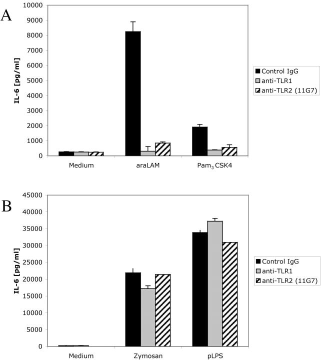
Figure 1.
Anti-TLR1 and -TLR2 mAbs block the IL-6 response of human PBMCs to araLAM and Pam 3 CSK 4 . Fresh human PBMCs were preincubated for 30 min with anti-TLR1 mAb (GD2.F4) or anti-TLR2 mAb (11G7) before ad...

Figure 2.
TLR1 cooperates with TLR2 on the cell surface to initiate IL-8 secretion. Control antibody (OKT8), anti-CD14 (26ic), anti-TLR1 (GD2.F4), and anti-TLR2 (TL2.1) mAbs were immobilized on sterile high pro...
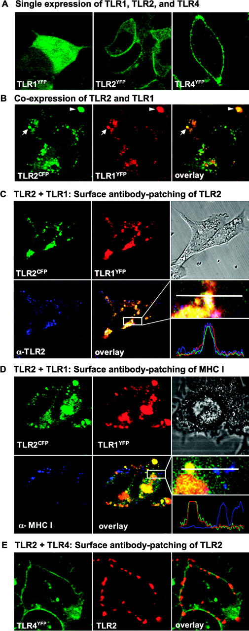
Figure 3.
Confocal imaging of live HEK cells expressing fluorescent proteinu2013tagged TLR1, TLR2, and TLR4. (A) Confocal microscopy of HEK cells stably expressing TLR1 YFP , TLR2 YFP , or TLR4 YFP . HEK cells ...

Figure 4.
TLR2 and TLR1 mutants inhibit NF u03ba B activation in HEK 293 cells in stimulation with araLAM and zymosan. (A) The response of HEK293-CD14 cells to araLAM and zymosan is inhibited by a single point ...
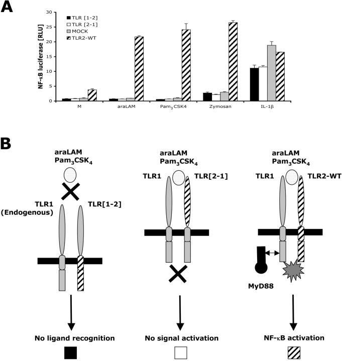
Figure 5.
Neither the intracellular nor the extracellular domain of TLR2 is sufficient to confer NF u03ba B signal activation. (A) HEK293-CD14 cells were transfected with either TLR [1u20132] or TLR [2u20131] D...
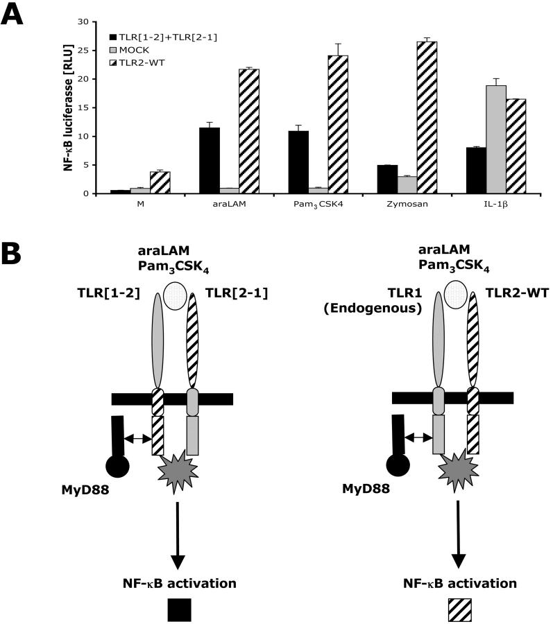
Figure 6.
NF u03ba B signal activation requires both extracellular and intracellular domains of TLR1 and TLR2. (A) HEK293-CD14 cells were cotransfected with TLR [1u20132] and TLR [2u20131] DNA encoding chimeric...
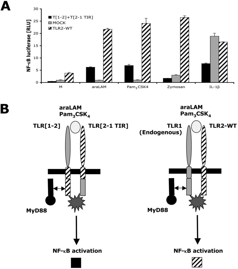
Figure 7.
Heterologous expression of the extracellular domains of TLR1 and TLR2 together with heterologous expression of the TIR domains of TLR1 and TLR2 is sufficient for NF u03ba B signal activation. (A) HEK2...
Figure images are served from the NIH/NLM PubMed Central Open Access Subset or Europe PMC; copyright remains with the publishers and authors.
💬 Discussion
0 commentsNo comments yet. Be the first to start a discussion!
Leave a Comment