Abstract
Many chemical mediators regulate neutrophil recruitment to inflammatory sites. Although the actions of each chemical mediator have been demonstrated with neutrophils in vitro, how such chemical mediators act cooperatively or counteractively in vivo remains largely unknown. Here, by in vivo two-photon excitation microscopy with transgenic mice expressing biosensors based on Förster resonance energy transfer, we time-lapse-imaged the activities of extracellular signal-regulated kinase (ERK) and protein kinase A (PKA) in neutrophils in inflamed intestinal tissue. ERK activity in neutrophils rapidly increased during spreading on the endothelial cells and showed positive correlation with the migration velocity on endothelial cells or in interstitial tissue. Meanwhile, in the neutrophils migrating in the interstitial tissue, high PKA activity correlated negatively with migration velocity. In contradiction to previous in vitro studies that showed ERK activation by prostaglandin E2 (PGE2) engagement with prostaglandin receptor EP4, intravenous administration of EP4 agonist activated PKA, inhibited ERK, and suppressed migration of neutrophils. The opposite results were obtained using nonsteroidal antiinflammatory drugs (NSAIDs). Therefore, NSAID-induced enteritis may be caused at least partially by the inhibition of EP4 receptor signaling of neutrophils. Our results demonstrate that ERK positively regulates the neutrophil recruitment cascade by promoting adhesion and migration steps.
🔬 Techniques
💻 Software
✨ Fluorophores
🧪 Sample Preparation
🔬 Cell Lines
🏭 Microscope Brands
🧪 Reagent Suppliers
🔴 Lasers
🔎 Objectives
🎨 Filters
💻 Software Details
🏛️ Research Organizations (ROR)
Affiliated research institutions:
📋 Methods
Transgenic mice expressing FRET biosensors. The generation of transgenic mice expressing the ERK FRET biosensor EKAREVnes (Eisuke mice), the PKA FRET biosensor AKAR3EVnes (PKAchu mice), and the negative control FRET biosensor AKAR3EV-NC was reported previously ( Kamioka et al., 2012 ). Founder animals were backcrossed more than five generations to C57BL/6N Jcl. Mice were housed in a specific pathogen–free facility and received a routine chow diet and water ad libitum. To date, no disease or anomaly has been associated with the expression of the FRET biosensors used in this study. 6–16-wk-old mice were used for the in vivo imaging. The animal protocols were reviewed and approved by the Animal Care and Use Committee of Kyoto University Graduate School of Medicine (No. 10584). Microscopy and image processing. 2PM was performed with an FV1000MVE inverted microscope (Olympus) equipped with a 30×/1.05 NA silicon oil-immersion objective lens (UPLSAPO 30xS; Olympus) and a Mai Tai DeepSee HP Ti:sapphire Laser (Spectra Physics) or with an FV1000MVE inverted microscope equipped with a 30×/1.05 NA silicon oil-immersion objective lens and an InSight DeepSee Laser (Spectra Physics). The laser power used for observation was between 4 and 8%. Scan speed was set between 12.5 and 20 µs/pixel. Images were recorded every 10–30 s for long-term imaging or every 1.5 s for short-term imaging. The excitation wavelength for CFP was 840 nm. We used an IR-cut filter, BA685RIF-3, two dichroic mirrors, DM505 and DM570, and three emission filters, BA460-500 (Olympus) for CFP, BA520-560 (Olympus) for YFP, and 645/60 (Chroma Technology Corp.) for Qtracker 655. Acquired images were analyzed with MetaMorph software (Universal Imaging) as described previously ( Aoki and Matsuda, 2009 ; Kamioka et al., 2012 ). In brief, the level of FRET was represented by the FRET/CFP ratio image in intensity modulated display mode; eight colors from red to blue are used to represent the FRET/CFP ratio, and the 32 grades of color intensity are used to represent the signal intensity of the CFP image. The warm and cold colors indicate high and low FRET levels, respectively. Neutrophils were distinguished from the other cell types by their characteristic segmented nuclei. Neutrophil recruitment and extravasation were divided arbitrarily into the following five steps: rolling, adhesion, crawling, transmigration, and chemotaxis in the interstitial tissue. The term “rolling” was used in the case of round-shape neutrophils that rolled over the endothelial cells by the frame by frame comparison. Under the current scanning conditions, cells flowing in the venules could not be imaged clearly and were excluded from the designation of rolling neutrophils. “Adhesion” was the term used for neutrophils that had ceased rolling and remained in the same position for at least 30 s. “Crawling” was used in the case of neutrophils that adopted an amoeboid shape and crawled over the endothelial cells. “Transmigration” was defined as neutrophils invading into or between endothelial cells to emigrate. Finally, the term “chemotaxis” was applied when neutrophils migrated in the interstitial tissue. Under our experimental conditions, we could not separate stochastic migration and chemotactic migration, so we referred to both as “chemotaxis.” The flux of rolling neutrophils and the number of adherent neutrophils were quantified as described previously ( Kubes et al., 2003 ) with time-lapse images acquired every 1.5 s. The number of rolling neutrophils (flux) was counted using frame by frame analysis for 10 min. A neutrophil was defined as adherent to endothelial cells if it remained stationary for >30 s. The numbers of adherent, crawling, or transmigrating neutrophils were averaged per 400–700 µm endothelial cells during a 10-min observation period. In vivo observation of the small intestine. Mice were anesthetized with 1.5–2% isoflurane (Abbott) inhalation and placed in the supine position on an electric heat pad maintained at 37°C. Before surgery, the abdominal area of the mouse was disinfected using 70% ethanol. A small vertical incision was made in the right side of the abdominal wall. The small intestine was pulled out of the abdominal cavity, and both proximal and distal sides of the small intestine of interest were ligated using 5-0 silk surgical sutures (Nesco Suture). With a 29-gauge needle, 1 µg/ml LPS (Sigma-Aldrich) and 100 nM fMLP (Sigma-Aldrich) were administered into the intestinal cavity. The small intestine was returned to the abdominal cavity before closing the wound with 6-0 silk sutures (Ethicon). After 2 h, the abdominal cavity was reopened to pull out the small intestine on a cover-glass placed in a heat-stage maintained at 37°C. The small intestine was fixed with surgical sutures to minimize peristalsis and observed by 2PM. Time-lapse imaging was performed with a two-photon microscope running FluoView software (Olympus). To observe extravasation of neutrophils, we observed postcapillary venules with a diameter of ∼30–50 µm. In some experiments, 5 mg/kg PD0325901 MEK inhibitor (EMD Millipore) in PBS, 0.3 g/kg dbcAMP (EMD Millipore), 7.5 mg/kg flurbiprofen axetil (KAKEN Pharmaceutical), 15 mg/kg SB203580 p38 inhibitor (Cayman Chemical), 7.5 µg/kg LTB 4 (Cayman Chemical), 4 mg/kg LY293111 LTB 4 inhibitor (Cayman Chemical), 0.5 mg/kg ONO-AE-1-259-01, 0.25 mg/kg ONO-AE1-329, or 5 mg/kg ONO-AE3-208 (Ono Pharmaceutical Co.) was administered via the jugular vein during the course of in vivo imaging. The plasma half-life of ONO-AE3-208 measured in an experiment of intravenous injection was 0.2 h ( Kabashima et al., 2002 ). The plasma half-lives of ONO-AE-1-259-01 and 0.25 mg/kg ONO-AE1-329 are not available, but a compound with similar structure is inactivated in the lung within 20 min. The reagents were administered for 30 s except for LTB 4 , which was administered for 3–5 min. To visualize the blood vessels, 3 µl Qtracker 655 (Invitrogen) was coinjected with the drugs. Mice were euthanized after the experiments. Bone marrow transplantation. Bone marrow was harvested from the femur and tibia of 14-wk-old female PKAchu mice. Whole bone marrow nucleated cells were transplanted into 10-wk-old C57BL/6 female mice (10 7 cells/mouse), which had been subjected to 9.6 Gy irradiation. Mice were observed 7 wk after transplantation. NSAID enteritis. 10-wk-old C57BL/6 mice were administered subcutaneously with 7.5 mg/kg flurbiprofen axetil with or without 0.25 mg/kg ONO-AE1-329 or 5 mg/kg ONO-AE3-208 as described previously ( Kabashima et al., 2002 ). For the mice administered with ONO-AE3-208, mice were provided with drinking water containing 37.5 µg/ml ONO-AE3-208. 24 h later, mice were sacrificed to count intestinal ulcers under a stereoscopic microscope. Statistical analysis. P-values for normal distributed data were calculated with the Student’s t test or paired Student’s t test for the evaluation of statistically significant differences. Otherwise, the Mann–Whitney U test was used. Data analysis was performed using Prism software (GraphPad Software). *, P < 0.05; **, P < 0.01; ***, P < 0.001. Online supplemental material. Video 1 shows the activation of ERK in neutrophils during adhesion to endothelial cells of the inflamed small intestine. Video 2 shows the requirement of ERK activity during the neutrophil recruitment cascade. Video 3 shows the activation of PKA during the neutrophil recruitment cascade. Video 4 shows the PKA activity in neutrophils derived from a PKAchu mouse and transplanted into a wild-type mouse. Video 5 shows the PKA inhibition of ERK activity, recruitment to endothelial cells, and migration of neutrophils. Video 6 shows the accelerated migration and activation of ERK of neutrophils after NSAID injection. Video 7 shows the inhibition of the rolling step and the migration of neutrophils by an EP4 agonist. Video 8 shows the promoting effect of NSAID on neutrophil recruitment was lost in the presence of an EP4 agonist. Online supplemental material is available at http://www.jem.org/cgi/content/full/jem.20132112/DC1 .
Show full methods section
Transgenic mice expressing FRET biosensors. The generation of transgenic mice expressing the ERK FRET biosensor EKAREVnes (Eisuke mice), the PKA FRET biosensor AKAR3EVnes (PKAchu mice), and the negative control FRET biosensor AKAR3EV-NC was reported previously ( Kamioka et al., 2012 ). Founder animals were backcrossed more than five generations to C57BL/6N Jcl. Mice were housed in a specific pathogen–free facility and received a routine chow diet and water ad libitum. To date, no disease or anomaly has been associated with the expression of the FRET biosensors used in this study. 6–16-wk-old mice were used for the in vivo imaging. The animal protocols were reviewed and approved by the Animal Care and Use Committee of Kyoto University Graduate School of Medicine (No. 10584). Microscopy and image processing. 2PM was performed with an FV1000MVE inverted microscope (Olympus) equipped with a 30×/1.05 NA silicon oil-immersion objective lens (UPLSAPO 30xS; Olympus) and a Mai Tai DeepSee HP Ti:sapphire Laser (Spectra Physics) or with an FV1000MVE inverted microscope equipped with a 30×/1.05 NA silicon oil-immersion objective lens and an InSight DeepSee Laser (Spectra Physics). The laser power used for observation was between 4 and 8%. Scan speed was set between 12.5 and 20 µs/pixel. Images were recorded every 10–30 s for long-term imaging or every 1.5 s for short-term imaging. The excitation wavelength for CFP was 840 nm. We used an IR-cut filter, BA685RIF-3, two dichroic mirrors, DM505 and DM570, and three emission filters, BA460-500 (Olympus) for CFP, BA520-560 (Olympus) for YFP, and 645/60 (Chroma Technology Corp.) for Qtracker 655. Acquired images were analyzed with MetaMorph software (Universal Imaging) as described previously ( Aoki and Matsuda, 2009 ; Kamioka et al., 2012 ). In brief, the level of FRET was represented by the FRET/CFP ratio image in intensity modulated display mode; eight colors from red to blue are used to represent the FRET/CFP ratio, and the 32 grades of color intensity are used to represent the signal intensity of the CFP image. The warm and cold colors indicate high and low FRET levels, respectively. Neutrophils were distinguished from the other cell types by their characteristic segmented nuclei. Neutrophil recruitment and extravasation were divided arbitrarily into the following five steps: rolling, adhesion, crawling, transmigration, and chemotaxis in the interstitial tissue. The term “rolling” was used in the case of round-shape neutrophils that rolled over the endothelial cells by the frame by frame comparison. Under the current scanning conditions, cells flowing in the venules could not be imaged clearly and were excluded from the designation of rolling neutrophils. “Adhesion” was the term used for neutrophils that had ceased rolling and remained in the same position for at least 30 s. “Crawling” was used in the case of neutrophils that adopted an amoeboid shape and crawled over the endothelial cells. “Transmigration” was defined as neutrophils invading into or between endothelial cells to emigrate. Finally, the term “chemotaxis” was applied when neutrophils migrated in the interstitial tissue. Under our experimental conditions, we could not separate stochastic migration and chemotactic migration, so we referred to both as “chemotaxis.” The flux of rolling neutrophils and the number of adherent neutrophils were quantified as described previously ( Kubes et al., 2003 ) with time-lapse images acquired every 1.5 s. The number of rolling neutrophils (flux) was counted using frame by frame analysis for 10 min. A neutrophil was defined as adherent to endothelial cells if it remained stationary for >30 s. The numbers of adherent, crawling, or transmigrating neutrophils were averaged per 400–700 µm endothelial cells during a 10-min observation period. In vivo observation of the small intestine. Mice were anesthetized with 1.5–2% isoflurane (Abbott) inhalation and placed in the supine position on an electric heat pad maintained at 37°C. Before surgery, the abdominal area of the mouse was disinfected using 70% ethanol. A small vertical incision was made in the right side of the abdominal wall. The small intestine was pulled out of the abdominal cavity, and both proximal and distal sides of the small intestine of interest were ligated using 5-0 silk surgical sutures (Nesco Suture). With a 29-gauge needle, 1 µg/ml LPS (Sigma-Aldrich) and 100 nM fMLP (Sigma-Aldrich) were administered into the intestinal cavity. The small intestine was returned to the abdominal cavity before closing the wound with 6-0 silk sutures (Ethicon). After 2 h, the abdominal cavity was reopened to pull out the small intestine on a cover-glass placed in a heat-stage maintained at 37°C. The small intestine was fixed with surgical sutures to minimize peristalsis and observed by 2PM. Time-lapse imaging was performed with a two-photon microscope running FluoView software (Olympus). To observe extravasation of neutrophils, we observed postcapillary venules with a diameter of ∼30–50 µm. In some experiments, 5 mg/kg PD0325901 MEK inhibitor (EMD Millipore) in PBS, 0.3 g/kg dbcAMP (EMD Millipore), 7.5 mg/kg flurbiprofen axetil (KAKEN Pharmaceutical), 15 mg/kg SB203580 p38 inhibitor (Cayman Chemical), 7.5 µg/kg LTB 4 (Cayman Chemical), 4 mg/kg LY293111 LTB 4 inhibitor (Cayman Chemical), 0.5 mg/kg ONO-AE-1-259-01, 0.25 mg/kg ONO-AE1-329, or 5 mg/kg ONO-AE3-208 (Ono Pharmaceutical Co.) was administered via the jugular vein during the course of in vivo imaging. The plasma half-life of ONO-AE3-208 measured in an experiment of intravenous injection was 0.2 h ( Kabashima et al., 2002 ). The plasma half-lives of ONO-AE-1-259-01 and 0.25 mg/kg ONO-AE1-329 are not available, but a compound with similar structure is inactivated in the lung within 20 min. The reagents were administered for 30 s except for LTB 4 , which was administered for 3–5 min. To visualize the blood vessels, 3 µl Qtracker 655 (Invitrogen) was coinjected with the drugs. Mice were euthanized after the experiments. Bone marrow transplantation. Bone marrow was harvested from the femur and tibia of 14-wk-old female PKAchu mice. Whole bone marrow nucleated cells were transplanted into 10-wk-old C57BL/6 female mice (10 7 cells/mouse), which had been subjected to 9.6 Gy irradiation. Mice were observed 7 wk after transplantation. NSAID enteritis. 10-wk-old C57BL/6 mice were administered subcutaneously with 7.5 mg/kg flurbiprofen axetil with or without 0.25 mg/kg ONO-AE1-329 or 5 mg/kg ONO-AE3-208 as described previously ( Kabashima et al., 2002 ). For the mice administered with ONO-AE3-208, mice were provided with drinking water containing 37.5 µg/ml ONO-AE3-208. 24 h later, mice were sacrificed to count intestinal ulcers under a stereoscopic microscope. Statistical analysis. P-values for normal distributed data were calculated with the Student’s t test or paired Student’s t test for the evaluation of statistically significant differences. Otherwise, the Mann–Whitney U test was used. Data analysis was performed using Prism software (GraphPad Software). *, P < 0.05; **, P < 0.01; ***, P < 0.001. Online supplemental material. Video 1 shows the activation of ERK in neutrophils during adhesion to endothelial cells of the inflamed small intestine. Video 2 shows the requirement of ERK activity during the neutrophil recruitment cascade. Video 3 shows the activation of PKA during the neutrophil recruitment cascade. Video 4 shows the PKA activity in neutrophils derived from a PKAchu mouse and transplanted into a wild-type mouse. Video 5 shows the PKA inhibition of ERK activity, recruitment to endothelial cells, and migration of neutrophils. Video 6 shows the accelerated migration and activation of ERK of neutrophils after NSAID injection. Video 7 shows the inhibition of the rolling step and the migration of neutrophils by an EP4 agonist. Video 8 shows the promoting effect of NSAID on neutrophil recruitment was lost in the presence of an EP4 agonist. Online supplemental material is available at http://www.jem.org/cgi/content/full/jem.20132112/DC1 .
Online supplemental material. Video 1 shows the activation of ERK in neutrophils during adhesion to endothelial cells of the inflamed small intestine. Video 2 shows the requirement of ERK activity during the neutrophil recruitment cascade. Video 3 shows the activation of PKA during the neutrophil recruitment cascade. Video 4 shows the PKA activity in neutrophils derived from a PKAchu mouse and transplanted into a wild-type mouse. Video 5 shows the PKA inhibition of ERK activity, recruitment to endothelial cells, and migration of neutrophils. Video 6 shows the accelerated migration and activation of ERK of neutrophils after NSAID injection. Video 7 shows the inhibition of the rolling step and the migration of neutrophils by an EP4 agonist. Video 8 shows the promoting effect of NSAID on neutrophil recruitment was lost in the presence of an EP4 agonist. Online supplemental material is available at http://www.jem.org/cgi/content/full/jem.20132112/DC1 .
📊 Figures
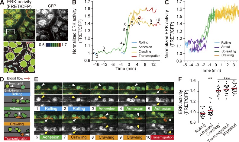
Figure 1.
Activation of ERK in neutrophils during adhesion to endothelial cells of the inflamed small intestine. (A) In vivo imaging of the lamina propria of the intestinal mucosa from an Eisuke mouse, which wa...
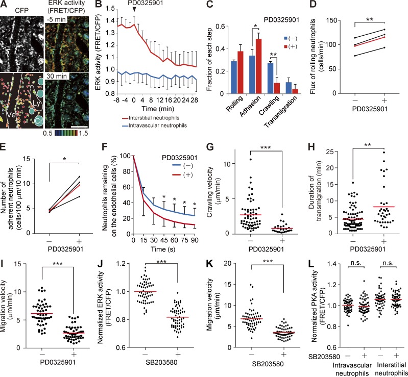
Figure 2.
Requirement of ERK activity for the neutrophil recruitment. (A) FRET images of the lamina propria of the intestinal mucosa in Eisuke mice 2 h after LPS and fMLP treatment. At time 0, 5 mg/kg of the PD...
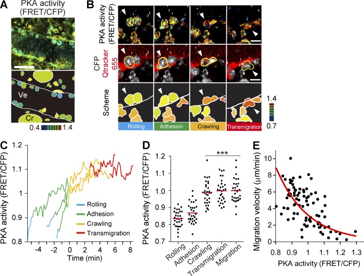
Figure 3.
Activation of PKA during the neutrophil recruitment cascade. (A) In vivo imaging of the lamina propria of the intestinal mucosa of PKAchu mice. A representative FRET/CFP ratio image from Video 3 and a...
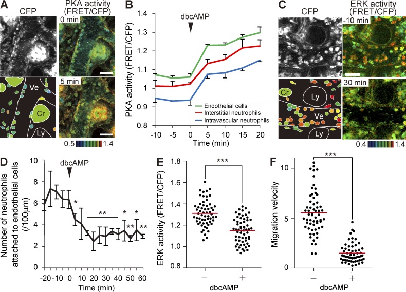
Figure 4.
PKA inhibition of ERK activity, recruitment to endothelial cells, and migration of neutrophils. (A) FRET images of the lamina propria of the intestinal mucosa in PKAchu mice pretreated with LPS and fM...
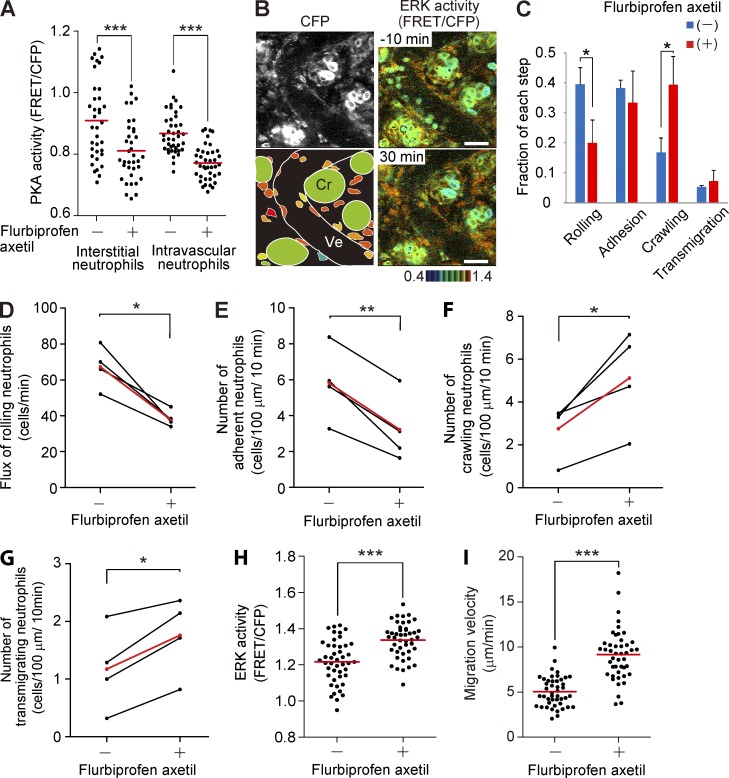
Figure 5.
Accelerated migration and activation of ERK in neutrophils treated with NSAID. (A) PKA activity of intravascular and interstitial neutrophils before and after 7.5 mg/kg flurbiprofen axetil injection. ...
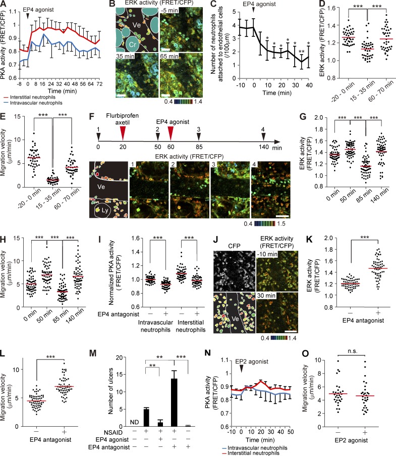
Figure 6.
Inhibition of the recruitment to endothelial cells and the migration of neutrophils by an EP4 agonist. (A) Time courses of the PKA activity of intravascular and interstitial neutrophils are plotted ag...
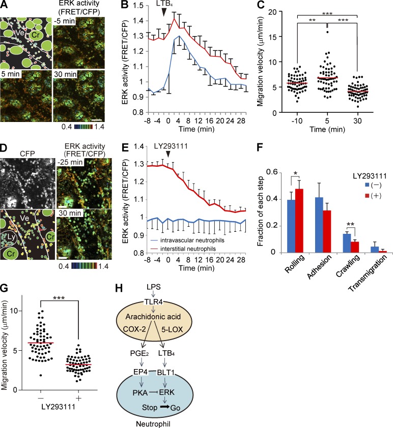
Figure 7.
Effect of LTB 4 and an LTB 4 receptor antagonist on the ERK activity and the neutrophil recruitment. (A) FRET and CFP images of the lamina propria of the intestinal mucosa in Eisuke mice. The top left...
Figure images are served from the NIH/NLM PubMed Central Open Access Subset or Europe PMC; copyright remains with the publishers and authors.
💬 Discussion
0 commentsNo comments yet. Be the first to start a discussion!
Leave a Comment