Abstract
BACKGROUND: Hyperglycemia is acknowledged as an independent risk factor for developing diabetes-associated atherosclerosis. At present, most therapeutic approaches are targeted at a tight glycemic control in diabetic patients, although this fails to prevent macrovascular complications of the disease. Indeed, it remains highly controversial whether or not the mere elevation of extracellular D-glucose can directly promote vascular inflammation, which favors early pro-atherosclerotic events. METHODS AND FINDINGS: In the present work, increasing extracellular D-glucose from 5.5 to 22 mmol/L was neither sufficient to induce intercellular adhesion molecule-1 (ICAM-1) and vascular cell adhesion molecule-1 (VCAM-1) expression, analyzed by flow cytometry, nor to promote leukocyte adhesion to human umbilical vein endothelial cells (HUVEC) in vitro, measured by flow chamber assays. Interestingly, the elevation of D-glucose levels potentiated ICAM-1 and VCAM-1 expression and leukocyte adhesion induced by a pro-inflammatory stimulus, such as interleukin (IL)-1beta (5 ng/mL). In HUVEC, high D-glucose augmented the activation of extracellular signal-regulated kinase 1/2 (ERK 1/2) and nuclear transcription factor-kappaB (NF-kappaB) elicited by IL-1beta, measured by Western blot and electromobility shift assay (EMSA), respectively, but had no effect by itself. Both ERK 1/2 and NF-kappaB were necessary for VCAM-1 expression, but not for ICAM-1 expression. In vivo, leukocyte trafficking was evaluated in the rat mesenteric microcirculation by intravital microscopy. In accordance with the in vitro data, the acute intraperitoneal injection of D-glucose increased leukocyte rolling flux, adhesion and migration, but only when IL-1beta was co-administered. CONCLUSIONS: These results indicate that the elevation of extracellular D-glucose levels is not sufficient to promote vascular inflammation, and they highlight the pivotal role of a pro-inflammatory environment in diabetes, as a critical factor conditioning the early pro-atherosclerotic actions of hyperglycemia.
🔬 Techniques
✨ Fluorophores
🧪 Sample Preparation
🔬 Cell Lines
🏭 Microscope Brands
🧪 Reagent Suppliers
🏛️ Research Organizations (ROR)
Affiliated research institutions:
📋 Methods
Methods and Findings In the present work, increasing extracellular D-glucose from 5.5 to 22 mmol/L was neither sufficient to induce intercellular adhesion molecule-1 (ICAM-1) and vascular cell adhesion molecule-1 (VCAM-1) expression, analyzed by flow cytometry, nor to promote leukocyte adhesion to human umbilical vein endothelial cells (HUVEC) in vitro, measured by flow chamber assays. Interestingly, the elevation of D-glucose levels potentiated ICAM-1 and VCAM-1 expression and leukocyte adhesion induced by a pro-inflammatory stimulus, such as interleukin (IL)-1β (5 ng/mL). In HUVEC, high D-glucose augmented the activation of extracellular signal-regulated kinase 1/2 (ERK 1/2) and nuclear transcription factor-κB (NF-κB) elicited by IL-1β, measured by Western blot and electromobility shift assay (EMSA), respectively, but had no effect by itself. Both ERK 1/2 and NF-κB were necessary for VCAM-1 expression, but not for ICAM-1 expression. In vivo , leukocyte trafficking was evaluated in the rat mesenteric microcirculation by intravital microscopy. In accordance with the in vitro data, the acute intraperitoneal injection of D-glucose increased leukocyte rolling flux, adhesion and migration, but only when IL-1β was co-administered.
Materials and Methods Ethics statement
The investigation conforms with the principles outlined in the Declaration of Helsinki. Experiments with human cells were reviewed and approved by the ethics committee of Universidad Autónoma de Madrid and Hospital Universitario de Getafe, and written informed consent was obtained from all donors. The investigation with animals conforms to the Guide for the Care and Use of Laboratory Animals published by the US National Institutes of Health (NIH Publication No. 85–23, revised 1996) and was approved by the ethics committee of Universidad de Valencia. Materials Culture plastic ware was from TPP (Tragadingen, Switzerland). M199 and fetal calf serum were from Biological Industries (Beit-Haemek, Israel). Human recombinant IL-1β was purchased from Peprotech (London, UK), with an endotoxin level below 0.1 ng per µg. D-glucose was supplied by Serva (Heidelberg, Germany). Endothelial cell growth supplement, heparin, L-glucose, pyrrolidine dithiocarbamate, PD 98059 and, unless otherwise stated, all other reagents were purchased from Sigma Chemical Co. (St. Louis, MO).
Show full methods section
Methods and Findings In the present work, increasing extracellular D-glucose from 5.5 to 22 mmol/L was neither sufficient to induce intercellular adhesion molecule-1 (ICAM-1) and vascular cell adhesion molecule-1 (VCAM-1) expression, analyzed by flow cytometry, nor to promote leukocyte adhesion to human umbilical vein endothelial cells (HUVEC) in vitro, measured by flow chamber assays. Interestingly, the elevation of D-glucose levels potentiated ICAM-1 and VCAM-1 expression and leukocyte adhesion induced by a pro-inflammatory stimulus, such as interleukin (IL)-1β (5 ng/mL). In HUVEC, high D-glucose augmented the activation of extracellular signal-regulated kinase 1/2 (ERK 1/2) and nuclear transcription factor-κB (NF-κB) elicited by IL-1β, measured by Western blot and electromobility shift assay (EMSA), respectively, but had no effect by itself. Both ERK 1/2 and NF-κB were necessary for VCAM-1 expression, but not for ICAM-1 expression. In vivo , leukocyte trafficking was evaluated in the rat mesenteric microcirculation by intravital microscopy. In accordance with the in vitro data, the acute intraperitoneal injection of D-glucose increased leukocyte rolling flux, adhesion and migration, but only when IL-1β was co-administered.
Materials and Methods Ethics statement
The investigation conforms with the principles outlined in the Declaration of Helsinki. Experiments with human cells were reviewed and approved by the ethics committee of Universidad Autónoma de Madrid and Hospital Universitario de Getafe, and written informed consent was obtained from all donors. The investigation with animals conforms to the Guide for the Care and Use of Laboratory Animals published by the US National Institutes of Health (NIH Publication No. 85–23, revised 1996) and was approved by the ethics committee of Universidad de Valencia. Materials Culture plastic ware was from TPP (Tragadingen, Switzerland). M199 and fetal calf serum were from Biological Industries (Beit-Haemek, Israel). Human recombinant IL-1β was purchased from Peprotech (London, UK), with an endotoxin level below 0.1 ng per µg. D-glucose was supplied by Serva (Heidelberg, Germany). Endothelial cell growth supplement, heparin, L-glucose, pyrrolidine dithiocarbamate, PD 98059 and, unless otherwise stated, all other reagents were purchased from Sigma Chemical Co. (St. Louis, MO).
Cell isolation and culture Human umbilical vein endothelial cells
(HUVEC) were enzymatically isolated, as previously described [30] , and cultured in M199 medium supplemented with 20% fetal calf serum (FCS), 25 µg/mL endothelial cell growth supplement (ECGS), 100 µg/mL heparin and antibiotics [30] . For experiments, HUVEC at passages 1–5 were used. For adhesion under flow assays, HL60 leukocytes were obtained from American Type Culture Collection (ATCC; Rockville, MD) and grown in RPMI-1640 medium (Biowhittaker, Walkersville, MD) supplemented with 10% FCS and antibiotics.
Flow cytometry Confluent
HUVEC monolayers were treated with different concentrations of D-glucose (5.5 and 22 mmol/L), either alone or in combination with increasing concentrations of IL-1β. The non-metabolizable analogue of D-glucose, L-glucose, was used as an osmotic control. After 18 h, the expression of VCAM-1 and ICAM-1 at the cell surface was measured by flow cytometry. In brief, HUVEC were gently detached with phosphate-buffered saline (PBS) containing 0.05% trypsin, fixed with 2% paraformaldehyde and blocked with PBS containing 3% bovine serum albumin (BSA) for 15 min. Cells were then suspended in PBS containing 0.5% BSA and incubated for 30 min with primary antibodies against VCAM-1 (clone IE5) or ICAM-1 (clone 6.5B5; Chemicon, Temecula, CA), both at a 1/100 dilution. Detection of primary antibodies was performed using an appropriate Alexa Fluor 488-secondary antibody (Molecular Probes-Invitrogen Corporation, Carlsbad, CA; dilution 1/250). Fluorescence was measured in a FACScan flow cytometer (Beckton-Dickinson, Franklin Lakes, NJ), and data analyzed using Cell Quest software (Beckton-Dickinson, Franklin Lakes, NJ). The expression of CD11b/CD18 integrins was determined on human leukocytes in heparinized whole blood. Blood samples were obtained from buffy coats of four healthy donors by Ficoll Hypaque density gradient centrifugation. Samples (basal glucose concentration: 5.6±0.3 mmol/L) were incubated at 37°C with PBS or D-glucose (16.5 mmol/L to achieve a final concentration of around 22 mmol/L), either alone or in combination with IL-1β (5 ng/mL) for 18 h. Samples were then incubated for 20 min on ice in the dark with saturating amounts (10 µL) of the conjugated mAb anti-human-CD11b/CD18-FITC (clone ICRF 44; Serotec, Madrid, Spain). Red blood cells were lysed and leukocytes fixed using an automated EPICS Q-PREP system (Coulter Electronics, Hialeah, FL). Samples were run in an EPICS XL-MCL flow cytometer (Beckman-Coulter, Hialeah, FL) [31] . Indirect immunofluorescence ICAM-1, VCAM-1 and NF-κB were visualized in HUVEC by indirect immunofluorescence, accordingly to a previously described protocol [32] . Primary antibodies against VCAM-1 (dilution 1/250), ICAM-1 (dilution 1/25) or the NF-κB p65 subunit (dilution 1/100; Transduction Laboratories, Lexington, KY) were used, followed by incubation for 1 h at room temperature with an Alexa Fluor 488-conjugated secondary antibody (1/250). Cell nuclei were counterstained with 4′-6-diamidino-2-phenylindole (DAPI; Molecular Probes-Invitrogen). HUVEC were observed with an Eclipse TE300 epifluorescence microscope (Nikon, Tokyo, Japan).
Nuclear extracts and electrophoretic mobility shift assay
HUVEC were exposed to the different treatments during 1, 4, 6 and 18 h and nuclear extracts were prepared as described before [32] . A commercial oligonucleotide (Promega, Madison, WI) encoding the NF-κB consensus sequence ( 5′-AGTTGAGGGGACTTTCCCAGGC-3′ ) was 5′-end labeled using [γ- 32 P]ATP and T4 polynucleotide kinase (Promega, Madison, WI) and purified using MicroSpin™ G-25 columns (GE Healthcare, Chicago, IL). For binding reactions, nuclear extracts (5 µg) were incubated on ice for 15 min in a reaction buffer [40 mmol/L HEPES (pH 7.0), 140 mmol/L NaCl, 5 mmol/L dithiothreitol, 10 µg/mL BSA, 0.01% Nonidet P-40, 4% Ficoll and 0.05 µg/mL poly(dI-dC).poly(dI-dC)]. After addition of the labeled oligonucleotide (∼50,000 cpm) the reaction mix was further incubated for 20 min at room temperature. For competition experiments a 100-fold excess of unlabeled doubled-stranded oligonucleotide was added to the binding reaction. DNA-protein complexes were resolved on 4% nondenaturing polyacrylamide gels in 0.5x TBE (45 mmol/L Tris-borate, 1 mmol/L EDTA, pH 8.0) at 4°C. Gels were dried and exposed to autoradiography at −80°C.
Western blotting
Extracellular signal-regulated kinase 1/2 (ERK1/2) activation was determined by immunoblotting as previously described [32] . Polyclonal antibodies against both the phosphorylated (activated) and total forms of ERK 1/2 (Cell Signaling Technology, Inc., Danvers, MA; dilution 1/1,000) were used, followed by incubation with a horseradish peroxidase-conjugated secondary antibody (dilution 1/10,000; Chemicon, Temecula, California, USA). ERK 1/2 activity was expressed as the phospho-ERK 1/2:total ERK1/2 ratio.
Flow chamber assays
The in vitro adhesion of HL60 to HUVEC monolayers was analyzed using a previously described live imaging flow model [33] . Briefly, HUVEC monolayers grown on glass coverslips were exposed for 18 h to 5.5 or 22 mmol/L D-glucose either alone or in combination with IL-1β (5 ng/mL). Coverslips were placed in a parallel plate flow chamber maintained at 37°C and HL60 leukocytes (1.5×10 6 cells/mL) were drawn for 2 minutes across the monolayers at a flow rate of 0.26 mL/min, corresponding to at a shear stress of 0.5 dynes/cm 2 . Monolayers were visualized with an inverted microscope (Nikon TE2000, Nikon Inc., Melville, NY), and at least 5 fields were recorded during 10 seconds each using a phase contrast objective and VideoLab software (Ed Marcus Lbs, Newton, MA).
Intravital microscopy
Non-diabetic male Sprague-Dawley rats (200–250 g) with a basal glycemia of 4.9±0.1 mmol/L (results from 5 animals) were employed. Animals were sedated with ether and intraperitoneally injected with 10 mL of either PBS alone, PBS with D-glucose (40 mg/kg), PBS with IL-1β (200 ng/kg), or PBS with D-glucose plus IL-1β. Parallel experiments were performed replacing D-glucose by L-glucose (40 mg/kg), which was used as an osmotic control. After 18 h, the mesentery was exposed in preparation for intravital microscopy, following a previously described protocol [31] . Single unbranched mesenteric venules (25–40 µm in diameter) were selected, and the diameters measured on-line using a video caliper (Microcirculation Research Institute, Texas A&M University, TX). The number of rolling, adherent, and emigrated leukocytes was determined off-line during playback analysis of videotaped images.
Immunohistochemistry
After the completion of the intravital microscopy measurements, the mesentery was isolated, fixed in 4% paraformaldehyde, dehydrated using graded acetone washes at 4°C, and embedded in paraffin wax for localization of ICAM-1 and VCAM-1, using a modified avidin and biotin immunoperoxidase technique as described previously [31] . Anti-rat-VCAM-1 (clone 5F10, kindly donated by Biogen Inc., Cambridge, MA) or anti-rat-ICAM-1 (clone 1A29, Serotec) monoclonal antibodies, or their isotype-matched control murine antibodies (UPC 10, IgG 2a and MOPC21 IgG 1 , Sigma Chemical Co.) were used. Positive staining was defined as a venule displaying brown reaction product.
Statistical analysis
Results are expressed as mean±SEM from three to six independent experiments. Student's t-test was used for data points and one-way ANOVA with Fisher's post-test correction was used for curves. A P value ≤0.05 was considered statistically significant.
Materials Culture plastic ware was from TPP (Tragadingen, Switzerland). M199 and fetal calf serum were from Biological Industries (Beit-Haemek, Israel). Human recombinant IL-1β was purchased from Peprotech (London, UK), with an endotoxin level below 0.1 ng per µg. D-glucose was supplied by Serva (Heidelberg, Germany). Endothelial cell growth supplement, heparin, L-glucose, pyrrolidine dithiocarbamate, PD 98059 and, unless otherwise stated, all other reagents were purchased from Sigma Chemical Co. (St. Louis, MO).
Supporting Information Video S1 Confluent HUVEC monolayers were exposed to 5.5 mmol/L extracellular D-glucose for 18 h prior to leukocyte perfusion (1.5×10⁶ cells/mL) drawn at 0.5 dynes/cm2 (x200). (2.77 MB MOV) Click here for additional data file. Video S2 Confluent HUVEC monolayers were exposed to 22 mmol/L extracellular D-glucose for 18 h prior to leukocyte perfusion (1.5×10⁶ cells/mL) drawn at 0.5 dynes/cm2 (x200). (2.76 MB MOV) Click here for additional data file. Video S3 Confluent HUVEC monolayers were exposed to 5.5 mmol/L extracellular D-glucose in presence of IL-1β (5 ng/mL) for 18 h prior to leukocyte perfusion (1.5×10⁶ cells/mL) drawn at 0.5 dynes/cm2 (x200). (2.66 MB MOV) Click here for additional data file. Video S4 Confluent HUVEC monolayers were exposed to 22 mmol/L extracellular D-glucose in presence of IL-1β (5 ng/mL) for 18 h prior to leukocyte perfusion (1.5×10⁶ cells/mL) drawn at 0.5 dynes/cm2 (x200). (2.61 MB MOV) Click here for additional data file.
📊 Figures
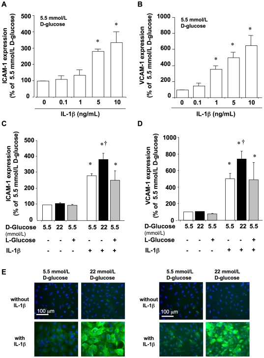
Figure 1
Effect of IL-1u03b2 and D-glucose on the expression of adhesion molecules in HUVEC.
Cells incubated in a medium containing 5.5 mmol/L D-glucose were challenged for 18 h with IL-1u03b2 (0.1 to 10 ng/mL) and the levels of (A) ICAM-1 and (B) VCAM-1 were determined at the cell surface by...
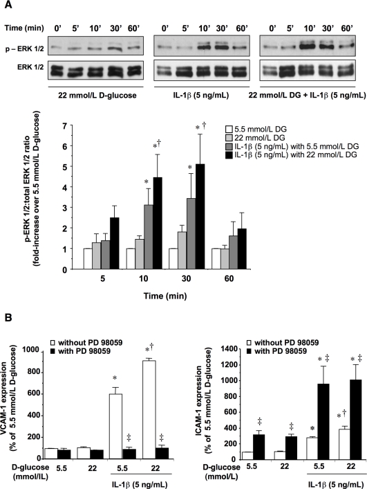
Figure 2
ERK 1/2 activation and its impact on ICAM-1 and VCAM-1 levels in HUVEC.
(A) Cells were incubated in medium containing 5.5 mmol/L or 22 mmol/L D-glucose with or without IL-1u03b2 (5 ng/mL) for 5u201360 min, after which ERK 1/2 activation was determined by Western blotting....
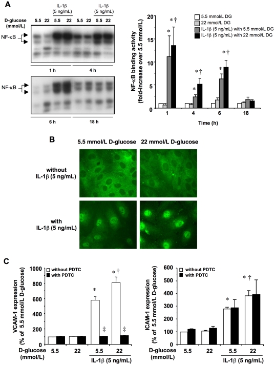
Figure 3
NF-u03baB activation in HUVEC and its impact on ICAM-1 and VCAM-1 levels.
(A) HUVEC were incubated in medium containing 5.5 mmol/L or 22 mmol/L D-glucose in the presence or absence of IL-1u03b2 (5 ng/mL) during 1, 4, 6 and 18 h, after which NF-u03baB binding activity was qu...
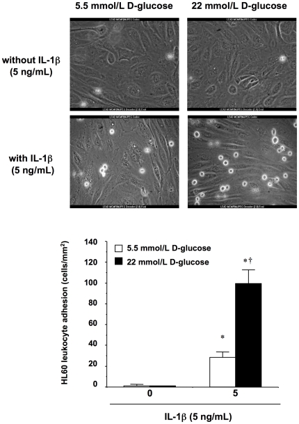
Figure 4
Adhesion of HL60 leukocytes to HUVEC under flow conditions in vitro .
HUVEC monolayers were exposed to either 5.5 or 22 mmol/L extracellular D-glucose in the presence or absence of IL-1u03b2 (5 ng/mL) for 18 h prior to leukocyte perfusion. * P <0.05 versus 5.5 mmol/L...
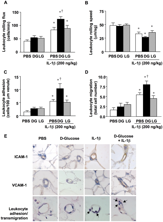
Figure 5
In vivo leukocyte trafficking in rat mesenteric post-capillary venules.
Animals were i.p. injected with 10 mL of PBS alone (C) or supplemented with D-glucose (DG; 40 mg/kg), either in the absence or presence of IL-1u03b2 (200 ng/kg). L-glucose (LG; 40 mg/kg) was used as a...
Figure images are served from the NIH/NLM PubMed Central Open Access Subset or Europe PMC; copyright remains with the publishers and authors.
💬 Discussion
0 commentsNo comments yet. Be the first to start a discussion!
Leave a Comment