Abstract
Abstract Background Disruption of the blood–brain barrier (BBB) after a stroke can lead to brain injury and neurological impairment. Previous work confirmed the involvement of the immunoproteasome subunit of low molecular mass peptide 2 (LMP2) in the pathophysiology of ischemia stroke. However, the relationship between the immunoproteasome LMP2 and the BBB remains unclear. Methods Adult male Sprague–Dawley rats were subjected to transient middle cerebral artery occlusion/reperfusion (MCAO/R). Three days before MCAO, the rats were treated with lentivirus-mediated LMP2 shRNA preparations by stereotactical injection into the ipsilateral hemispheric region. The rat brain microvascular endothelial cell (RBMVEC) line was exposed to oxygen–glucose deprivation/reperfusion (OGD/R) to mimic ischemic conditions in vitro. The RNA interference-mediated knockdown of LMP2 or β-catenin was analysed in vivo and in vitro. Analysis of the quantity of extravasated Evans blue (EB) and cerebral fluorescent angiography were performed to evaluate the integrity of the BBB. Immunofluorescence and Western blotting were employed to detect the expression of target proteins. Cell migration was evaluated using a scratch migration assay. The results of immunofluorescence, Western blotting and cell migration were quantified using the software ImageJ (Version 1.53m). Parametric data from different groups were compared using one-way ANOVA followed by the least significant difference (LSD) test. Results Cerebral ischemia led to lower levels of structural components of the BBB such as tight junction proteins (occludin, claudin-1 and ZO-1) in the MCAO/R group compared with the sham group ( P < 0.001). However, inhibition of the immunoproteasome LMP2 restored the expression of these proteins, resulting in higher levels of occludin, claudin-1 and ZO-1 in the LMP2-shRNA group compared with the control-shRNA group ( P < 0.001). In addition, inhibition of the immunoproteasome LMP2 contributed to higher microvascular density and decreased BBB permeability [e.g., the quantity of extravasated EB: LMP2-shRNA group (58.54 ± 7.37) µg/g vs. control-shRNA group (103.74 ± 4.32) µg/g, P < 0.001], and promoted the upregulation of Wnt-3a and β-catenin proteins in rats following MCAO/R. In vitro experiments, OGD/R induced marked upregulation of LMP2, proapoptotic protein Bax and cleaved caspase-3, and downregulation of occludin, claudin-1, ZO-1 and Bcl-2, as well as inhibition of the Wnt/β-catenin pathway Wnt-3a and β-catenin proteins in RBMVECs, compared with the control group under normal culture conditions ( P < 0.001). However, silencing of LMP2 gene expression reversed these protein changes and promoted proliferation and migration of RBMVECs following OGD/R. Silencing of β-catenin by transfection of RBMVECs with β-catenin-siRNA aggravated the downregulation of tight junction proteins, and reduced the proliferation and migration of RBMVECs following OGD/R, compared with the control-siRNA group ( P < 0.001). LMP2-siRNA and β-catenin-siRNA co-transfection partly counteracted the beneficial effects of silencing LMP2-siRNA on the levels of tight junction proteins in RBMVECs exposed to OGD/R. Conclusion This study suggests that inhibition of the immunoproteasome LMP2 ameliorates ischemia/hypoxia-induced BBB injury, and that the molecular mechanism involves the immunoproteasome-regulated activation of the Wnt/β-catenin signalling pathway under ischemic conditions.
🔬 Techniques
💻 Software
✨ Fluorophores
🧪 Sample Preparation
🔬 Cell Lines
🏭 Microscope Brands
🧪 Reagent Suppliers
💻 Software Details
🏛️ Research Organizations (ROR)
Affiliated research institutions:
📋 Methods
Adult male Sprague–Dawley rats were subjected to transient middle cerebral artery occlusion/reperfusion (MCAO/R). Three days before MCAO, the rats were treated with lentivirus-mediated LMP2 shRNA preparations by stereotactical injection into the ipsilateral hemispheric region. The rat brain microvascular endothelial cell (RBMVEC) line was exposed to oxygen–glucose deprivation/reperfusion (OGD/R) to mimic ischemic conditions in vitro. The RNA interference-mediated knockdown of LMP2 or β-catenin was analysed in vivo and in vitro.
Analysis of the quantity of extravasated Evans blue
(EB) and cerebral fluorescent angiography were performed to evaluate the integrity of the BBB. Immunofluorescence and Western blotting were employed to detect the expression of target proteins. Cell migration was evaluated using a scratch migration assay. The results of immunofluorescence, Western blotting and cell migration were quantified using the software ImageJ (Version 1.53m). Parametric data from different groups were compared using one-way ANOVA followed by the least significant difference (LSD) test.
Methods Ethical approval and experimental animals
All experiments were approved by the Institutional Animal Ethical Committee of Fujian Medical University (No. FJMUIACUC2020-0059) and performed according to the guidelines of the US Department of Health for the Use and Care of Laboratory Animals. Adult male Sprague–Dawley rats (weight 230–250 g) were included in the study. Rats were randomly assigned into three groups (each group n = 8): sham group, LMP2-shRNA group [rats were injected with lentivirus-mediated LMP2 short hairpin RNA (shRNA)] and control-shRNA group (rats were injected with control lentivirus vector carrying scrambled shRNA). Middle cerebral artery occlusion (MCAO) model Rats were anesthetized and subjected to MCAO as described previously, with minor modifications [ 9 ]. In brief, a midline neck incision was made, and the right common carotid artery, external carotid artery and internal carotid artery were isolated. The external carotid artery was tied. A 4–0 monofilament nylon suture (Beijing Sunbio Biotech Co. Ltd., Beijing, China) with a rounded tip was aseptically inserted from the right common carotid artery to the internal carotid artery through the stump of the external carotid artery and gently advanced to occlude the middle cerebral artery. Recirculation/reperfusion of the cerebral blood flow was allowed by gently removing the monofilament after one hour of ischemia, followed by 14 days of reperfusion. In sham-operated animals, all procedures except occlusion of the MCA were performed.
Show full methods section
Adult male Sprague–Dawley rats were subjected to transient middle cerebral artery occlusion/reperfusion (MCAO/R). Three days before MCAO, the rats were treated with lentivirus-mediated LMP2 shRNA preparations by stereotactical injection into the ipsilateral hemispheric region. The rat brain microvascular endothelial cell (RBMVEC) line was exposed to oxygen–glucose deprivation/reperfusion (OGD/R) to mimic ischemic conditions in vitro. The RNA interference-mediated knockdown of LMP2 or β-catenin was analysed in vivo and in vitro.
Analysis of the quantity of extravasated Evans blue
(EB) and cerebral fluorescent angiography were performed to evaluate the integrity of the BBB. Immunofluorescence and Western blotting were employed to detect the expression of target proteins. Cell migration was evaluated using a scratch migration assay. The results of immunofluorescence, Western blotting and cell migration were quantified using the software ImageJ (Version 1.53m). Parametric data from different groups were compared using one-way ANOVA followed by the least significant difference (LSD) test.
Methods Ethical approval and experimental animals
All experiments were approved by the Institutional Animal Ethical Committee of Fujian Medical University (No. FJMUIACUC2020-0059) and performed according to the guidelines of the US Department of Health for the Use and Care of Laboratory Animals. Adult male Sprague–Dawley rats (weight 230–250 g) were included in the study. Rats were randomly assigned into three groups (each group n = 8): sham group, LMP2-shRNA group [rats were injected with lentivirus-mediated LMP2 short hairpin RNA (shRNA)] and control-shRNA group (rats were injected with control lentivirus vector carrying scrambled shRNA). Middle cerebral artery occlusion (MCAO) model Rats were anesthetized and subjected to MCAO as described previously, with minor modifications [ 9 ]. In brief, a midline neck incision was made, and the right common carotid artery, external carotid artery and internal carotid artery were isolated. The external carotid artery was tied. A 4–0 monofilament nylon suture (Beijing Sunbio Biotech Co. Ltd., Beijing, China) with a rounded tip was aseptically inserted from the right common carotid artery to the internal carotid artery through the stump of the external carotid artery and gently advanced to occlude the middle cerebral artery. Recirculation/reperfusion of the cerebral blood flow was allowed by gently removing the monofilament after one hour of ischemia, followed by 14 days of reperfusion. In sham-operated animals, all procedures except occlusion of the MCA were performed.
Lentiviral construction preparation and injection
According to our previous study [ 9 ], four shRNA sequences targeting rat LMP2 (GenBank, Psmb9, NM_012708 ) and a negative control sequence were constructed by Genechem (Shanghai, China). Lentivirus-mediated LMP2 shRNA preparations were constructed and infused stereotactically into the ipsilateral hemispheric region 3 days before MCAO. Briefly, rats were anesthetized as above and placed on a stereotactic apparatus. A total volume of 10 µl of lentivirus suspension was delivered into the right ischemia region using a 15 µl syringe at the following coordinates: bregma backward 1 mm, 1.5 mm lateral, 4 mm dorsoventral.
Evaluation of BBB disruption
The integrity of the BBB was measured using Evans blue (EB) solution (Solarbio, Beijing, China). Before being euthanized, the rats were injected via the tail vein with 2% EB (4 ml/kg). After 2 h circulation, the rats were transcardially perfused with 0.9% NaCl until the outflow fluid from the right atrium was clear. Then, the injured hemisphere was dissected, weighed and incubated in formamide solution in a 37 °C water bath. After 48 h, the supernatant was obtained by centrifugation of the tissue at 1000 × g for 15 min. Finally, the quantity of extravasated EB in the sample was detected by a spectrophotometer at a wavelength of 632. Cerebral fluorescent angiography Cerebral fluorescent angiography was performed as described in detail elsewhere. Briefly, FITC-dextran (150 kD, 0.1 ml 50 mg/ml in double-distilled water; Sigma, USA) was administered intravenously via the rat tail vein. After 60 min, rats were euthanized. Brains were rapidly removed and placed in 4% paraformaldehyde (PFA) in 0.01 mol/L phosphate-buffered saline (PBS) at 4 °C for 24 h and then incubated in 30% sucrose in PBS for another 48 h at 4 °C. Sequential coronal Sects. (40-µm thick) were cut and observed under a fluorescence microscope.
Cell culture Rat brain microvascular endothelial cells
(RBMVECs) involved in this study were purchased from the cell bank of Shanghai Zishi Biotechnology Co., Ltd. (Shanghai, China). Normal RBMVECs were cultured in Dulbecco’s modified Eagle’s medium (DMEM)/high glucose medium (Solarbio) containing 10% fetal bovine serum (FBS, AusGeneX, Australia) and 1% penicillin–streptomycin solution (Solarbio), and maintained in an incubator at 37 °C with a humidified atmosphere containing 5% CO 2 . Medium was exchanged every 3–4 days. RBMVECs were identified by immunofluorescence of vascular von Willebrand factor (VWF). Oxygen–glucose deprivation and reoxygenation (OGD/R) The OGD/R model using RBMVECs was performed to mimic ischemic/hypoxic/reperfusion conditions in vitro as described previously with minor modifications [ 16 ]. Briefly, cells were washed with PBS (pH 7.4) twice and cultured in glucose-free DMEM (Solarbio) without FBS. Then, cells were placed into a hypoxic incubator chamber (Changjing Biotech Co. Ltd., Changsha, China) containing a gas mixture composed of 5% CO 2 and 95% N 2 at 37 °C for 1 h of hypoxia followed by a return to normoxic conditions with glucose-containing DMEM supplemented with 10% FBS for 24 h reoxygenation in a humidified atmosphere containing 5% CO 2 at 37 °C. Control cells were cultured in DMEM and treated similarly to those of the experimental groups. siRNA transfection in RBMVECs RBMVECs were seeded into 6-well plates in 2 ml of antibiotic-free DMEM supplemented with FBS and then maintained at 37 °C for 24 h until they reached 70–80% confluence. During transfection, Lipofectamine® RNAiMAX (Invitrogen, Carlsbad, CA, USA) was used according to the manufacturer’s instructions. The siRNA duplexes targeting LMP2 (LMP2-siRNA) or β-catenin (β-catenin-siRNA) were purchased from RiboBio, Co., Ltd. (Guangzhou, China) and their sequences were as follows: LMP2-siRNA: Si1 (TGAAGAACATCTCCTACAA), Si2 (TAGTGAACCGCGTGTTTGA), Si3 (GCACCTATATTTACGGTTA); β-catenin-siRNA: Si1 (ACATCGAAGACTCTACAAT), Si2 (TAGTGATTGAACCGCGTGT), Si3 (ACCGCGTGTAGACTCAATG). After 48 h transfection, cells were used in subsequent experiments.
Scratch migration assay
The scratch or wound healing assay, which involves measuring cell migration across a gap induced by a scratch injury to the monolayer of cells, is the method of choice for studying cell migration due to its simplicity and low cost. Cells were seeded into 6-well plates, and the monolayer was then gently scratched with a sterile 200-µl pipette tip. Cell migration into the gap was monitored at 0, 24 and 48 h by phase-contrast microscopy. Cell migration was evaluated by a widely used quantification method, which we term the area method, as previously described [ 17 ]. Briefly, to assess migration in an indirect manner, the wound healing (WH) percentage was tracked: WH = [A(t) − A(0)]/A(0) × 100%, where A(t) is the wound area at time t and A(0) is its initial area. The area was quantified using the software ImageJ (Version 1.53 m) and followed the literature [ 18 ].
Immunofluorescence
Briefly, cells grown on coverslips in 12-well plates were fixed with freshly prepared 4% paraformaldehyde in 0.01 mol/L PBS for 30 min. After washing with PBS, the cells were blocked with 10% normal goat serum (Solarbio) for 1 h at room temperature. Then, cells were incubated with the following primary and secondary antibodies: rabbit anti-von Willebrand factor (1:400, Abcam, Cambridge, MA, USA), rabbit anti-CD31 (1:200, Abcam), mouse anti-LMP2, mouse anti-occludin and mouse anti-claudin-1 (1:100, Santa Cruz Biotechnology, USA), rabbit anti-β-catenin (1:200, Cell Signaling Technology, Danvers, MA, USA), rabbit anti-ZO-1 (1:100, Invitrogen), Alexa Fluor® 594 conjugated goat anti-rabbit IgG or Alexa Fluor® 488 conjugated goat anti-mouse IgG (1:1000, Cell Signaling Technology). Finally, slides were mounted in antifade mountant with DAPI antifade reagent (Invitrogen) prior to imaging. The mean immunofluorescent intensity of each target protein was calculated using the software ImageJ (Version 1.53 m) and followed the literature [ 18 ].
RNA extraction and real-time fluorescence quantitative PCR
In brief, after washing with 0.01 PBS (pH 7.4), the cells were lysed by TRIzol for 15 min and then transferred to a fresh 1.5-ml centrifugal tube. After the addition of 200 µl of chloroform, the tube was shaken for 1 min, and then centrifuged for 15 min (14,000× g ). The supernatant was collected and mixed with an equal volume of isopropanol in an RNase-free centrifuge tube and the samples were centrifuged for 10 min (14,000× g ). The supernatant was collected and mixed with 75% cold ethanol. Finally, RNA sediments were collected after centrifuging for 5 min (14,000× g ), and were diluted using RNase-free H 2 O. Then, RNAs were reverse-transcribed into cDNAs using the PrimeScript RT kit (Takara, Dalian, China), according to the reference instructions. Gene expression was detected by reverse transcription-polymerase chain reaction (RT-PCR) assays in the ABI 7500 Fast Real-Time PCR System (Applied Biosystems, CA, USA). RNAs were quantified by the 2 − ΔΔCT method. The primer sequences used were as follows: LMP2 upstream: CATCTACTGTGCCCTCTCGG, LMP2 downstream: CAGCTACCATGAGATGCGCT; β-actin upstream: CGCGAGTACAACCTTCTTGC, β-actin downstream: CCTTCTGACCCATACCCACC.
Western blotting analyses
Western blotting was performed as described previously [ 9 ]. Briefly, total cellular protein (20–30 µg) was separated by 12% gradient sodium dodecyl sulphate/polyacrylamide gel electrophoresis (SDS/PAGE) and then transferred onto polyvinylidene fluoride (PVDF) membrane (Millipore, USA). Membranes were blocked with Tris-buffered saline containing 0.1% Tween-20 (TBST) and 5% nonfat milk (Solarbio). The membranes were then incubated with the following primary antibodies: mouse anti-LMP2, mouse anti-occludin, mouse anti-claudin-1, mouse anti-caspase-3, mouse anti-Bax (1:500, Santa Cruz Biotechnology), rabbit anti-ZO-1 (1:1000, Invitrogen), rabbit anti-Bcl2 (1:1000, Abcam), rabbit anti-Wnt-3a (1:1000, Abcam), rabbit anti-β-catenin (1:1000, Cell Signaling Technology) and mouse anti-β-actin (1:3000, Cell Signaling Technology). The next day, horseradish peroxidase (HRP)-conjugated secondary antibodies, goat anti-mouse IgG secondary antibody (1:3000, Santa Cruz Biotechnology) and goat anti-rabbit IgG secondary antibody (1:3000, Cell Signaling Technology), were incubated with the membranes for 1 h at room temperature. Finally, immunoreactivity was detected with SuperSignal West Pico Chemiluminescent Substrate (Thermo Fisher Scientific, MA, USA) in the ChemiDoc MP Gel Imaging System (Bio-Rad, USA). The optical densities were normalized to those of β-actin and calculated as target protein expression/β-actin expression ratios [using ImageJ (Version 1.53m)]. If necessary, the blots were stripped with Restore™ Western Blot Stripping Buffer (Thermo Fisher Scientific, MA, USA) according to manufacturer’s instructions, and then the western blot procedure was repeated as described above.
Statistical analysis
Data were analysed with software SPSS (version 20, IBM Corp., Armonk, NY, USA) and expressed as the mean ± standard deviation (SD). Parametric data from different groups were compared using one-way ANOVA followed by the least significant difference (LSD) test. P < 0.05 was considered statistically significant.
Ethical approval and experimental animals
All experiments were approved by the Institutional Animal Ethical Committee of Fujian Medical University (No. FJMUIACUC2020-0059) and performed according to the guidelines of the US Department of Health for the Use and Care of Laboratory Animals. Adult male Sprague–Dawley rats (weight 230–250 g) were included in the study. Rats were randomly assigned into three groups (each group n = 8): sham group, LMP2-shRNA group [rats were injected with lentivirus-mediated LMP2 short hairpin RNA (shRNA)] and control-shRNA group (rats were injected with control lentivirus vector carrying scrambled shRNA).
Supplementary Information Additional file 1: Fig. S1 .
Rat brain microvascular endothelial cells
(RBMVECs) were cultured and identified. a Rat brain microvascular endothelial cells (RBMVECs) were observed under the inverted microscope (Scale bars, left 250 µm, right 50 µm). b RBMVECs were confirmed with immunofluorescence of vascular von Willebrand factor (VWF). Scale bars = 50 µm. Additional file 2: Fig. S2 . Immunofluorescence staining showed the expressions of LMP2 and CD31 in rat brain cortex tissue. Some LMP2 positive cells were colocalized with CD31-positive vascular endothelial cell (arrow). Scale bars = 50 µm.
📊 Figures
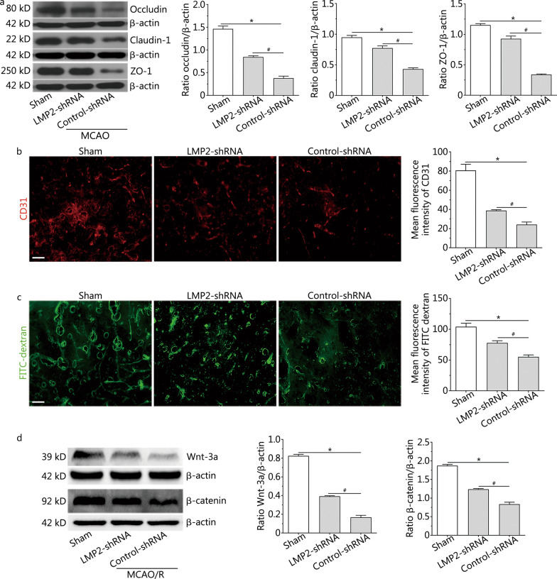
Fig. 1
LMP2 inhibition increased the levels of tight junction proteins, improved BBB integrity and upregulated Wnt/u03b2-catenin signalling after MCAO. a Western blotting showed the levels of tight junction ...
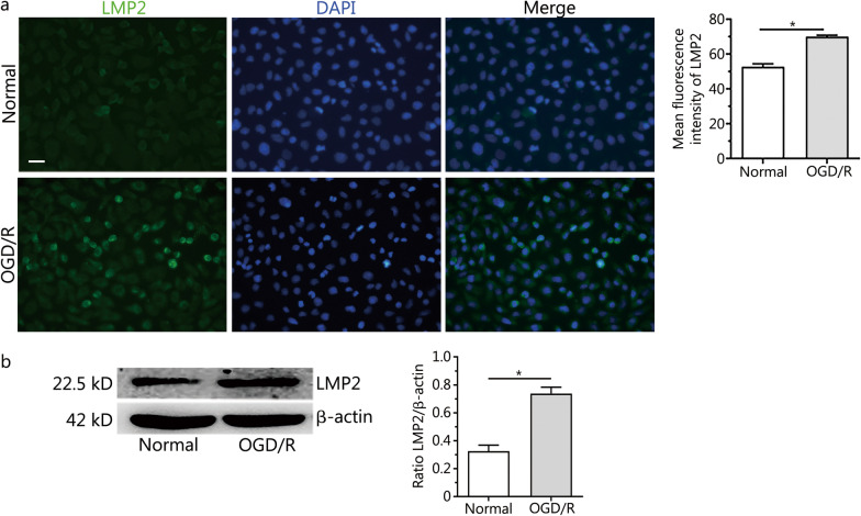
Fig. 2
Expression of LMP2 protein in RBMVECs exposed to OGD/R. a Immunofluorescence staining and quantitative analysis of LMP2 in RBMVECs exposed to OGD/R. b Western blotting showed that the expression of LM...
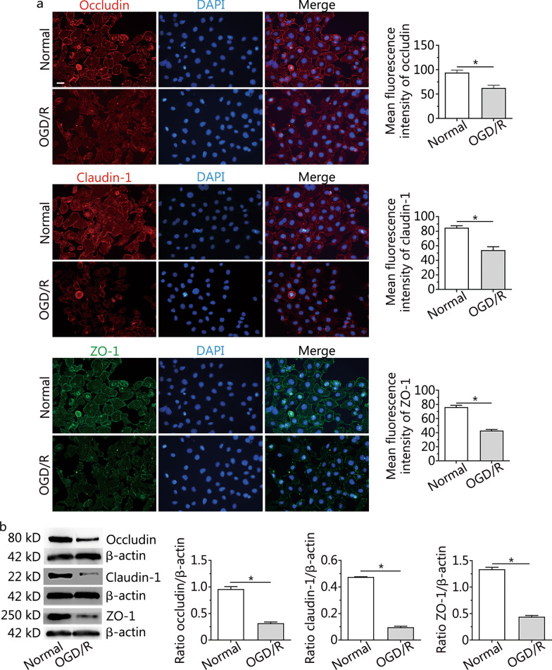
Fig. 3
Changes to occludin, claudin-1 and ZO-1 proteins in RBMVECs after OGD/R. a Immunofluorescence staining and quantitative analysis of occludin, claudin-1 and ZO-1 proteins in RBMVECs in each group. b Re...
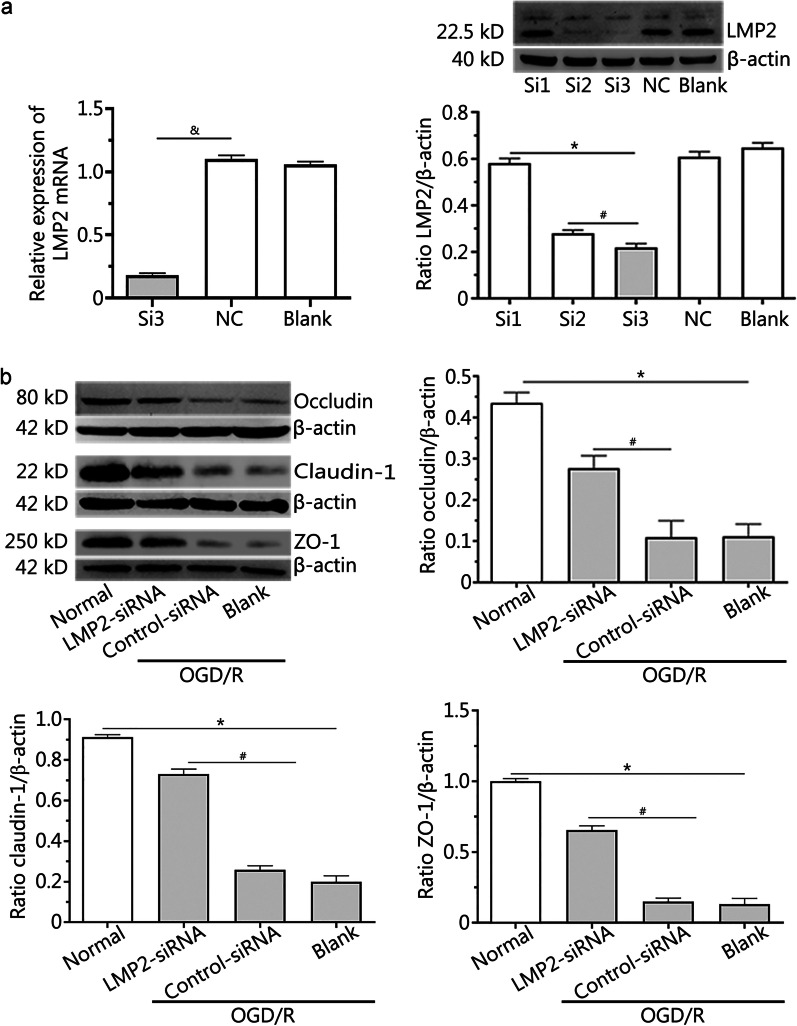
Fig. 4
Silencing LMP2 reversed the downregulated expression of the occludin, claudin-1 and ZO-1 proteins in RBMVECs following OGD/R. a Transfection efficiency of LMP2-siRNA was verifiedu00a0by RT-PCR and Wes...
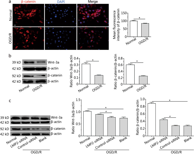
Fig. 5
Silencing LMP2 rescued the downregulation of the Wnt-3a and u03b2-catenin proteins in RBMVECs following OGD/R. a Immunofluorescence and quantitative analysis of u03b2-catenin protein in RBMVECs under ...
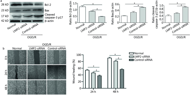
Fig. 6
Silencing LMP2 decreased the expression of apoptosis-related proteins and promoted the proliferation and migration of RBMVECs following OGD/R. a Western blotting and quantitative analysis the levels o...
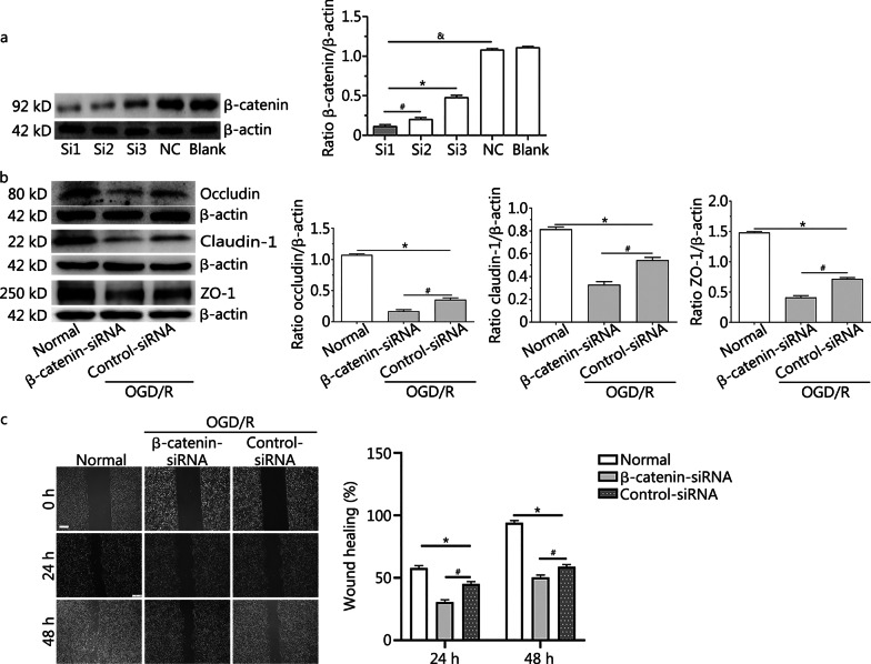
Fig. 7
Silencing of u03b2-catenin aggravated downregulation of the occludin, claudin-1 and ZO-1 proteins, and reduced the proliferation and migration of RBMVECs following OGD/R . a Western blotting showed th...
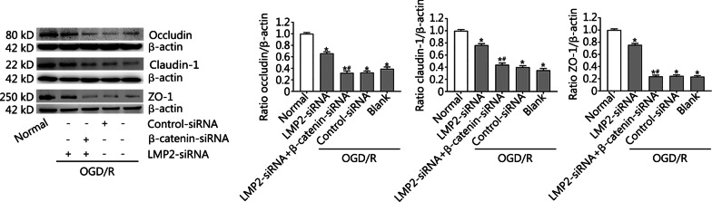
Fig. 8
Effects of combined silencing of the u03b2-catenin and LMP2 genes on the expression of occludin, claudin-1 and ZO-1 proteins in RBMVECs following OGD/R . Western blotting and quantitative analysis of ...
Figure images are served from the NIH/NLM PubMed Central Open Access Subset or Europe PMC; copyright remains with the publishers and authors.
💬 Discussion
0 commentsNo comments yet. Be the first to start a discussion!
Leave a Comment