Abstract
During cell migration, integrins are redistributed from focal adhesions undergoing disassembly at the cell's trailing edges to new focal adhesions assembling at leading edges. The initial step of integrin redistribution is thought to require clathrin-mediated endocytosis. However, whether clathrin-mediated endocytosis functions in different contexts, such as basal versus stimulated migration, has not been determined. In this paper, we examine the spatial and temporal redistribution of integrins from focal adhesions upon stimulation by growth factors. Four-dimensional confocal live-cell imaging along with functional analysis reveals that surface integrins do not undergo significant endocytosis at ventral focal adhesions upon cell stimulation with the platelet-derived growth factor. Rather, they abruptly redistribute to dorsal circular ruffles, where they are internalized through macropinocytosis. The internalized integrins then transit through recycling endosomal compartments to repopulate newly formed focal adhesions on the ventral surface. These findings explain why integrins have long been observed to redistribute through both surface-based and internal routes and identify a new function for macropinocytosis during growth factor-induced cell migration.
🔬 Techniques
🔭 Microscopes
💻 Software
✨ Fluorophores
🧪 Sample Preparation
🔬 Cell Lines
🏭 Microscope Brands
🧪 Reagent Suppliers
🔎 Objectives
💻 Software Details
🏛️ Research Organizations (ROR)
Affiliated research institutions:
📋 Methods
Mice and cell culture Wild-type
C57BL/6 mice were maintained as homozygous inbred lines in the Dana-Farber Cancer Institute animal facility. All mice were male and 8–12 wk old at the time of the study.
Mouse experiments followed Institutional Animal
Care and Use Committee guidelines. Primary wild-type mouse synovial fibroblasts were recovered from normal mouse joints and were cultured in DME supplemented with 10% FBS (Hyclone), l -glutamine, penicillin/streptomycin, 2-mercaptoethanol, and essential and nonessential amino acids at 37°C under 10% CO 2 . Cells had fibroblast-like morphology and were VCAM-1 positive but lacked F4/80 and were CD45 negative by flow cytometry ( Lee et al., 2007 ; Agarwal et al., 2008 ). Cell culture reagents were purchased from Invitrogen unless otherwise indicated. Retrieved mouse synovial fibroblasts were used between passages 5 and 10. NIH3T3 cells were purchased from American Type Culture Collection and cultured in DME supplemented with 10% bovine calf serum (Hyclone), l -glutamine, and penicillin/streptomycin at 37°C under 10% CO 2 . MDA-MB-231 human breast cancer cells were purchased from American Type Culture Collection and cultured in American Type Culture Collection–formulated Leibovitz’s L-15 medium supplemented with 10% FBS, l -glutamine, and penicillin/streptomycin at 37°C without CO 2 . HUV-EC-C human umbilical vein endothelial cells were purchased from American Type Culture Collection and cultured in American Type Culture Collection–formulated F-12K medium supplemented with 10% FBS (Hyclone), 0.1 mg/ml heparin (Sigma-Aldrich), 0.03–0.05 mg/ml endothelial cell growth supplement (Sigma-Aldrich), l -glutamine, and penicillin/streptomycin at 37°C under 5% CO 2 . Plasmids, siRNA, and transfection cDNA encoding the full-length mouse integrin β3–EGFP fusion protein in a pcDNA3 expression vector was a gift from B. Wehrle-Haller (University of Geneva, Geneva, Switzerland; Ballestrem et al., 2001 ). siRNA against BARS ( Yang et al., 2005 ), CHC ( Li et al., 2007 ), and WAVE1 ( Suetsugu et al., 2003 ) were previously described. siRNAs against CAV1, Rab4A, and Rab4B were obtained from Santa Cruz Biotechnology, Inc. Nonsilencing negative control siRNA was purchased from QIAGEN. Plasmids were transfected with Lipofectamine LTX reagent (Invitrogen), and siRNAs were transfected with Lipofectamine RNAiMAX reagent (Invitrogen).
Show full methods section
Mice and cell culture Wild-type
C57BL/6 mice were maintained as homozygous inbred lines in the Dana-Farber Cancer Institute animal facility. All mice were male and 8–12 wk old at the time of the study.
Mouse experiments followed Institutional Animal
Care and Use Committee guidelines. Primary wild-type mouse synovial fibroblasts were recovered from normal mouse joints and were cultured in DME supplemented with 10% FBS (Hyclone), l -glutamine, penicillin/streptomycin, 2-mercaptoethanol, and essential and nonessential amino acids at 37°C under 10% CO 2 . Cells had fibroblast-like morphology and were VCAM-1 positive but lacked F4/80 and were CD45 negative by flow cytometry ( Lee et al., 2007 ; Agarwal et al., 2008 ). Cell culture reagents were purchased from Invitrogen unless otherwise indicated. Retrieved mouse synovial fibroblasts were used between passages 5 and 10. NIH3T3 cells were purchased from American Type Culture Collection and cultured in DME supplemented with 10% bovine calf serum (Hyclone), l -glutamine, and penicillin/streptomycin at 37°C under 10% CO 2 . MDA-MB-231 human breast cancer cells were purchased from American Type Culture Collection and cultured in American Type Culture Collection–formulated Leibovitz’s L-15 medium supplemented with 10% FBS, l -glutamine, and penicillin/streptomycin at 37°C without CO 2 . HUV-EC-C human umbilical vein endothelial cells were purchased from American Type Culture Collection and cultured in American Type Culture Collection–formulated F-12K medium supplemented with 10% FBS (Hyclone), 0.1 mg/ml heparin (Sigma-Aldrich), 0.03–0.05 mg/ml endothelial cell growth supplement (Sigma-Aldrich), l -glutamine, and penicillin/streptomycin at 37°C under 5% CO 2 . Plasmids, siRNA, and transfection cDNA encoding the full-length mouse integrin β3–EGFP fusion protein in a pcDNA3 expression vector was a gift from B. Wehrle-Haller (University of Geneva, Geneva, Switzerland; Ballestrem et al., 2001 ). siRNA against BARS ( Yang et al., 2005 ), CHC ( Li et al., 2007 ), and WAVE1 ( Suetsugu et al., 2003 ) were previously described. siRNAs against CAV1, Rab4A, and Rab4B were obtained from Santa Cruz Biotechnology, Inc. Nonsilencing negative control siRNA was purchased from QIAGEN. Plasmids were transfected with Lipofectamine LTX reagent (Invitrogen), and siRNAs were transfected with Lipofectamine RNAiMAX reagent (Invitrogen).
Antibodies
Armenian hamster anti–integrin β3 (clone 2C9.G2), mouse anti-EEA1, mouse anti-Rab5, mouse anti-Rab4, mouse anti-Rab11, mouse anti-WAVE1, mouse anti-CAV1, and rat anti–integrin β1 (clone Ha2/5) antibodies were purchased from BD. Nonblocking rat anti–integrin β3 antibody (clone 1–55-4) was obtained from MBL International. The rat anti–integrin β1 antibody (clone MB1.2) was obtained from Millipore. The mouse anti–integrin β1 antibody (clone 12G10) was obtained from Abcam. The rabbit anticortactin antibody was purchased from Sigma-Aldrich. The rabbit anti-BARS antibody (clone p50-2) was raised against GST-BARS and purified as described previously ( Spanò et al., 1999 ). The mouse anti-CHC antibody was obtained from American Type Culture Collection. The mouse anti–β-actin antibody was obtained from Sigma-Aldrich. The mouse anti-MHC class I antibody was purchased from Santa Cruz Biotechnology, Inc. Alexa Fluor–conjugated anti–mouse, anti–rat, and anti–rabbit secondary antibodies and Alexa Fluor–conjugated phalloidin were purchased from Invitrogen. FITC-conjugated anti–Armenian hamster IgG secondary antibody and peroxidase-conjugated anti–mouse, anti–rat, and anti–rabbit secondary antibodies were obtained from Jackson ImmunoResearch Laboratories, Inc.
Immunofluorescence
(IF), dextran endocytosis assay, cell surface antibody-binding, acid-stripping endocytosis and recycling assay, and transferrin endocytosis assay For IF microscopy experiments, cells were cultured on 10-mm glass coverslips coated with 0.1 µg/cm 2 vitronectin (Sigma-Aldrich) for integrin β3 experiments or 1 µg/cm 2 fibronectin (Sigma-Aldrich) for integrin β1 experiments. PDGF-BB and VEGF were obtained from Sigma-Aldrich. EGF was obtained from Invitrogen. For IF staining, cells were serum starved for 16 h and stimulated with 20 ng/ml PDGF-BB, 30 ng/ml EGF, or 30 ng/ml VEGF in serum-free medium for various times as indicated in the following paragraphs. Cells were fixed in 4% paraformaldehyde (Electron Microscopy Sciences) for 15 min. For intracellular protein staining, cells were permeabilized with 0.3% Triton X-100 (Thermo Fisher Scientific) for 10 min. Permeabilized cells were labeled with primary antibodies for 1 h at RT or 16 h at 4°C followed by secondary antibody labeling for 1 h and mounted on slides with FluorSave reagent (EMD). Tetramethylrhodamine (TMR)-conjugated, 10,000-MW fixable dextran was obtained from Invitrogen. For the PDGF-BB– and TMR-dextran–chasing endocytosis assay, cells were serum starved for 16 h and then stimulated with 20 ng/ml PDGF-BB together with 200 µg/ml TMR-dextran in serum-free medium for various times. The cells were then washed three times to remove all cell surface–remaining TMR-dextran and fixed, and IF staining was performed. For the cell surface antibody-binding, acid-stripping endocytosis and recycling assay, surface integrin β3 on live cells was stained by a nonblocking rat anti–integrin β3 monoclonal antibody (clone 1–55-4) in serum-free medium containing 1% BSA as blocking reagent for 1 h at RT. Cells were washed in antibody-free medium three times. Then, cells were treated with 20 ng/ml PDGF-BB for 15 min to allow cells time to internalize the antibody-bound integrin β3 by CDR macropinocytosis. Then, cells were equilibrated to 4°C followed by a 1-min ice-cold acid (0.5% acetic acid and 0.5 M NaCl, pH 3.0)-washing step to remove leftover cell surface integrin β3–bound antibodies. After acid washing, cells were washed in ice-cold medium to bring the pH back to 7.4 as indicated by phenol red. Cells were then incubated at 37°C in 20 ng/ml PDGF-BB–containing medium for another 60 min to allow cells time to recycle the antibody-bound integrin β3 back to the cell surface. Cells were then fixed, and without cell membrane permeabilization, the recycled cell surface antibody-bound integrin β3 was detected by IF staining with an Alexa Fluor 488 goat anti–rat secondary antibody. After PBS wash, cell membranes were permeabilized with 0.3% Triton X-100 for 5 min. F-actin was stained by Alexa Fluor 568 phalloidin and visualized by confocal microscopy. For transferrin endocytosis assays, nonsilencing negative control siRNA or CHC siRNA-transfected primary mouse fibroblasts were incubated with Alexa Fluor 488 transferrin (Invitrogen) for 1 h at 4°C. Unbound transferrin was washed, and cells were then incubated at 37°C for various times. After incubation, cells were equilibrated to 4°C followed by a 1-min ice-cold acid (0.5% acetic acid and 0.5 M NaCl, pH 3.0)-washing step to remove leftover cell surface transferrin. After acid washing, cells were washed in ice-cold medium to bring the pH back to 7.4 as indicated by phenol red. Cells were then fixed and subjected to confocal microscopy to detect internalized Alexa Fluor 488 transferrin.
3D confocal microscopy and 4D time-lapse confocal live-cell imaging
IF images were captured on an inverted microscope (TE2000-U; Nikon) equipped with a confocal system (C1; Nikon) controlled by EZ-C1 software (Nikon) using a Plan Apochromat 60×/1.40 NA oil objective or a Plan Apochromat 20×/0.75 NA objective (Nikon). 3D confocal microscopy was performed by scanning multiple confocal layers along the z axis. For 4D time-lapse confocal live-cell imaging experiments, cells were grown in 35-mm glass-bottomed Petri dishes (World Precision Instruments). These dishes were mounted onto the confocal microscope with a heating chamber at 37°C and were superfused with 10% CO 2 . 4D confocal microscopy was performed by 3D confocal microscopy scanning over time. Fluorescent 3D or 4D image reconstructions and protein colocalization analysis were performed in MetaMorph Imaging software (version 7.6.1.0; Universal Imaging).
Flow cytometry
Cells were detached by a brief incubation with 0.05% trypsin-EDTA (Invitrogen) at RT to minimize endocytosis. Trypsin was quenched by a trypsin inhibitor from Glycine max (soybean; Sigma-Aldrich). Cells were then washed by 2% FBS in PBS and stained with anti–integrin β3 monoclonal antibody (1–55-4) or isotype control antibody for 1 h at RT followed by Alexa Fluor 488 secondary antibody staining. After staining, cells were fixed in 1% paraformaldehyde in PBS and subjected to flow cytometry analysis by a flow cytometer (FACS Canto; BD). FlowJo software (Tree Star) was used to analyze the flow cytometry data. Cell migration assay 56 h after siRNA transfection, cells were serum starved for another 16 h and detached by a brief incubation with 0.05% trypsin-EDTA at RT. Trypsin was quenched by a trypsin inhibitor from Glycine max (soybean). Cells were washed and seeded with or without antiintegrin-blocking antibodies into the upper wells of vitronectin-coated Transwell inserts (8.0-µm pore size; Corning) in serum-free media. After a 2-h incubation at 37°C, 50 ng/ml PDGF-BB–containing serum-free media were then added into the lower well to drive cell migration. After a 2-h migration, the upper filter membrane surface was wiped to remove cells that had not migrated through the filter, and then the filter was fixed and stained to detect cells on the lower filter membrane surface using a stain set (Diff-Quik; Dade Behring). The number of cells that had migrated through a 0.8-mm 2 Transwell membrane was counted. SDS-PAGE and Western blotting For SDS-PAGE experiments, cells were lysed and scraped at 4°C in a cell lysis buffer of the following composition: 1% Triton X-100, 50 mM Hepes, 150 mM NaCl, 5 mM EDTA, 5 mM EGTA, 20 mM NaF, 20 mM sodium pyrophosphate, 1 mM PMSF, and 1 mM Na 3 VO 4 , pH 7.4. DC protein assays (Bio-Rad Laboratories) were performed on cell lysate samples. Equal amounts of protein from each sample were run on each lane of 7.5% SDS-PAGE gels. After gel electrophoresis, proteins were transferred to a polyvinylidene fluoride membrane for Western blotting analysis. Proteins on the membranes were labeled with primary antibodies overnight at 4°C and then labeled by peroxidase-conjugated secondary antibodies and visualized by ECL detection reagents (Thermo Fisher Scientific). β-Actin was blotted as a protein loading control.
Statistics
Numerical data are presented as means ± SD. Student’s t test was used for the comparison of two means (P < 0.05 was considered significant as marked by asterisks in the figures). Online supplemental material Fig. S1 shows that integrin β3 and integrin β1 localize at CDRs in various cell types after growth factor stimulation. Fig. S2 shows that PDGF-BB–stimulated integrin β3 macropinocytosis is BARS dependent but clathrin and CAV1 independent. Video 1 shows a 3D view of CDRs with integrin β3, F-actin, and cortactin colocalization. Video 2 shows integrin β3–GFP translocation 4D tracing in a live NIH3T3 cell after PDGF-BB stimulation. Video 3 shows integrin β3–GFP translocation 4D tracing in a live NIH3T3 cell without PDGF-BB stimulation. Video 4 shows integrin β3–GFP translocation 4D tracing in a BARS siRNA-transfected live NIH3T3 cell after PDGF-BB stimulation. Online supplemental material is available at http://www.jcb.org/cgi/content/full/jcb.201007003/DC1 .
Online supplemental material Fig. S1 shows that integrin β3 and integrin β1 localize at CDRs in various cell types after growth factor stimulation. Fig. S2 shows that PDGF-BB–stimulated integrin β3 macropinocytosis is BARS dependent but clathrin and CAV1 independent. Video 1 shows a 3D view of CDRs with integrin β3, F-actin, and cortactin colocalization. Video 2 shows integrin β3–GFP translocation 4D tracing in a live NIH3T3 cell after PDGF-BB stimulation. Video 3 shows integrin β3–GFP translocation 4D tracing in a live NIH3T3 cell without PDGF-BB stimulation. Video 4 shows integrin β3–GFP translocation 4D tracing in a BARS siRNA-transfected live NIH3T3 cell after PDGF-BB stimulation. Online supplemental material is available at http://www.jcb.org/cgi/content/full/jcb.201007003/DC1 .
📊 Figures
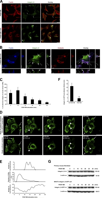
Figure 1.
Integrin u03b23 localizes at CDRs after PDGF-BB stimulation and integrin u03b23u2013GFP translocation 4D tracing in a live cell. (A) Primary mouse fibroblasts were stimulated with or without PDGF-BB, ...
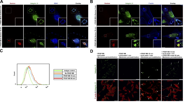
Figure 2.
PDGF-BB stimulates integrin u03b23 macropinocytosis at CDRs and recycling back to the ventral cell surface to form new focal adhesions. (A and B) Primary mouse fibroblasts were stimulated with PDGF-BB...
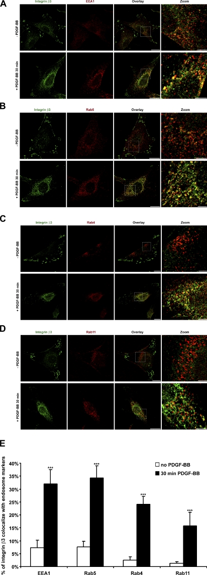
Figure 3.
After macropinosome disassembly, internalized integrin u03b23 colocalizes with early endosomes (EEA1, Rab5, and Rab4) and recycling endosomes (Rab11). (Au2212D) Primary mouse fibroblasts were stimulat...
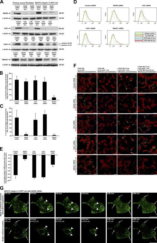
Figure 4.
PDGF-BBu2013stimulated integrin u03b23 macropinocytosis is BARS dependent but clathrin and CAV1 independent. (A) BARS, CHC, CAV1, and WAVE1 protein expression levels from whole-cell lysates were analy...
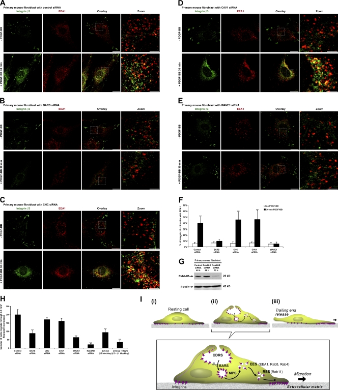
Figure 5.
PDGF-BBu2013stimulated fast cell migration is BARS dependent. (Au2212E) siRNA-transfected primary mouse fibroblasts were prepared as in Fig. 3 A . (F) Overlay images in Au2212E were analyzed by a colo...
Figure images are served from the NIH/NLM PubMed Central Open Access Subset or Europe PMC; copyright remains with the publishers and authors.
💬 Discussion
0 commentsNo comments yet. Be the first to start a discussion!
Leave a Comment