Abstract
BACKGROUND: The role of lymphatic vessels in tissue and organ transplantation as well as in tumor growth and metastasis has drawn great attention in recent years. METHODOLOGY/PRINCIPAL FINDINGS: We now developed a novel method using non-invasive two-photon microscopy to simultaneously visualize and track specifically stained lymphatic vessels and autofluorescent adjacent tissues such as collagen fibrils, blood vessels and immune cells in the mouse model of corneal neovascularization in vivo. The mouse cornea serves as an ideal tissue for this technique due to its easy accessibility and its inducible and modifiable state of pathological hem- and lymphvascularization. Neovascularization was induced by suture placement in corneas of Balb/C mice. Two weeks after treatment, lymphatic vessels were stained intravital by intrastromal injection of a fluorescently labeled LYVE-1 antibody and the corneas were evaluated in vivo by two-photon microscopy (TPM). Intravital TPM was performed at 710 nm and 826 nm excitation wavelengths to detect immunofluorescence and tissue autofluorescence using a custom made animal holder. Corneas were then harvested, fixed and analyzed by histology. Time lapse imaging demonstrated the first in vivo evidence of immune cell migration into lymphatic vessels and luminal transport of individual cells. Cells immigrated within 1-5.5 min into the vessel lumen. Mean velocities of intrastromal corneal immune cells were around 9 µm/min and therefore comparable to those of T-cells and macrophages in other mucosal surfaces. CONCLUSIONS: To our knowledge we here demonstrate for the first time the intravital real-time transmigration of immune cells into lymphatic vessels. Overall this study demonstrates the valuable use of intravital autofluorescence two-photon microscopy in the model of suture-induced corneal vascularizations to study interactions of immune and subsequently tumor cells with lymphatic vessels under close as possible physiological conditions.
🔬 Techniques
✨ Fluorophores
🧪 Sample Preparation
🔬 Cell Lines
🏭 Microscope Brands
🧪 Reagent Suppliers
🔴 Lasers
💻 Software Details
🏛️ Research Organizations (ROR)
Affiliated research institutions:
📋 Methods
Methodology/Principal Findings We now developed a novel method using non-invasive two-photon microscopy to simultaneously visualize and track specifically stained lymphatic vessels and autofluorescent adjacent tissues such as collagen fibrils, blood vessels and immune cells in the mouse model of corneal neovascularization in vivo. The mouse cornea serves as an ideal tissue for this technique due to its easy accessibility and its inducible and modifiable state of pathological hem- and lymphvascularization. Neovascularization was induced by suture placement in corneas of Balb/C mice. Two weeks after treatment, lymphatic vessels were stained intravital by intrastromal injection of a fluorescently labeled LYVE-1 antibody and the corneas were evaluated in vivo by two-photon microscopy (TPM). Intravital TPM was performed at 710 nm and 826 nm excitation wavelengths to detect immunofluorescence and tissue autofluorescence using a custom made animal holder. Corneas were then harvested, fixed and analyzed by histology. Time lapse imaging demonstrated the first in vivo evidence of immune cell migration into lymphatic vessels and luminal transport of individual cells. Cells immigrated within 1–5.5 min into the vessel lumen. Mean velocities of intrastromal corneal immune cells were around 9 µm/min and therefore comparable to those of T-cells and macrophages in other mucosal surfaces.
Materials and Methods
Mice and anesthesia Six to eight weeks-old female
Balb/C mice (Charles River Germany, Sulzfeld, Germany) were used. All animals were treated in accordance with the ARVO Statement for the Use of Animals in Ophthalmic and Vision Research. For surgical procedures, mice were anaesthetized using an intraperitoneal injection of Ketamine (Ketanest S, Parke-Davis, Germany) and Xylazine (Rompun, Bayer, Germany).
Show full methods section
Methodology/Principal Findings We now developed a novel method using non-invasive two-photon microscopy to simultaneously visualize and track specifically stained lymphatic vessels and autofluorescent adjacent tissues such as collagen fibrils, blood vessels and immune cells in the mouse model of corneal neovascularization in vivo. The mouse cornea serves as an ideal tissue for this technique due to its easy accessibility and its inducible and modifiable state of pathological hem- and lymphvascularization. Neovascularization was induced by suture placement in corneas of Balb/C mice. Two weeks after treatment, lymphatic vessels were stained intravital by intrastromal injection of a fluorescently labeled LYVE-1 antibody and the corneas were evaluated in vivo by two-photon microscopy (TPM). Intravital TPM was performed at 710 nm and 826 nm excitation wavelengths to detect immunofluorescence and tissue autofluorescence using a custom made animal holder. Corneas were then harvested, fixed and analyzed by histology. Time lapse imaging demonstrated the first in vivo evidence of immune cell migration into lymphatic vessels and luminal transport of individual cells. Cells immigrated within 1–5.5 min into the vessel lumen. Mean velocities of intrastromal corneal immune cells were around 9 µm/min and therefore comparable to those of T-cells and macrophages in other mucosal surfaces.
Materials and Methods
Mice and anesthesia Six to eight weeks-old female
Balb/C mice (Charles River Germany, Sulzfeld, Germany) were used. All animals were treated in accordance with the ARVO Statement for the Use of Animals in Ophthalmic and Vision Research. For surgical procedures, mice were anaesthetized using an intraperitoneal injection of Ketamine (Ketanest S, Parke-Davis, Germany) and Xylazine (Rompun, Bayer, Germany).
Suture induced corneal inflammation assay
We used the mouse model of suture induced inflammatory corneal neovascularization as described previously [6] . Briefly, mice were put under general anesthesia and 3 intrastromal 11-0 nylon sutures (70 µm diameter needles; Serag-Wiesner, Naila, Germany) were placed in the corneal stroma with two incursions extending over 120° of corneal circumference each. After two weeks corneal neovascularizations extended towards the sutures and animals were investigated by means of ex vivo and intravital two-photon microscopy. Corneal immune cell staining 14 days after suture placement, corneas were harvested and fixed in acetone for 15 min at 4°C. After three washing steps the specimens were blocked with 2% BSA in PBS for 15 min and afterwards with FC-Block (CD12/CD36; Invitrogen, USA) for 30 min at 4°C. The specimens where stained with the primary antibodies over night at 4°C. We used LYVE-1 (rb α ms, AngioBio, USA; preconjugated with FITC or with Cy3 goat α rabbit, Dianova, Germany) for lymphatic vessels, CD4 (rb α ms, Santa Cruz, USA) for T-cells, CD11b (FITC rt α ms, Serotec, USA) for tissue macrophages, CD11c (FITC hamster α ms, AbD serotec, UK) for dendritic cells, MHC II (PE rat α ms I-A/I-E; BD Pharmingen, USA) for antigen presenting cells and CD45 (FITC rat α ms, BD Pharmingen, USA) as leukocyte marker. According isotype controls: normal-rat (Santa Cruz, USA); normal-hamster (Santa Cruz, USA); normal-rabbit (Abcam, USA). At day two the secondary antibody was applied for 45 min at room temperature.
Intravital two-photon microscopy
We used a commercially available, modified two-photon microscope (DermaInspect JenLab GmbH, Neuengönna, Germany). The DermaInspect consisted of a solid-state, mode-locked 80 MHz Ti:sapphire laser (MaiTai, Spectra Physics, Darmstadt, Germany) with a tuning range of 710–920 nm and a mean laser output of >900 mW at 800 nm which delivered pulses with a width of approximately 150 fs to the sample. Setting of the excitation power and beam steering were done by a computer-controlled beam attenuator, a shutter, and a two axis galvoscanner. For in-vivo imaging of comparatively large image volumes with a high resolution a 20× objective (Plan-Apochromat DIC 20/1.0 W objective, Carl Zeiss GmbH, Jena, Germany), which was focused by a piezodriven holder, was chosen. Larger scale motions of the sample in x- and y-directions were performed by computer-controlled stepper-motors (Owis GmbH, Staufen, Germany). After passing through a specially designed main beam splitter with high transmission between 350 nm und 710 nm (Layertec, Mellingen, Germany) the fluorescence was detected in parallel in four spectral ranges (380–450 nm, 450–500 nm, 500–580 nm, 580–680 nm) by a combination of dichroic beam splitters (ST440CCXR, HCBS495 und ST580DCXR, AHF analysentechnik AG, Tübingen), a blocking filter (E680SP, AHF analysentechnik AG, Tübingen) and four photomultipliers (R1924 and R1925, Hamamatsu, Herrsching, Germany). Three of the four spectral ranges (channel 1–3) were used in these experiments. Channel 1 mainly detected autofluorescence and second-harmonic generation (SHG) signals from collagen, channel 2 detected autofluorescence signals from cells and structures except collagen and channel 3 detected the fluorescing antibody. Prior to examination, a LYVE-1 antibody (AngioBio Co., Del Mar, CA, USA) or the according isotype control (Rabbit IgG, Abcam, USA) was conjugated with the fluorochrome Alexa 488 (Alexa Fluor 488 Monoclonal Antibody Labeling Kit, Invitrogen, Paisley, UK) and a volume of 5 µl was injected into a stromal pocket in the prevascularized murine cornea. Intravital examination was conducted 24 hours after intrastromal antibody injection. Custom made animal holder For the intravital investigations, a custom made animal holder was used to secure and orientate the mice ( Fig. 2A+B ). The animal holder was equipped with a heating device to maintain normal body temperature and additional monitoring of the blood oxygen level, pulse and breath rate was facilitated by a murine pulse oximeter system (MouseOx, Starr Life Sciences Corp., Pittsburgh, PA, USA). The animals were anesthetized by constantly infusing 0.2–0.3 ml/h of a solution of 300 µl fentanyl, 400 µl midazolame, 200 µl domitor and 3.5 ml sodiumchloride via an intraperitoneal catheter. Controlled ventilation was conducted via tracheotomy followed by intubation at settings of 180–250 strokes/min and 180–220 µl/stroke (MouseVent, Harvard Apparatus, Hugo Sachs Elektronik, Germany), maintaining SO 2 levels of 90–98%. The secured, heated, ventilated and monitored mouse was placed beneath the two-photon microscope and the eye was covered with an artificial tears gel (Vidisic Gel, Bausch &Lomb/Dr. Mann Pharma, Berlin, Germany) to bridge the working distance of the water immersion objective used.
Image analysis
Image stacks of the vascularized cornea were made at 730 nm excitation wavelength and comprised up to 80 images and volumes up to 200×200×70 µm. To obtain information about the cellular dynamics in living animals, time series were made in one image plain, consisting of up to 80 images (one image every 4.4, 11.7 or 13.4 seconds). Image stacks and series were analyzed using Imaris software 6.5 (Bitplane, Switzerland). Datasets of three spectral ranges were used for analysis and color coded as follows: 380–450 nm: blue, 450–500 nm: red, 500–580 nm: green. For statistical analysis of cellular dynamics, individual cells were tracked manually using Imaris and tracks were generated automatically. Minimum speed in each time series was 0, as at least one cell showed a rest of motility at a given timepoint, e.g. during cell-cell interaction. Maximum speed resembles the maximum dislocation of a single cell in one time series.
Supporting Information Video S1 3D-reconstruction of an image stack displaying intravital imaging of lymphatic vessels and immune cells in the cornea, recorded by 2-photon microscopy. Intrastromal sutures (bright red) induce lymphatic vessels (green) within the superficial stroma of the cornea, below the corneal epithelium (red). By injecting an Alexa 488-conjugated anti-LYVE-1-antibody into an intrastromal pocket, the draining lymphatics are labeled by the antibody and imaged by 2-photon microscopy to the point of their finest branches. Blue: collagen fibrils (grid spacing 10 µm). (MOV) Click here for additional data file. Video S2 Time series on corneal lymphatic vessels induced by intrastromal sutures detected by intravital 2-photon microscopy. Autofluorescent immune cells (red) migrate rapidly within the corneal stroma and around anti-LYVE-1-antibody labeled lymphatic vessels (green) and stationary cells with long dendrites (arrows). Some cells (red spheres) follow presumed preformed paths (grey lines). (Time series 15 min 11 s, acquisition time 13.4s/image). (MOV) Click here for additional data file. Video S3 Intravital 2-photon microscopy of suture-induced lymphatic vessels within the cornea. By using a 2-photon microscope, equipped with a four channel detector, epithelial cells, stromal collagen, individual immune cells, blood vessels and fluorochrome-conjugated antibody labeled lymphatic vessels can be investigated simultaneously. In this time series an individual cell (arrow) migrates into the lymphatic vessel via an opening in the vessel wall that is depicted by enhanced antibody labeling and consecutive enhanced fluorescence signal. (Time series: 16 min 46 s, acquisition time 11.7s/image). (MOV) Click here for additional data file.
📊 Figures
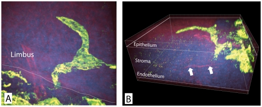
Figure 1
3D reconstruction of the lymphatic arcades ex vivo.
A) At the limbus, the junction between the epithelium of conjunctiva and cornea (dotted line), lymphatic vessels (green) invade into the corneal stroma (blue) and are primarily located directly below ...
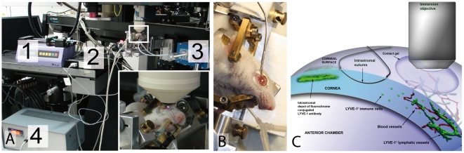
Figure 2
Experimental setup for intravital 2-photon microscopy.
A) 1: intraperitoneal anesthesia/infusion pump; 2: monitoring blood oxygen, pulse and breath rate (by MouseOx; 3: ventilation by tracheotomy followed by intubation; 4: body temperature control; Inset:...
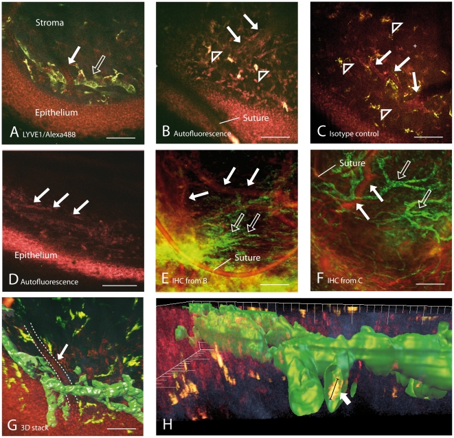
Figure 3
Intravital visualization of lymphatic vessels in the subepithelial stroma of the cornea.
A) Intravital image of a lymphatic vessel in an experimentally vascularized murine cornea. The fluorochrome labeled lymphatic vessel (open arrow) is shown in green and autofluorescence of epithelium, ...
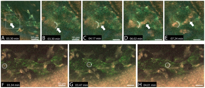
Figure 4
Intravital visualization of immune cell migration into and within a lymphatic vessel in a pathologically vascularized cornea.
Au2013E) Lymphatic vessel (green), crossing blood vessel (red), corneal epithelium (red) and stromal collagen fibrils (blue) are imaged simultaneously. An individual cell (arrow) migrates into the lym...
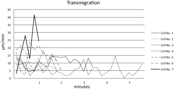
Figure 5
Velocity characteristics of the transmigrating cells.
Cell tracking from first contact of leading cell protrusion with the lymphatic vessel until last contact with rear protrusion after passage. Cells 1u20136 demonstrate velocities with min/max of 0u2013...
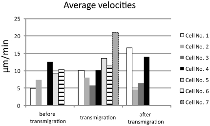
Figure 6
Average velocities of transmigrating cells correlated to transmigration process.
Classification: before transmigration: track until first contact of leading protrusion with the lymphatic vessel wall, transmigration: track from first until last contact of migrating cell with the ly...
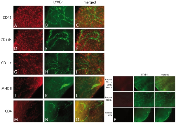
Figure 7
Presence of antigen presenting cells and lymphocytes near corneal lymphatic vessels.
Numerous leucocytes are located within the vascularized cornea (Au2013C: CD45), consisting of CD11b+ macrophages (Du2013F), CD11c+ dendritic cells (Gu2013I), MHCII+ antigen-presenting cells (Ju2013L) ...
Figure images are served from the NIH/NLM PubMed Central Open Access Subset or Europe PMC; copyright remains with the publishers and authors.
💬 Discussion
0 commentsNo comments yet. Be the first to start a discussion!
Leave a Comment