Abstract
Second and Third Harmonic Generation (SHG and THG) microscopy is based on optical effects which are induced by specific inherent physical properties of a specimen. As a multi-photon laser scanning approach which is not based on fluorescence it combines the advantages of a label-free technique with restriction of signal generation to the focal plane, thus allowing high resolution 3D reconstruction of image volumes without out-of-focus background several hundred micrometers deep into the tissue. While in mammalian soft tissues SHG is mostly restricted to collagen fibers and striated muscle myosin, THG is induced at a large variety of structures, since it is generated at interfaces such as refraction index changes within the focal volume of the excitation laser. Besides, colorants such as hemoglobin can cause resonance enhancement, leading to intense THG signals. We applied SHG and THG microscopy to murine (Mus musculus) muscles, an established model system for physiological research, to investigate their potential for label-free tissue imaging. In addition to collagen fibers and muscle fiber substructure, THG allowed us to visualize blood vessel walls and erythrocytes as well as white blood cells adhering to vessel walls, residing in or moving through the extravascular tissue. Moreover peripheral nerve fibers could be clearly identified. Structure down to the nuclear chromatin distribution was visualized in 3D and with more detail than obtainable by bright field microscopy. To our knowledge, most of these objects have not been visualized previously by THG or any label-free 3D approach. THG allows label-free microscopy with inherent optical sectioning and therefore may offer similar improvements compared to bright field microscopy as does confocal laser scanning microscopy compared to conventional fluorescence microscopy.
🔬 Techniques
🧬 Organisms
✨ Fluorophores
🧪 Sample Preparation
🔬 Cell Lines
🏭 Microscope Brands
🧪 Reagent Suppliers
🔴 Lasers
📷 Detectors
💻 Software Details
🏛️ Research Organizations (ROR)
Affiliated research institutions:
📋 Methods
Specimen preparation
Tissue extraction was performed according to German legislation for the protection of animals. Male C57BL/6 mice at the age of 10 – 12 weeks were purchased from Charles River (Sulzfeld, Germany). Animals were housed under conventional conditions with free access to food and water. The Institutional Board of the Walter-Brendel-Zentrum has approved the tissue extraction after euthanization of the animals (approval ID K09), according to the local law "Bayerisches Tierschutzgesetz, Paragraph 6 Abs. 1 Satz 2 Nr. 4". The animals were euthanized by an intraperitoneal pentobarbital overdose (Narcoren, Merial, Germany). Subsequently, the right or left cremaster muscle was exposed through a ventral incision of the scrotum. The muscle was opened ventrally, and epididymis and testicle were detached, subsequently the cremaster was explanted. Throughout the procedure as well as after surgical preparation during microscopy, the muscle was immersed in buffered saline or 0.9% sodium chloride solution. Structures and cells in isolated cremaster muscles were studied without fixation or other further treatment, except for those counterstained for DNA. For DRAQ5 staining, freshly excised, unfixed tissue was incubated for 15 – 60 min at room temperature in 0.9% NaCl with a 1∶1000 dilution of the 5 mM stock solution (Biostatus Ltd, Shepshed Leicestershire, UK). Microscopic observation was in 1∶10 diluted staining solution. For TO-PRO-3 (Invitrogen, Karlsruhe, Germany) staining, excised tissue was fixed for 10 min with buffered 4% paraformaldehyde (Microcos, Garching, Germany). Staining was for 1 hour in PBS with 2 µM TO-PRO-3 and 0.5% Triton X-100 (Sigma). TO-PRO-3-stained tissue was mounted in PermaFluor (Beckman Coulter, Fullerton, CA) on glass slides. For imaging of unstained, unfixed cremaster preparations at room temperature, the muscle was placed on a standard microscopic glass slide or on a round cover slip and covered with sodium chloride solution (0.9%) solution. Round cover slips were mounted with vacuum grease (Baysilone-Paste mittel-viskös, GE Bayer Silicones GmbH & Co. KG, Leverkusen) in a plastic Petri dish with a hole in the bottom. On the sides, the edges of the muscle were weighed down to avoid movement of the tissue. Such cremaster muscle preparations have a thickness of 160 – 200 µm. For observation of leukocyte motility the tissue was placed in a custom-made heating device, submerged in Hank's Balanced Salt Solution and maintained at physiological temperature. For the microscopy of thigh muscles, a longitudinal 10 mm incision through the skin of the right hind limb of a sacrificed mouse was made, beginning at the inguinal crease. The skin was removed to expose the musculus aductor magnus. The muscle was either directly immersed with saline, or excised and placed in a Petri dish filled with saline.
Show full methods section
Specimen preparation
Tissue extraction was performed according to German legislation for the protection of animals. Male C57BL/6 mice at the age of 10 – 12 weeks were purchased from Charles River (Sulzfeld, Germany). Animals were housed under conventional conditions with free access to food and water. The Institutional Board of the Walter-Brendel-Zentrum has approved the tissue extraction after euthanization of the animals (approval ID K09), according to the local law "Bayerisches Tierschutzgesetz, Paragraph 6 Abs. 1 Satz 2 Nr. 4". The animals were euthanized by an intraperitoneal pentobarbital overdose (Narcoren, Merial, Germany). Subsequently, the right or left cremaster muscle was exposed through a ventral incision of the scrotum. The muscle was opened ventrally, and epididymis and testicle were detached, subsequently the cremaster was explanted. Throughout the procedure as well as after surgical preparation during microscopy, the muscle was immersed in buffered saline or 0.9% sodium chloride solution. Structures and cells in isolated cremaster muscles were studied without fixation or other further treatment, except for those counterstained for DNA. For DRAQ5 staining, freshly excised, unfixed tissue was incubated for 15 – 60 min at room temperature in 0.9% NaCl with a 1∶1000 dilution of the 5 mM stock solution (Biostatus Ltd, Shepshed Leicestershire, UK). Microscopic observation was in 1∶10 diluted staining solution. For TO-PRO-3 (Invitrogen, Karlsruhe, Germany) staining, excised tissue was fixed for 10 min with buffered 4% paraformaldehyde (Microcos, Garching, Germany). Staining was for 1 hour in PBS with 2 µM TO-PRO-3 and 0.5% Triton X-100 (Sigma). TO-PRO-3-stained tissue was mounted in PermaFluor (Beckman Coulter, Fullerton, CA) on glass slides. For imaging of unstained, unfixed cremaster preparations at room temperature, the muscle was placed on a standard microscopic glass slide or on a round cover slip and covered with sodium chloride solution (0.9%) solution. Round cover slips were mounted with vacuum grease (Baysilone-Paste mittel-viskös, GE Bayer Silicones GmbH & Co. KG, Leverkusen) in a plastic Petri dish with a hole in the bottom. On the sides, the edges of the muscle were weighed down to avoid movement of the tissue. Such cremaster muscle preparations have a thickness of 160 – 200 µm. For observation of leukocyte motility the tissue was placed in a custom-made heating device, submerged in Hank's Balanced Salt Solution and maintained at physiological temperature. For the microscopy of thigh muscles, a longitudinal 10 mm incision through the skin of the right hind limb of a sacrificed mouse was made, beginning at the inguinal crease. The skin was removed to expose the musculus aductor magnus. The muscle was either directly immersed with saline, or excised and placed in a Petri dish filled with saline.
Microscopy
Microscopic observations were performed using a commercially available system, a TriMScope (LaVision BioTec, Bielefeld, Germany) built around an Olympus BX 51 microscope (Olympus, Hamburg, Germany) and equipped with a tunable Ultra II Titanium:Sapphire Laser (Coherent, Dieburg, Germany) and an Optical Parametric Oscillator (Chameleon OPO; APE, Berlin, Germany; typical pulse width 200 fs, repetition rate 80 MHz) which is pumped by the Ti:Sa. The OPO generated 1270 nm or 1275 nm light with 640–700 mW output. The unattenuated intensity at the sample (i.e. after the objective) was ∼250 mW. Scanning cremaster tissue with full intensity and high resolution (400x400 µm with 1680×1680 pixels and 200 lines per second) did not produce any visible damage, except if the area of observation contained light absorbing dirt which would induce plasma formation. An Olympus XLUMPlanFl 20×/0.95W objective was used (working distance 2 mm). The following detection channels were used: backward (epi) detection blue (417 – 477 nm), red (604 – 644 nm) and far red (645 – 695 nm), forward detection blue and longpass 488 nm. 700 nm short pass filters blocked out excitation light. Light collection in forward direction was performed by an Olympus WI-UCD condenser, NA 0.8. Photomultiplier tubes (PMTs) were Hamamatsu H6780-01 for the blue channels and H6780-20 for the others. Where mentioned (see main text) more sensitive gallium arsenide phosphide detectors (Hamamatsu H7422-40) were used for backward detection. To characterize the performance of the system we measured the full width half maximum (FWHM) of signals from subresolution particles embedded in 1% agarose with 1275 nm excitation. Two-photon excitation fluorescence of deep red beads (size 175 nm, exc 633, em. 660, Invitrogen P7220) provided an FWHM of 1.1 µm laterally and 6.2 µm axially. This compares to theoretical values of (0.51*λ) / ((√2)*NA) = 484 nm laterally and (0.64* λ) / (n-√(n2-NA2)) = 1.7 µm axially [21] , [22] . Latex beads did not generate a suitable THG signal. The THG-FWHM was measured with titanium dioxide nanoparticles (anatase configuration,
📊 Figures
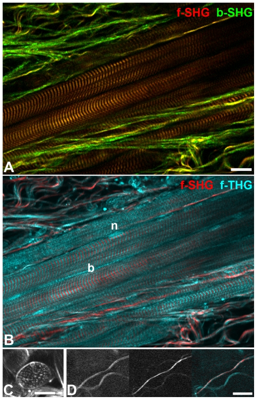
Figure 1
Collagen fibers and striated muscle.
A) The relative SHG signal intensity ratio collagen/myosin is higher in backward SHG (green) than in forward SHG (red). B) While in striated muscle SHG visualizes myosin and thus A-bands of the sarcom...
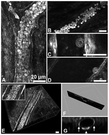
Figure 2
THG of blood vessels and blood cells in explanted, unfixed cremaster muscles recorded in forward direction.
A) Projection of optical sections. Without flow, red blood cells (RBCs) sink to the bottom of the vessel, forming a tightly packed mass. B) Visualization of typical RBC shape. C,D) Leukocytes adhering...
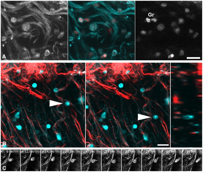
Figure 3
Migrating leukocytes.
A) Fixed cremaster muscle, optical section about 50 u00b5m from the surface. THG (left, cyan) revealed shapes reminiscent of migrating leukocytes deep in the tissue. DNA counterstaining (right, red) c...
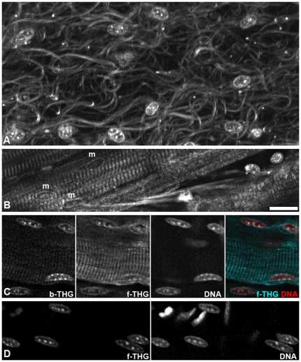
Figure 4
Nuclei and chromatin structure.
A,B) Single optical forward THG sections from unstained, explanted cremaster. A, Section near the surface, nuclei most likely belong to macrophages. B, In deeper regions, nuclei of muscle fibers (m) c...
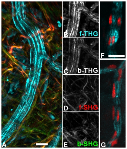
Figure 5
Nerve fibers in the cremaster.
A) Unfixed, explanted muscle, projection of several optical sections covering 50 u00b5m depth. Peripheral nerve (forward THG, cyan) surrounded by collagen fibers (forward SHG in red, backward SHG in g...
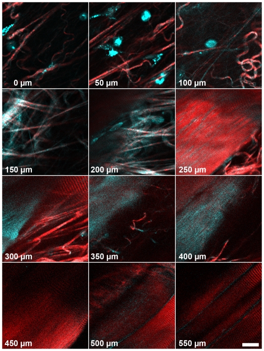
Figure 6
Deep tissue imaging in the mouse thigh muscle with backward GaAsP-detectors.
The first 100 u00b5m consist of connective tissue, followed by a dense meshwork of collagen fibers (150 and 200 u00b5m) and finally striated muscle fibers with interspersed collagen. For this display,...
Figure images are served from the NIH/NLM PubMed Central Open Access Subset or Europe PMC; copyright remains with the publishers and authors.
💬 Discussion
0 commentsNo comments yet. Be the first to start a discussion!
Leave a Comment