Abstract
Nodes of Ranvier are specialized, highly polarized axonal domains crucial to the propagation of saltatory action potentials. In the peripheral nervous system, axo–glial cell contacts have been implicated in Schwann cell (SC) differentiation and formation of the nodes of Ranvier. SC microvilli establish axonal contact at mature nodes, and their components have been observed to localize early to sites of developing nodes. However, a role for these contacts in node formation remains controversial. Using a myelinating explant culture system, we have observed that SCs reorganize and polarize microvillar components, such as the ezrin-binding phosphoprotein 50 kD/regulatory cofactor of the sodium-hydrogen exchanger isoform 3 (NHERF-1), actin, and the activated ezrin, radixin, and moesin family proteins before myelination in response to inductive signals. These components are targeted to the SC distal tips where live cell imaging reveals novel, dynamic growth cone–like behavior. Furthermore, localized activation of the Rho signaling pathway at SC tips gives rise to these microvillar component–enriched “caps” and influences the efficiency of node formation.
🔬 Techniques
🔭 Microscopes
💻 Software
✨ Fluorophores
🧪 Sample Preparation
🔬 Cell Lines
🏭 Microscope Brands
🧪 Reagent Suppliers
📷 Detectors
💻 Software Details
💾 Data Repositories
🏛️ Research Organizations (ROR)
Affiliated research institutions:
📋 Methods
DRG explant and SC cultures
DRG explants were cultured essentially as described previously ( Howe and McCarthy, 1998 ). In brief, spinal cords and associated DRGs were dissected out of Wistar rat embryos (embryonic day 16; Charles River) and rinsed in HBSS with 1.2 mM calcium and 0.8 mM magnesium (Sigma-Aldrich) supplemented with 5% FBS, 50 U/ml penicillin, and 50 μg/ml streptomycin. Ganglia were trimmed of spinal roots, removed, and rinsed with HBSS (no serum) and plated into standard feed (basal medium Eagle's [BME]; Sigma-Aldrich) including ITS + supplements (Discovery Labware, Inc.), 0.2% BSA, 10 mM Hepes, 498 mg/dl d -glucose, 100 ng/ml 2.5 S NGF (Discovery Labware, Inc.), 50 U/ml penicillin, and 50 μg/ml streptomycin. Coverslips (Carolina Biologicals) were coated with 0.4 mg/ml Matrigel in BME (Discovery Labware, Inc.) and 10 μg/ml poly- d -lysine (Sigma-Aldrich), and maintained on Teflon circles (American Durafilm). After ∼21 d, cultures were supplemented with 15% FBS and 50 μg/ml ascorbate to induce myelination ( Eldridge et al., 1987 ). Cultures were incubated at 37°C in humidified 5% CO 2 /95% air with regular feeding every other day. In some instances, media also included various combinations of 1 μM LPA (Sigma-Aldrich) and 2–20 μM Rho-kinase inhibitor, Y-27632 (Calbiochem-Novabiochem). Isolated SCs were cultured according to the Brockes method ( Brockes et al., 1979 ). P1 rat pups were killed and their sciatic nerves removed. The nerves were incubated with 0.1% collagenase (Sigma-Aldrich) in L-15 for 30 min followed by 0.1% collagenase with 0.25% trypsin for 30 min. After rinsing with DME + 10% FBS, the nerves were triturated and plated into the same media in an untreated 100-mm culture dish. The next day, 10 −5 M 1-(β- d -arabinofuranosyl) cytosine (AraC; Calbiochem) was added. After 3 d of anti-mitotic treatment, the AraC was washed out and the cells were allowed to recover for 24 h. Anti-Thy1.1 (clone TN-26; 1:100 in DME) and rabbit complement sera HLA-ABC (Sigma-Aldrich; 1:1 in DME) treatment followed to remove residual fibroblasts. Cultures were maintained in Primaria tissue culture flasks (BD Biosciences) with twice weekly feedings of DME + 10% FBS. EBP50 antibody An antibody against EBP50 was raised in rabbits for the following study (Covance). A recombinant EBP50-GST fusion was generated (EBP50 cDNA; gift from R. Brian Doctor, University of Colorado Health Sciences Center, Denver, CO) using the pET-41a + vector (Novagen), expressed in bacteria, purified, and used as the immunogen. Antisera were purified using a Talon kit (CLONTECH Laboratories, Inc.), isolated, His-tagged EBP50 (generated using pET-28b + [Novagen]) which was coupled to CNBr Sepharose (Amersham Biosciences). Eluted antibody was dialyzed against PBS followed by 50% glycerol in PBS.
Show full methods section
DRG explant and SC cultures
DRG explants were cultured essentially as described previously ( Howe and McCarthy, 1998 ). In brief, spinal cords and associated DRGs were dissected out of Wistar rat embryos (embryonic day 16; Charles River) and rinsed in HBSS with 1.2 mM calcium and 0.8 mM magnesium (Sigma-Aldrich) supplemented with 5% FBS, 50 U/ml penicillin, and 50 μg/ml streptomycin. Ganglia were trimmed of spinal roots, removed, and rinsed with HBSS (no serum) and plated into standard feed (basal medium Eagle's [BME]; Sigma-Aldrich) including ITS + supplements (Discovery Labware, Inc.), 0.2% BSA, 10 mM Hepes, 498 mg/dl d -glucose, 100 ng/ml 2.5 S NGF (Discovery Labware, Inc.), 50 U/ml penicillin, and 50 μg/ml streptomycin. Coverslips (Carolina Biologicals) were coated with 0.4 mg/ml Matrigel in BME (Discovery Labware, Inc.) and 10 μg/ml poly- d -lysine (Sigma-Aldrich), and maintained on Teflon circles (American Durafilm). After ∼21 d, cultures were supplemented with 15% FBS and 50 μg/ml ascorbate to induce myelination ( Eldridge et al., 1987 ). Cultures were incubated at 37°C in humidified 5% CO 2 /95% air with regular feeding every other day. In some instances, media also included various combinations of 1 μM LPA (Sigma-Aldrich) and 2–20 μM Rho-kinase inhibitor, Y-27632 (Calbiochem-Novabiochem). Isolated SCs were cultured according to the Brockes method ( Brockes et al., 1979 ). P1 rat pups were killed and their sciatic nerves removed. The nerves were incubated with 0.1% collagenase (Sigma-Aldrich) in L-15 for 30 min followed by 0.1% collagenase with 0.25% trypsin for 30 min. After rinsing with DME + 10% FBS, the nerves were triturated and plated into the same media in an untreated 100-mm culture dish. The next day, 10 −5 M 1-(β- d -arabinofuranosyl) cytosine (AraC; Calbiochem) was added. After 3 d of anti-mitotic treatment, the AraC was washed out and the cells were allowed to recover for 24 h. Anti-Thy1.1 (clone TN-26; 1:100 in DME) and rabbit complement sera HLA-ABC (Sigma-Aldrich; 1:1 in DME) treatment followed to remove residual fibroblasts. Cultures were maintained in Primaria tissue culture flasks (BD Biosciences) with twice weekly feedings of DME + 10% FBS. EBP50 antibody An antibody against EBP50 was raised in rabbits for the following study (Covance). A recombinant EBP50-GST fusion was generated (EBP50 cDNA; gift from R. Brian Doctor, University of Colorado Health Sciences Center, Denver, CO) using the pET-41a + vector (Novagen), expressed in bacteria, purified, and used as the immunogen. Antisera were purified using a Talon kit (CLONTECH Laboratories, Inc.), isolated, His-tagged EBP50 (generated using pET-28b + [Novagen]) which was coupled to CNBr Sepharose (Amersham Biosciences). Eluted antibody was dialyzed against PBS followed by 50% glycerol in PBS.
Explant transfection
Explants were transfected using Lipofectamine PLUS (Invitrogen). Cultures were treated for 24 h before transfection with media of choice ( Pedraza and Colman, 2000 ). 1 μg DNA per coverslip was used, and the DNA/PLUS (3.5 μ l )/Lipofectamine (2.8 μ l ) complexes were incubated with the cultures for 3–4 h in unsupplemented BME. The transfection mixture was then removed. The cultures were rinsed once with BME and returned to the appropriate medium. Immunostaining Explants were fixed for 10 min at 4°C in 4% PFA/4% sucrose in PBS. After washing with PBS, cultures were blocked with 10% BSA in PBS-T (PBS + 0.2% Triton X-100) for 1 h at 37°C. Primary antibodies were diluted in 2% BSA in PBS-T and incubated for 1–2 h at 37°C. Coverslips were washed with PBS-T; then secondary antibodies, also diluted in 2% BSA in PBS-T (1:1,000), were added and incubated for 1 h at 37°C. After washing with PBS-T, followed by PBS alone and a water rinse, coverslips were mounted using a Vectashield mounting medium (Vector Laboratories). SCs were processed similarly with the exceptions of fixation for 20 min at room temperature followed by a 7-min permeabilization using PBS-T and the omission of Triton X-100 from the proceeding steps. Images were captured using IPLab spectrum software (Signal Analytics Corporation) on an upright Axioplan (Carl Zeiss MicroImaging, Inc.) with a Sensys digital camera (Photometrics Limited), and processed using Adobe Photoshop. Sources of primary antibodies are as follows: anti-neurofilament (Sternberger Monoclonals, Inc.); anti–α-tubulin (Sigma-Aldrich); anti-ERM and anti–phospho-ezrin (Thr567)/radixin(Thr564)/moesin (Thr558) (Cell Signaling Technology, Inc.); anti-MAG (CHEMICON International, Inc.); anti-MBP (Boehringer); anti-RhoA (Santa Cruz Biotechnology, Inc.); Alexa-Fluor 594 phalloidin (Molecular Probes); anti-βIV spectrin (a gift from M. Komada, Tokyo Institute of Technology, Tokyo, Japan; Komada and Soriano, 2002 ); anti-ERM (a gift from E. Luna, University of Massachusetts Medical School, Worcester, MA); anti-ankyrin G ( Lambert et al., 1997 ); and anti-EBP50 as described above (EBP50 antibody). All secondary antibodies were purchased from Molecular Probes.
Live cell imaging Explants or dispersed
SCs were grown on either 22-mm square coverslips or 30-mm Rose dishes (World Precision Instruments) and treated as indicated. Preparations were mounted for imaging on an inverted microscope (DMIRE 2; Leica) in a 37°C-heated 5% CO 2 /95% room air plexiglass environmental chamber. Image acquisition and processing was performed using an extended range cooled CCD camera (ORCA ER; Hamamatsu) and OpenLab software (Improvision). Online supplemental material The original time-lapse videos from which the still images presented in Fig. 3 were taken are available for viewing as supplemental material. Video 1 contains time-lapse fluorescence microscopy of an EBP50-GFP–transfected SC maintained in standard feed. Video 2 specifically shows the distal tip of this cell. Video 3 shows time-lapse fluorescence microscopy of an EBP50-GFP–transfected SC after the induction of myelination using serum and ascorbate. Video 4 shows the remodeling EBP50 enriched tip of this cell. Videos are available at http://www.jcb.org/cgi/content/full/jcb.200303039/DC1 .
Online supplemental material The original time-lapse videos from which the still images presented in Fig. 3 were taken are available for viewing as supplemental material. Video 1 contains time-lapse fluorescence microscopy of an EBP50-GFP–transfected SC maintained in standard feed. Video 2 specifically shows the distal tip of this cell. Video 3 shows time-lapse fluorescence microscopy of an EBP50-GFP–transfected SC after the induction of myelination using serum and ascorbate. Video 4 shows the remodeling EBP50 enriched tip of this cell. Videos are available at http://www.jcb.org/cgi/content/full/jcb.200303039/DC1 .
Supplemental Material [Supplemental Material Index]
📊 Figures
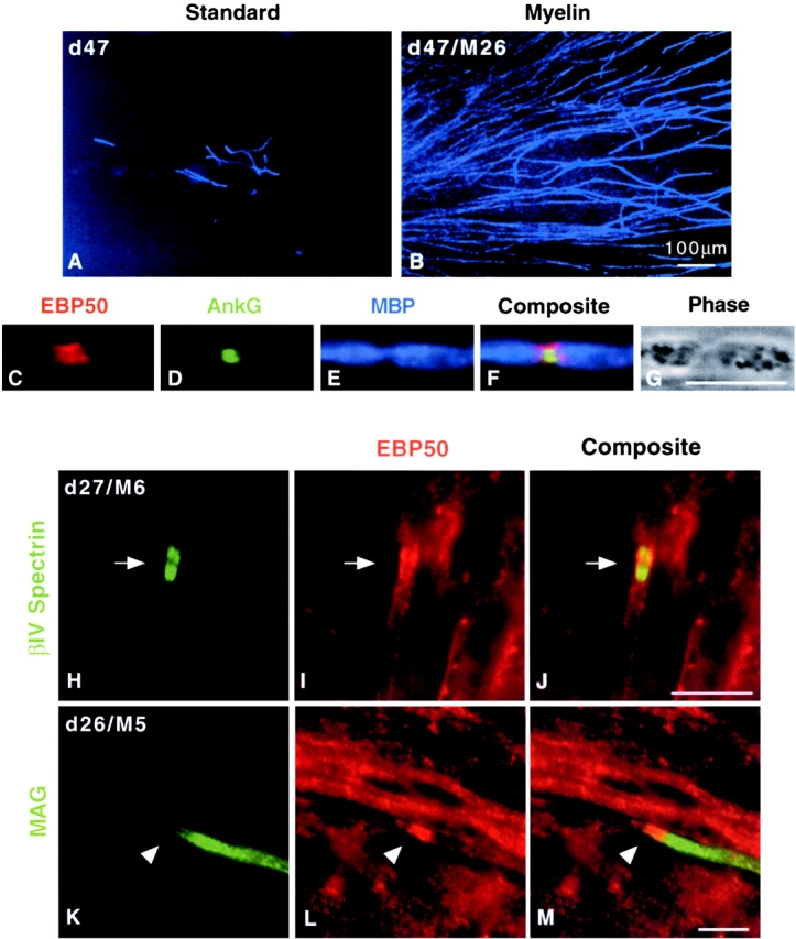
Figure 1.
EBP50/ERM localization to early and mature nodes in myelinating DRG explant cultures. (A, ST:d47; B, MY:d47/M26) Myelinated and comparable control cultures were stained for MBP to show mature myelin. ...
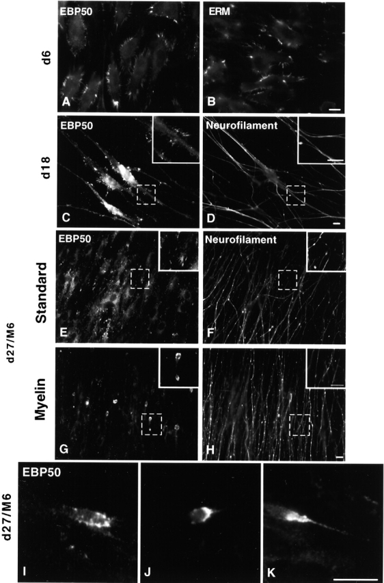
Figure 2.
EBP50/ERM localization in premyelinating/induced DRG explant cultures changes from cell surface microvilli to a focal concentration at the SC tip. After 6 d in vitro, cultures were stained for (A) EBP...
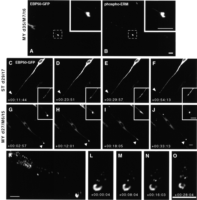
Figure 3.
Dynamic EBP50/ERM-positive SC distal tips. Cultures were transfected after 1 d of induction. After 5 d of transfection, (A) EBP50-GFP was seen to colocalize with (B) endogenous phospho-ERM staining at...
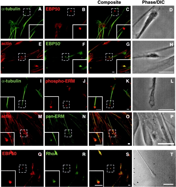
Figure 4.
EBP50, actin, activated ERMs, and RhoA localize to SC tips. Isolated SCs were stained to identify other proteins residing at the tips. (Au2013C) EBP50 was found at the tip of u03b1-tubulin containing ...
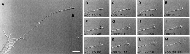
Figure 5.
SC tips display novel growth coneu2013like behavior. Isolated SCs were studied using time-lapse live cell imaging. (A, arrow) SC tips displayed dynamic, active remodeling similar to that seen in an ax...
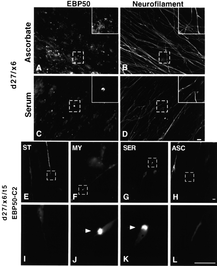
Figure 6.
Cap formation is dependent on serum and can be promoted via Rho stimulation. At d 21, cultures were supplemented with either serum or ascorbate alone, maintained for 6 d (d27/x6), and stained for (A a...
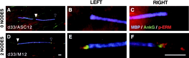
Figure 7.
Efficient node formation is linked to cap formation. Cultures maintained with either ascorbate alone or complete myelin feed were stained for MBP, AnkG, and phospho-ERM. Myelin segments were identifie...
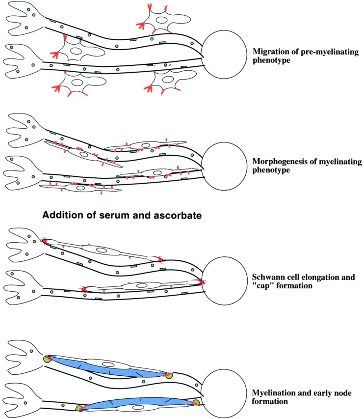
Figure 8.
Model: SC progression toward myelination in DRG explant culture system. Highly motile, premyelinating SC migrate along axons extending from the DRG. These SC harbor numerous cell surface microvilli (r...
Figure images are served from the NIH/NLM PubMed Central Open Access Subset or Europe PMC; copyright remains with the publishers and authors.
💬 Discussion
0 commentsNo comments yet. Be the first to start a discussion!
Leave a Comment