Abstract
Limbal epithelial stem cells (LESCs) are essential to maintain the transparent ocular surface required for vision. Despite great advances in our understanding of ocular stem cell biology over the last decade, the exact location of the LESC niche remains unclear. In the present study we have used in vitro clonal analysis to confirm that limbal crypts provide a niche for the resident LESCs. We have used high-resolution imaging of the basal epithelial layer at the limbus to identify cells with a morphology consistent with stem cells that were only present within the basal layer of the limbal crypts. These cells are proximal to limbal stromal cells suggesting direct cell-to-cell interaction. Serial block-face scanning electron microscopy (SBFSEM) confirmed that the putative LESCs are indeed in direct contact with cells in the underlying stroma, a contact that is facilitated by focal basement membrane interruptions. Limbal mesenchymal cells previously identified in the human limbus collocate in the crypt-rich limbal stromal area in the vicinity of LESCs and may be involved in the cell-to-cell contact revealed by SBFSEM. We also observed a high population of melanocytes within the basal layer of the limbal crypts. From these observations we present a three dimensional reconstruction of the LESC niche in which the stem cell is closely associated and maintained by both dendritic pigmented limbal melanocytes and elongated limbal stromal cells.
🔬 Techniques
🔭 Microscopes
✨ Fluorophores
🧪 Sample Preparation
🔬 Cell Lines
🏭 Microscope Brands
🧪 Reagent Suppliers
📷 Detectors
💻 Software Details
🏛️ Research Organizations (ROR)
Affiliated research institutions:
📋 Methods
1.
Ethics Statement
All human tissue was handled according to the tenets of the Declaration of Helsinki and written consent was acquired from next of kin of all deceased donors regarding eye donation for research. Research consent was obtained via the Moorfields Eye Hospital Lions Eye Bank (U.K) http://www.moorfields.nhs.uk/Aboutus/Clinicalsupportservices/Eyebank and Lions Eye Institute (Florida, U.S) http://www.fleb.org/ . All experiments were approved by the National Research Ethics Service, South west 3 REC, reference 10/H0106/57. 2.
Isolation and Culture of Limbal Epithelial Cells
Limbal palisades, which surround the LCs were macroscopically identified under a dissecting microscope. After dissection, crypt-rich (from superior and inferior limbus) and non-crypt rich (from temporal and nasal limbus) limbal biopsies isolated from the same donor were transferred separately into a solution containing 1.2 U/mL dispase II (Roche diagnostics GmbH, Mannheim, Germany) in corneal epithelial cell culture medium (CECM) containing a 1∶1 ratio of DMEM:F12, 10% (v/v) fetal bovine serum, 100 U/mL penicillin, 100 μg/mL streptomycin, 0.25 μg/mL Fungizone, epidermal growth factor (EGF) 10 ng/mL (Life technologies, Paisley, UK), hydrocortisone (0.4 μg/mL), insulin (5 μg/mL), adenine (0.18 mM), transferrin (5 μg/mL), T3 (2 nM), cholera toxin (0.1 nM) (Sigma-Aldrich, Dorset, UK) and incubated for 2 hours at 37°C.
Limbal epithelial cells
(LECs) were isolated from the crypt-rich and non-crypt rich biopsies by gently scraping the epithelium using the point of thin forceps. LECs were then pre-expanded for 7 days in T25 flasks on a 3T3 feeder layer that had been previously growth arrested with 4 μg/mL mitomycin C (Sigma-Aldrich, Dorset, UK) for 2 hours. CECM culture medium was changed three times a week and the co-cultures maintained at 37°C in a humidified atmosphere containing 5% CO 2 in air. 3.
Show full methods section
1.
Ethics Statement
All human tissue was handled according to the tenets of the Declaration of Helsinki and written consent was acquired from next of kin of all deceased donors regarding eye donation for research. Research consent was obtained via the Moorfields Eye Hospital Lions Eye Bank (U.K) http://www.moorfields.nhs.uk/Aboutus/Clinicalsupportservices/Eyebank and Lions Eye Institute (Florida, U.S) http://www.fleb.org/ . All experiments were approved by the National Research Ethics Service, South west 3 REC, reference 10/H0106/57. 2.
Isolation and Culture of Limbal Epithelial Cells
Limbal palisades, which surround the LCs were macroscopically identified under a dissecting microscope. After dissection, crypt-rich (from superior and inferior limbus) and non-crypt rich (from temporal and nasal limbus) limbal biopsies isolated from the same donor were transferred separately into a solution containing 1.2 U/mL dispase II (Roche diagnostics GmbH, Mannheim, Germany) in corneal epithelial cell culture medium (CECM) containing a 1∶1 ratio of DMEM:F12, 10% (v/v) fetal bovine serum, 100 U/mL penicillin, 100 μg/mL streptomycin, 0.25 μg/mL Fungizone, epidermal growth factor (EGF) 10 ng/mL (Life technologies, Paisley, UK), hydrocortisone (0.4 μg/mL), insulin (5 μg/mL), adenine (0.18 mM), transferrin (5 μg/mL), T3 (2 nM), cholera toxin (0.1 nM) (Sigma-Aldrich, Dorset, UK) and incubated for 2 hours at 37°C.
Limbal epithelial cells
(LECs) were isolated from the crypt-rich and non-crypt rich biopsies by gently scraping the epithelium using the point of thin forceps. LECs were then pre-expanded for 7 days in T25 flasks on a 3T3 feeder layer that had been previously growth arrested with 4 μg/mL mitomycin C (Sigma-Aldrich, Dorset, UK) for 2 hours. CECM culture medium was changed three times a week and the co-cultures maintained at 37°C in a humidified atmosphere containing 5% CO 2 in air. 3.
Clonal Analysis
Cadaveric human corneas were obtained from three donors (age range 51–71 years; mean 58.3 years). In total, 124 clones isolated from the crypt-rich and non-crypt limbal biopsies from three donors were analyzed for their in vitro growth potential. When they had reached 50% confluence in primary 3T3-LECs co-cultures, the LECs were isolated using serial trypsinization. The 3T3 feeder cells were detached and removed using 0.05% trypsin-0,02% EDTA, before 0.5% trypsin-0.2% EDTA (Life technologies, Paisley, UK) was used to detach and prepare a single cell suspension of LECs. For each donor, 250 single LECs initially isolated from crypt-rich or from non-crypt limbal biopsies were seeded onto 55 mm 2 plates containing 1.4×10 6 growth-arrested 3T3s. Plates were then checked under an inverted microscope to confirm seeding of a single LECs suspension on top of growth arrested 3T3s. After 7 days in culture, small colonies (clones) of approximately 1 mm diameter were randomly selected and isolated using 8 mm sterile cloning cylinders (Sigma-Aldrich, Dorset, UK) and 0.5% trypsin-0.2% EDTA. Limbal epithelial cells from these single cell colonies (clone) were again seeded onto a 55 mm 2 plate containing 1.4×10 6 growth-arrested 3T3s. Epithelial cells were expanded for up to 12 days and plates fixed with 4% paraformaldehyde (PFA) (VWR International Ltd. Leicestershire, UK) and stained with 2% rhodamine. Each plate was then scored as holoclone, meroclone or paraclone depending on the percentage of aborted colonies [20] . When 0–5% of the total colonies were terminally differentiated, the clone was scored as a holoclone. When more than 95% of colonies were terminally differentiated, the clone was scored as a paraclone. Finally, when >5% but
📊 Figures
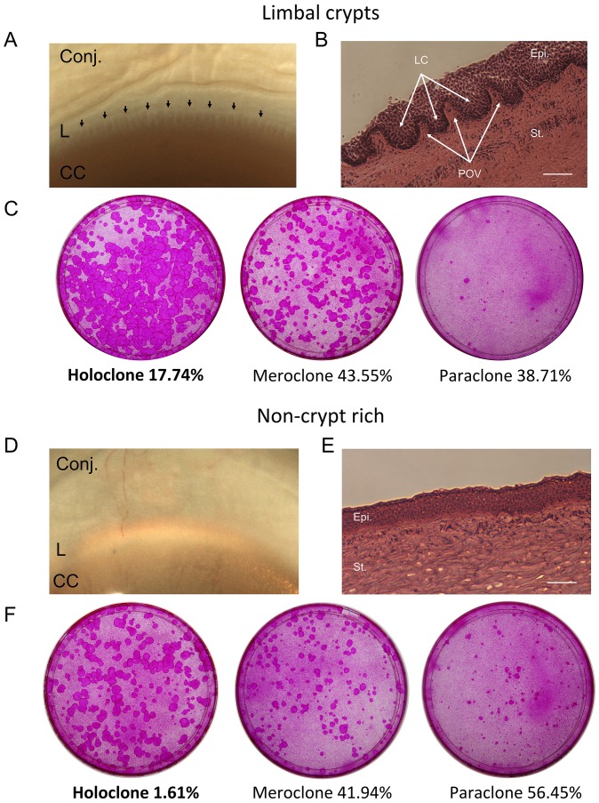
Figure 1
Clonal analysis.
Crypt-rich (A) and non-crypt rich (D) limbal biopsies observed under the dissecting microscope and targeted for clonal analysis. Sections were cut tangentially to the limbal circumference and stained ...
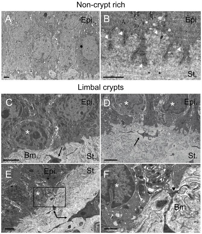
Figure 2
Observation of putative LESCs by TEM within the LCs.
Transmission electron micrographs highlighting the interface between the limbal stroma and the limbal basal epithelial layer within the non-crypt rich limbus (A, B) and the limbal crypts (C, D, E, F)....
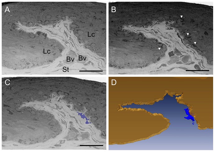
Figure 3
Limbal crypt ultrastructure observed by SBFSEM.
Limbal crypt tangentially imaged through 70 u03bcm from the corneal to the conjunctival side of the limbus. A, B and C represent non-sequential micrographs of the same 3D dataset. Manual segmentation ...
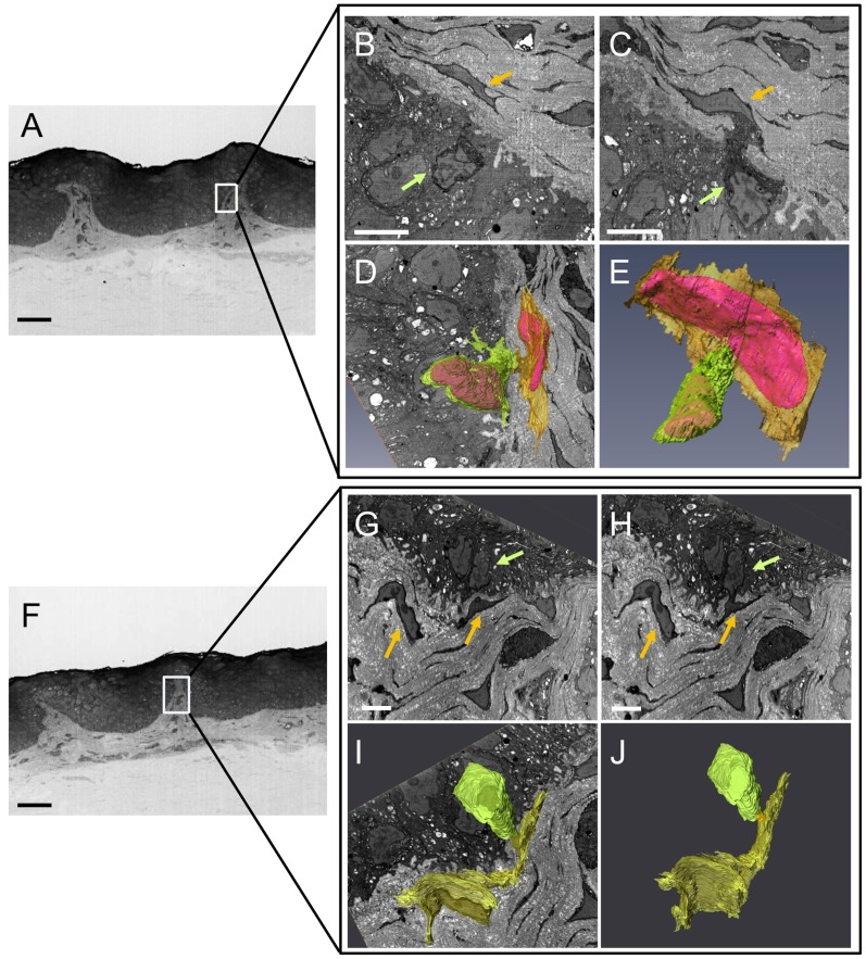
Figure 4
Cell-to-cell contacts imaged within the limbal crypts by SBFSEM.
Box in A and F represents the area in which contact between putative LESC (green arrows in B, C, G, H and green volumes in D, E, I, J after 3D reconstruction) and limbal stromal cells (yellow arrows i...
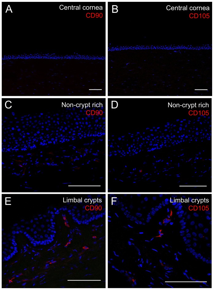
Figure 5
Results of immunohistochemistry staining for limbal mesenchymal cell markers CD90 and CD105 within the central cornea, the non-crypt rich limbus and the limbal crypts.
Immunofluorescence suggests CD90 and CD105 expression is markedly increased by limbal stromal cells underlying the limbal crypts (E, F) compared to the non-crypt rich limbus (C, D). Central corneal se...
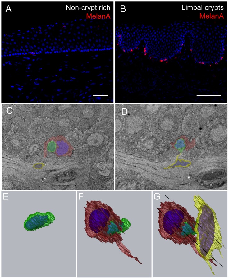
Figure 6
Melanocytes interact with putative LESCs in their niche.
Immunohistochemistry reveals much more MelanA positive cells within the LCs than within the non-crypt rich limbal areas (A, B). Serial-block face scanning electron micrographs showing the edge of one ...
Figure images are served from the NIH/NLM PubMed Central Open Access Subset or Europe PMC; copyright remains with the publishers and authors.
💬 Discussion
0 commentsNo comments yet. Be the first to start a discussion!
Leave a Comment