Abstract
AbstractLocalization-based imaging has revolutionized fluorescence optical microscopy and has also enabled unprecedented ultrasound images of microvascular structures in deep tissues. Herein, we introduce a new concept of localization optoacoustic tomography (LOT) that employs rapid sequential acquisition of three-dimensional optoacoustic images from flowing absorbing particles. We show that the new method enables breaking through the spatial resolution barrier of acoustic diffraction while further enhancing the visibility of structures under limited-view tomographic conditions. Given the intrinsic sensitivity of optoacoustics to multiple hemodynamic and oxygenation parameters, LOT may enable a new level of performance in studying functional and anatomical alterations of microcirculation.
🔬 Techniques
🧪 Sample Preparation
💻 Software Details
🏛️ Research Organizations (ROR)
Affiliated research institutions:
📋 Methods
Three dimensional optoacoustic imaging system LOT can in principle be performed with any OAT system providing real-time imaging capability. In order to demonstrate true 3D localization capacity, our previously reported real-time volumetric OAT system was used in the current work 21 . It employs a spherical matrix detection array uniformly populated by 256 individual piezocomposite elements having 4 MHz central frequency and >100% bandwidth in receive mode ( Figure 1 ). The spherical aperture has a radius of curvature of 40 mm and covers an angle of 90°. The array was fixed pointing upwards and the volume between its active detection aperture and the imaged sample was filled with agar, which served as an acoustic coupling medium and was further used to hold the imaged samples. Short-duration (
📊 Figures
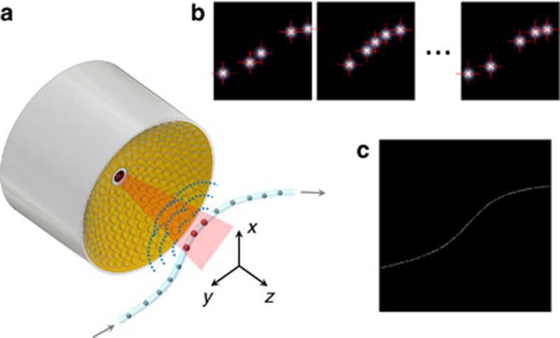
Figure 1
Imaging principle of localization optoacoustic tomography. ( a ) A spherical array of ultrasound transducers is used to acquire a three-dimensional optoacoustic image of flowing absorbers for each las...
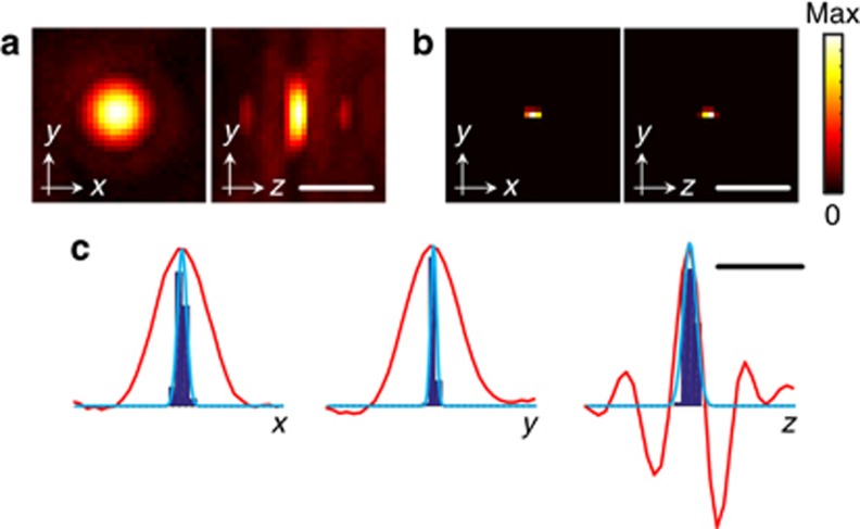
Figure 2
Localization accuracy. ( a ) Maximum intensity projections of the three-dimensional optoacoustic image of a 30u2009u03bcm absorbing microsphere. ( b ) Equivalent image obtained as the three-dimensiona...
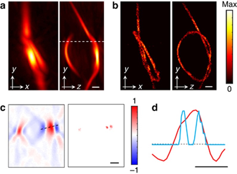
Figure 3
Resolution enhancement in localization optoacoustic tomography. ( a ) Maximum intensity projections of the three-dimensional optoacoustic image of a ~220u2009u03bcm diameter pipette tip bent to form a...
Figure images are served from the NIH/NLM PubMed Central Open Access Subset or Europe PMC; copyright remains with the publishers and authors.
💬 Discussion
0 commentsNo comments yet. Be the first to start a discussion!
Leave a Comment