Abstract
The negatively charged nitrogen vacancy (NV-) center in diamond is the focus of widespread attention for applications ranging from quantum information processing to nanoscale metrology. Although most work so far has focused on the NV- optical and spin properties, control of the charge state promises complementary opportunities. One intriguing possibility is the long-term storage of information, a notion we hereby introduce using NV-rich, type 1b diamond. As a proof of principle, we use multicolor optical microscopy to read, write, and reset arbitrary data sets with two-dimensional (2D) binary bit density comparable to present digital-video-disk (DVD) technology. Leveraging on the singular dynamics of NV- ionization, we encode information on different planes of the diamond crystal with no cross-talk, hence extending the storage capacity to three dimensions. Furthermore, we correlate the center's charge state and the nuclear spin polarization of the nitrogen host and show that the latter is robust to a cycle of NV- ionization and recharge. In combination with super-resolution microscopy techniques, these observations provide a route toward subdiffraction NV charge control, a regime where the storage capacity could exceed present technologies.
🔬 Techniques
🏭 Microscope Brands
🧪 Reagent Suppliers
🏛️ Research Organizations (ROR)
Affiliated research institutions:
📋 Methods
Diamond crystal A type 1b [111] diamond from Diamond Delaware Knives was used as the sample. Prior characterization via infrared spectroscopy ( 14 ) was consistent with the presence of substitutional nitrogen atoms at a concentration of approximately 40 ppm; the estimated NV content was 0.4 ppm. The absorption near 1282 cm −1 suggests that A-centers—formed by two adjacent nitrogen atoms—were, if at all present, at trace concentrations. Optical spectroscopy confirms that the collected fluorescence originated almost exclusively from NV centers. A distinctive peak at ~737 nm reveals the presence of silicon vacancy (SiV) centers; from the peak amplitude, we estimated the SiV-NV ratio to be about 0.6%.
NV magnetic resonance and optical microscopy
For our experiments, we used a custom-made, multicolor microscope. A 13-mW helium-neon laser and a 2 W continuous-wave solid-state laser served as the sources of red (632 nm) and green (532 nm) light, respectively. Excitation in the blue (450 nm) light was provided by a tunable ultrafast laser (Coherent Mira) and a frequency doubler generating 120-fs-long pulses at a repetition rate of 76 MHz; the average power at 450 nm was 400 μW. All laser beams were coupled into a 0.42–numerical aperture objective, which also collected the outgoing sample fluorescence. The illumination timing was set independently with the aid of acousto-optic modulators; a servo-controlled, two-mirror galvo system was used for sample scanning. Sample fluorescence ranging from 650 to 850 nm was detected after a dichroic mirror and notch filters by a solid-state avalanche photodetector. Control of the NV − electronic and nuclear spin was carried out via the use of MW and RF pulses produced by four signal generators: R&S SMB100A, R&S SMV03, Agilent E4433B, and Tektronix AFG3102. A 25-μm-diameter copper wire overlaid on the diamond surface served as the simultaneous source of the MW and RF fields. Upon amplification, the typical duration of an MW (RF) inversion pulse was 500 ns (30 μs). All magnetic resonance experiments were carried out in the presence of a 5.5-mT magnetic field emanating from a permanent magnet in the sample vicinity. The magnetic field was oriented so as to coincide with the sample crystal normal, that is, the [111] axis. A pulse generator (PulseBlasterESR-PRO) controlled the timing of all laser, MW, and RF pulses. All experiments were carried out under ambient conditions.
Show full methods section
Diamond crystal A type 1b [111] diamond from Diamond Delaware Knives was used as the sample. Prior characterization via infrared spectroscopy ( 14 ) was consistent with the presence of substitutional nitrogen atoms at a concentration of approximately 40 ppm; the estimated NV content was 0.4 ppm. The absorption near 1282 cm −1 suggests that A-centers—formed by two adjacent nitrogen atoms—were, if at all present, at trace concentrations. Optical spectroscopy confirms that the collected fluorescence originated almost exclusively from NV centers. A distinctive peak at ~737 nm reveals the presence of silicon vacancy (SiV) centers; from the peak amplitude, we estimated the SiV-NV ratio to be about 0.6%.
NV magnetic resonance and optical microscopy
For our experiments, we used a custom-made, multicolor microscope. A 13-mW helium-neon laser and a 2 W continuous-wave solid-state laser served as the sources of red (632 nm) and green (532 nm) light, respectively. Excitation in the blue (450 nm) light was provided by a tunable ultrafast laser (Coherent Mira) and a frequency doubler generating 120-fs-long pulses at a repetition rate of 76 MHz; the average power at 450 nm was 400 μW. All laser beams were coupled into a 0.42–numerical aperture objective, which also collected the outgoing sample fluorescence. The illumination timing was set independently with the aid of acousto-optic modulators; a servo-controlled, two-mirror galvo system was used for sample scanning. Sample fluorescence ranging from 650 to 850 nm was detected after a dichroic mirror and notch filters by a solid-state avalanche photodetector. Control of the NV − electronic and nuclear spin was carried out via the use of MW and RF pulses produced by four signal generators: R&S SMB100A, R&S SMV03, Agilent E4433B, and Tektronix AFG3102. A 25-μm-diameter copper wire overlaid on the diamond surface served as the simultaneous source of the MW and RF fields. Upon amplification, the typical duration of an MW (RF) inversion pulse was 500 ns (30 μs). All magnetic resonance experiments were carried out in the presence of a 5.5-mT magnetic field emanating from a permanent magnet in the sample vicinity. The magnetic field was oriented so as to coincide with the sample crystal normal, that is, the [111] axis. A pulse generator (PulseBlasterESR-PRO) controlled the timing of all laser, MW, and RF pulses. All experiments were carried out under ambient conditions.
Supplementary Material http://advances.sciencemag.org/cgi/content/full/2/10/e1600911/DC1
📊 Figures
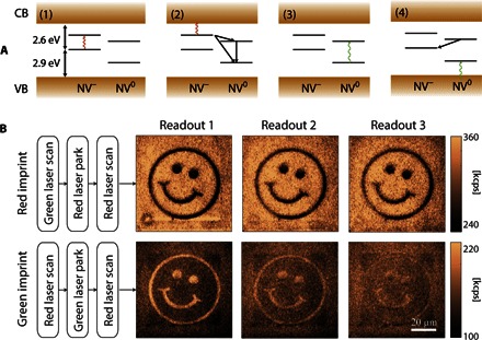
Fig. 1
Charge manipulation and readout in diamond.
( A ) Energy diagram for NV u2212 and NV 0 . In (1) and (2), the successive absorption of two photons (wavy arrows) of energy equal or greater than 1.95 eV (637 nm) propels the excess electron of an N...
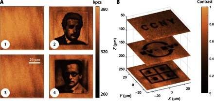
Fig. 2
Diamond as a 3D read/write memory.
( A ) Starting from a blank ensemble of NV u2212 centers (1), information can be written (2), erased (3), and rewritten (4). In (1) and (3), a green laser scan (1 mW at 1 ms per pixel) was used to res...
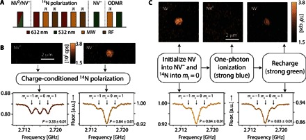
Fig. 3
Combined NV charge and spin manipulation.
( A ) Charge-conditional initialization of the 14 N nuclear spin host into m I = 0 is attained via spin transfer from the optically polarized NV u2212 electronic spin (see also figs. S4 and S5). Initi...
Figure images are served from the NIH/NLM PubMed Central Open Access Subset or Europe PMC; copyright remains with the publishers and authors.
💬 Discussion
0 commentsNo comments yet. Be the first to start a discussion!
Leave a Comment