Abstract
Point and regional centromeres specify a unique site on each chromosome for kinetochore assembly. The point centromere in budding yeast is a unique 150-bp DNA sequence, which supports a kinetochore with only one microtubule attachment. In contrast, regional centromeres are complex in architecture, can be up to 5 Mb in length, and typically support many kinetochore-microtubule attachments. We used quantitative fluorescence microscopy to count the number of core structural kinetochore protein complexes at the regional centromeres in fission yeast and Candida albicans. We find that the number of CENP-A nucleosomes at these centromeres reflects the number of kinetochore-microtubule attachments instead of their length. The numbers of kinetochore protein complexes per microtubule attachment are nearly identical to the numbers in a budding yeast kinetochore. These findings reveal that kinetochores with multiple microtubule attachments are mainly built by repeating a conserved structural subunit that is equivalent to a single microtubule attachment site.
🔬 Techniques
🧬 Organisms
💻 Software
✨ Fluorophores
🧪 Sample Preparation
🔬 Cell Lines
🏭 Microscope Brands
🧪 Reagent Suppliers
📷 Detectors
🔎 Objectives
💻 Software Details
💾 Data Repositories
🏛️ Research Organizations (ROR)
Affiliated research institutions:
📋 Methods
Strains and media
All the strains (listed in Table I ) were grown in YPD at 32°C (with the exception of the budding yeast strain expressing ScCse4p-GFP, which was grown at 25°C). YE5S, YPD, and YPD supplemented with 50 μg/ml of uridine (Sigma-Aldrich) were used to grow fission yeast, budding yeast, and C. albicans , respectively. GFP fusions were made by PCR amplification of a GFP–KAN r cassette (from pFA6a–GFP(S65T) KAN r MX6) flanked with 60 bp of homology to the site of integration at the 3′ end of the gene. The fission yeast strains with constitutive Cnp1p and Cnp1p-GFP overexpression were supplied by K. Takahashi and M. Yanagida (National BioResource Project, Okinawa Institute of Science and Technology, Okinawa, Japan). Table I. List of strains used in this study Genotype Source KBY7006: S. cerevisiae 473a CSE4-GFP:KAN KB YWY277: S. pombe h − Cnp1-GFP-KanMX6, ade6-m12/0, leu1-32, ura4D XH XHE255: S. pombe native promoter Cnp1-6GFP(lys1+) h − XH XL403: S. pombe NDC80:GFP XH XL101: S. Pombe MAL2:GFP XH XL174: S. pombe h − , mif2-GFP-KanMx6, ade6-m210, leu1-32, ura4D XH XL103: S. pombe h − , spc7-GFP-KanMx6, ade6-m210, leu1-32, ura4D XH XL358: S. pombe h + , mis12-GFP-KanMx6, ade6-m210, leu1-32, ura4D XH XL017: S. pombe h − , ask1-GFP-KanMx6, ade6-m210, leu1-32, ura4D XH XL101: S. pombe h − , mal2-GFP-KanMx6, ade6-m210, leu1-32, ura4D XH XL099: S. pombe h − , sim4-GFP-KanMx6, ade6-m210, leu1-32, ura4D XH XL059: S. pombe h − , fta1-GFP-KanMx6, ade6-m210, leu1-32, ura4D XH XL069: S. pombe h − , fta2-GFP-KanMx6, ade6-m210, leu1-32, ura4D XH XL067: S. pombe h − , fta3-GFP-KanMx6, ade6-m210, leu1-32, ura4D XH XL097: S. pombe h − , Dam1-GFP-KanMX6, ade6-m210, leu1-32, ura4D XH XL127: S. pombe h + , Dad1-GFP-KanMX6, ade6-m210, leu1-32, ura4D, DAD1 XH XL071: S. pombe h − , Dad2-GFP-KanMX6, ade6-m210, leu1-32, ura4D XH XL332: S. pombe h − , Nuf2-GFP-KanMX6, ade6-m210, leu1-32, ura4D XH XL462: S. pombe ask1-GFP-KANMX6, klp5∷ura4, leu1-32, ura4-D18, ade6- XH XL464: S. pombe ask1-GFP-KANMX6, klp6∷ura4, leu1-32, ura4-D18 XH XL468: S. pombe ndc80-GFP-KANMX6, nda3cs, ade6-m210, leu1-32, ura4- XH XL487: S. pombe h − , fta3-GFP-KANMX6, ade6-m210, leu1-32, ura4D, native promoter-cnp1+-3xHA6xHis[lys1+] XH XL489: S. pombe h − , mal2-GFP-KANMX6, ade6-m210, leu1-32, ura4D, native promoter-cnp1+-3xHA6xHis[lys1+] XH XL491: S. pombe h − , mis12-GFP-KANMX6, ade6-m210, leu1-32, ura4D, native promoter-cnp1+-3xHA6xHis[lys1+] XH XL493: S. pombe h − , ndc80-GFP-KANMX6, ade6-m210, leu1-32, ura4D, native promoter-cnp1+-3xHA6xHis[lys1+] XH XL 497: S. pombe mif2-GFP-KANMX6, ade6-m210, leu1-32, ura4D, native promoter cnp1+-3xHA-6xHis [lys1+] XH S. pombe XL 506: mis6GFP[leu+], native promoter-cnp1+-3xHA-6xHis [lys1+] XH CAJS1-1: C. albicans arg4∷hisG/ arg4∷hisG his1∷hisG/ his1∷hisG ura3Δ∷λimm434/ ura3Δ∷λimm434 CSE4:GFP:CSE4/cse4Δ∷hisG∷URA3∷hisG JB 10118: C. albicans ura3Δ∷λimm434/ura3Δimm434 his1∷hisG/his1∷hisG arg4∷hisG/arg4∷hisG cse4∷dpl200-URA3/CSE4:GFP:CSE4 JB 8675: C. albicans ura3Δ∷λimm434/ura3Δimm434 his1∷hisG/his1∷hisG arg4∷hisG/arg4∷hisG CSE4/CSE4:GFP:CSE4 JB 8676: C. albicans ura3Δ∷λimm434/ura3Δimm434 his1∷hisG/his1∷hisG arg4∷hisG/arg4∷hisG CSE4/CSE4:GFP:CSE4 JB 10116: C. albicans ura3Δ∷_imm434/ura3Δimm434 his1∷hisG/his1∷hisG arg4∷hisG/arg4∷hisG CSE4:GFP:CSE4/CSE4:GFP:CSE4 JB 10418: C. albicans ura3∷imm434/ura3∷imm434, Nuf2-GFP-URA3/Nuf2-GFP-NAT JB 10702 ura3Δ∷λimm434/ura3Δ∷λimm434 his1∷hisG/his1∷hisG arg4∷hisG/arg4∷hisG MTW1-GFP-URA3/MTW1-GFP-NAT1 JB 10635 ura3∷imm434/ura3∷imm434, Nuf2-GFP:NAT1/Nuf2-GFP/NAT1, CSE4/cse4∷URA3 JB YMG5629: C. albicans ura3Δ∷λimm434/ura3Δimm434 his1∷hisG/his1∷hisG arg4∷hisG/arg4∷hisG TUB1/TUB1:GFP-URA3 JB KB, K. Bloom laboratory; XH, X. He laboratory; JB, J. Berman Laboratory.
Show full methods section
Strains and media
All the strains (listed in Table I ) were grown in YPD at 32°C (with the exception of the budding yeast strain expressing ScCse4p-GFP, which was grown at 25°C). YE5S, YPD, and YPD supplemented with 50 μg/ml of uridine (Sigma-Aldrich) were used to grow fission yeast, budding yeast, and C. albicans , respectively. GFP fusions were made by PCR amplification of a GFP–KAN r cassette (from pFA6a–GFP(S65T) KAN r MX6) flanked with 60 bp of homology to the site of integration at the 3′ end of the gene. The fission yeast strains with constitutive Cnp1p and Cnp1p-GFP overexpression were supplied by K. Takahashi and M. Yanagida (National BioResource Project, Okinawa Institute of Science and Technology, Okinawa, Japan). Table I. List of strains used in this study Genotype Source KBY7006: S. cerevisiae 473a CSE4-GFP:KAN KB YWY277: S. pombe h − Cnp1-GFP-KanMX6, ade6-m12/0, leu1-32, ura4D XH XHE255: S. pombe native promoter Cnp1-6GFP(lys1+) h − XH XL403: S. pombe NDC80:GFP XH XL101: S. Pombe MAL2:GFP XH XL174: S. pombe h − , mif2-GFP-KanMx6, ade6-m210, leu1-32, ura4D XH XL103: S. pombe h − , spc7-GFP-KanMx6, ade6-m210, leu1-32, ura4D XH XL358: S. pombe h + , mis12-GFP-KanMx6, ade6-m210, leu1-32, ura4D XH XL017: S. pombe h − , ask1-GFP-KanMx6, ade6-m210, leu1-32, ura4D XH XL101: S. pombe h − , mal2-GFP-KanMx6, ade6-m210, leu1-32, ura4D XH XL099: S. pombe h − , sim4-GFP-KanMx6, ade6-m210, leu1-32, ura4D XH XL059: S. pombe h − , fta1-GFP-KanMx6, ade6-m210, leu1-32, ura4D XH XL069: S. pombe h − , fta2-GFP-KanMx6, ade6-m210, leu1-32, ura4D XH XL067: S. pombe h − , fta3-GFP-KanMx6, ade6-m210, leu1-32, ura4D XH XL097: S. pombe h − , Dam1-GFP-KanMX6, ade6-m210, leu1-32, ura4D XH XL127: S. pombe h + , Dad1-GFP-KanMX6, ade6-m210, leu1-32, ura4D, DAD1 XH XL071: S. pombe h − , Dad2-GFP-KanMX6, ade6-m210, leu1-32, ura4D XH XL332: S. pombe h − , Nuf2-GFP-KanMX6, ade6-m210, leu1-32, ura4D XH XL462: S. pombe ask1-GFP-KANMX6, klp5∷ura4, leu1-32, ura4-D18, ade6- XH XL464: S. pombe ask1-GFP-KANMX6, klp6∷ura4, leu1-32, ura4-D18 XH XL468: S. pombe ndc80-GFP-KANMX6, nda3cs, ade6-m210, leu1-32, ura4- XH XL487: S. pombe h − , fta3-GFP-KANMX6, ade6-m210, leu1-32, ura4D, native promoter-cnp1+-3xHA6xHis[lys1+] XH XL489: S. pombe h − , mal2-GFP-KANMX6, ade6-m210, leu1-32, ura4D, native promoter-cnp1+-3xHA6xHis[lys1+] XH XL491: S. pombe h − , mis12-GFP-KANMX6, ade6-m210, leu1-32, ura4D, native promoter-cnp1+-3xHA6xHis[lys1+] XH XL493: S. pombe h − , ndc80-GFP-KANMX6, ade6-m210, leu1-32, ura4D, native promoter-cnp1+-3xHA6xHis[lys1+] XH XL 497: S. pombe mif2-GFP-KANMX6, ade6-m210, leu1-32, ura4D, native promoter cnp1+-3xHA-6xHis [lys1+] XH S. pombe XL 506: mis6GFP[leu+], native promoter-cnp1+-3xHA-6xHis [lys1+] XH CAJS1-1: C. albicans arg4∷hisG/ arg4∷hisG his1∷hisG/ his1∷hisG ura3Δ∷λimm434/ ura3Δ∷λimm434 CSE4:GFP:CSE4/cse4Δ∷hisG∷URA3∷hisG JB 10118: C. albicans ura3Δ∷λimm434/ura3Δimm434 his1∷hisG/his1∷hisG arg4∷hisG/arg4∷hisG cse4∷dpl200-URA3/CSE4:GFP:CSE4 JB 8675: C. albicans ura3Δ∷λimm434/ura3Δimm434 his1∷hisG/his1∷hisG arg4∷hisG/arg4∷hisG CSE4/CSE4:GFP:CSE4 JB 8676: C. albicans ura3Δ∷λimm434/ura3Δimm434 his1∷hisG/his1∷hisG arg4∷hisG/arg4∷hisG CSE4/CSE4:GFP:CSE4 JB 10116: C. albicans ura3Δ∷_imm434/ura3Δimm434 his1∷hisG/his1∷hisG arg4∷hisG/arg4∷hisG CSE4:GFP:CSE4/CSE4:GFP:CSE4 JB 10418: C. albicans ura3∷imm434/ura3∷imm434, Nuf2-GFP-URA3/Nuf2-GFP-NAT JB 10702 ura3Δ∷λimm434/ura3Δ∷λimm434 his1∷hisG/his1∷hisG arg4∷hisG/arg4∷hisG MTW1-GFP-URA3/MTW1-GFP-NAT1 JB 10635 ura3∷imm434/ura3∷imm434, Nuf2-GFP:NAT1/Nuf2-GFP/NAT1, CSE4/cse4∷URA3 JB YMG5629: C. albicans ura3Δ∷λimm434/ura3Δimm434 his1∷hisG/his1∷hisG arg4∷hisG/arg4∷hisG TUB1/TUB1:GFP-URA3 JB KB, K. Bloom laboratory; XH, X. He laboratory; JB, J. Berman Laboratory.
Microscopy
An inverted microscope (TE-2000U; Nikon) with a 100× 1.4 NA differential interference contrast objective (Nikon) was used for imaging cells at 25°C. Cells suspended in filter sterile SD complete media were immobilized on coverslips coated with concanavaline A (Sigma-Aldrich) for imaging. A standard HQ EGFP long pass filter set (Chroma Technology Corp.) was used for fluorescence imaging. Images were acquired with a cooled charge-coupled device camera (Orca II ER; Hamamatsu Photonics) with 2 × 2 binning (1 pixel, ∼133 nm). The microscope shutters and the camera were operated by Metamorph 6.1 (MDS Analytical Technologies). A stack of 13 images was obtained for each chosen field (300 × 300 camera pixels in the center of the field to minimize excitation intensity irregularities), with an exposure time of 400 ms and a 200-nm axial separation between successive images in the stack.
Data analysis
Image analysis was performed with a custom-written graphical user interface in MATLAB (MathWorks) in the in-focus image plane for each kinetochore cluster (this plane also contains the pixel with maximum intensity value). Measurements of fluorescence signal from kinetochore clusters in fission yeast cells in G2/M were performed by placing a 6 × 6 pixel box on the signal region. It was placed so that the central 2 × 2 pixel region in the box had the maximum cumulative signal. For fission yeast and C. albicans cells in anaphase, a 5 × 5 pixel box was used to measure the fluorescence signal. The number of pixels for signal measurement for GFP-tagged kinetochore proteins was determined by fitting 1-D Gaussian curves to line scans through kinetochore clusters in the in-focus plane (the maximum intensity plane), with the standard deviation of the Gaussian as a free parameter. Fluorescence signal was measured over an area corresponding to the 4× (SD) around the maxima. For fission yeast cells expressing Ndc80p-GFP, this fitting procedure yielded a mean spot size of 600 ± 60 nm for anaphase cells and 700 ± 44 nm for G2 cells corresponding to an area defined by a 5 × 5 and 6 × 6 pixel square, respectively, in the acquired images. Measurements from C. albicans cells expressing Cse4p-GFP in anaphase prompted the use of a 5 × 5 pixel square for signal measurement. Background correction was applied by measuring the background from a larger square region (8 × 8 pixels for fission yeast and 7 × 7 pixels for budding yeast and C. albicans ) that is concentric with the signal region, following the scheme detailed in Hoffman et al. (2001) . Online supplemental material Fig. S1 shows quantification of the number of microtubules in the budding yeast and C. albicans spindles. Fig. S2 shows that a four- to fivefold excess of CENP-A nucleosomes does not alter the number of kinetochore proteins at the centromere in fission yeast.
Table
S1 contains a summary of fluorescence signal measurements for fission yeast. Online supplemental material is available at http://www.jcb.org/cgi/content/full/jcb.200803027/DC1 .
Online supplemental material Fig. S1 shows quantification of the number of microtubules in the budding yeast and C. albicans spindles. Fig. S2 shows that a four- to fivefold excess of CENP-A nucleosomes does not alter the number of kinetochore proteins at the centromere in fission yeast.
Table
S1 contains a summary of fluorescence signal measurements for fission yeast. Online supplemental material is available at http://www.jcb.org/cgi/content/full/jcb.200803027/DC1 .
Supplementary Material [Supplemental Material Index]
📊 Figures
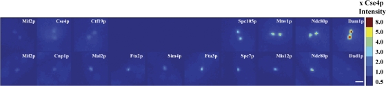
Figure 1.
Pseudo-colored images for a comparison of kinetochore protein fluorescence in metaphase budding yeast and G2/M fission yeast cells. Images were obtained under identical imaging and image acquisition c...
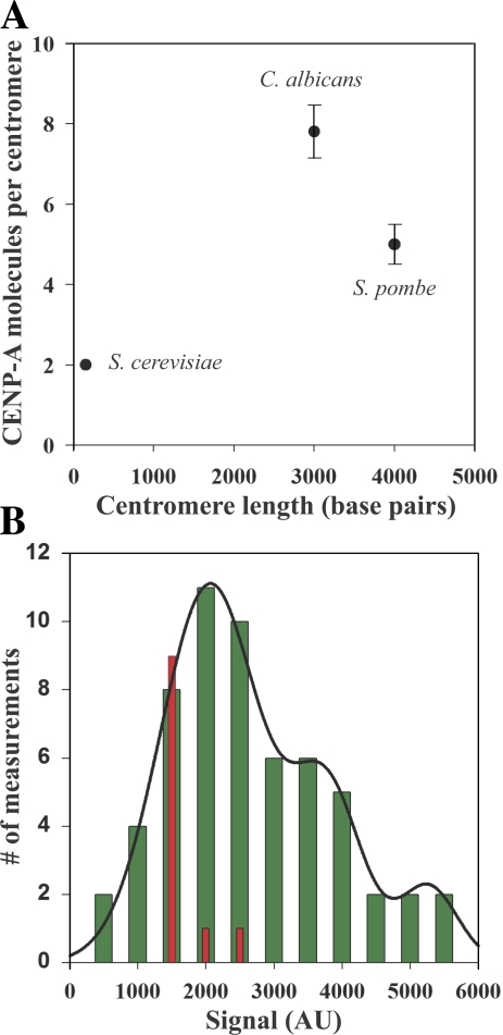
Figure 2.
The number of CENP-A molecules does not scale with the centromere DNA length. (A) The number of CENP-A molecules in the three fungi plotted as a function of centromere length. The number of CENP-A nuc...
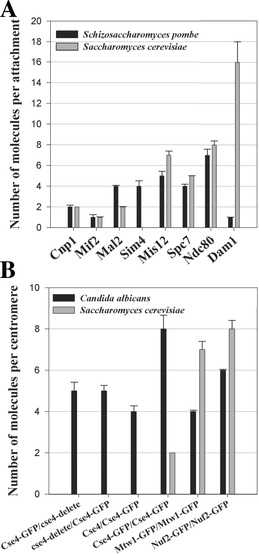
Figure 3.
Comparison of kinetochore protein numbers in the three fungi. (A) The number of core structural kinetochore proteins per microtubule attachment in fission yeast is very similar to the number of corres...
Figure images are served from the NIH/NLM PubMed Central Open Access Subset or Europe PMC; copyright remains with the publishers and authors.
💬 Discussion
0 commentsNo comments yet. Be the first to start a discussion!
Leave a Comment