Abstract
Vaccines based on mRNA have emerged as potent systems to elicit CD8+ T cell responses against various cancers and viral infectious diseases. The efficient intracellular delivery of mRNA molecules encoding antigens into the cytosol of antigen-presenting cells (APCs) is still challenging, requiring cell attachment, active uptake, and subsequent endosomal escape. Here, we report a facile approach for the formulation of peptide-functionalized mRNA polyplexes using copper-free click chemistry to promote presentation of mRNA antigen by dendritic cells (DCs). After screening different membrane active peptides, GALA modified mRNA polyplexes (PPx-GALA) with a size around 350 nm and with a slightly negative surface charge (-7 mV), exhibited the highest EGFP-mRNA transfection in RAW 246.7 macrophages (∼36%) and D1 dendritic cells (∼50%) as compared to polyplexes decorated with melittin or LEDE peptides. Interestingly, we found that PPx-GALA enters DCs through sialic acid mediated endo/phagocytosis, which was not influenced by DC maturation. The PPx-GALA formulation exhibited 18-fold higher cellular uptake compared to a lipofectamine mRNA formulation without inducing cytotoxicity. Live cell imaging showed that PPx-GALA that were taken up by endocytosis induced calcein release from endosomes into the cytosol. DCs treated with PPx-GALA containing mRNA encoding for OVA displayed enhanced T cell responses and DC maturation. Collectively, these data provide a strong rationale for further study of this PPx-GALA formulation in vivo as a promising mRNA vaccine platform.
🔬 Techniques
✨ Fluorophores
🧪 Sample Preparation
🔬 Cell Lines
🏭 Microscope Brands
🧪 Reagent Suppliers
💻 Software Details
🏛️ Research Organizations (ROR)
Affiliated research institutions:
📋 Methods
Materials All chemicals were purchased in the highest purity and used without further purification. Carbonic acid 2-dimethylamino-ethyl ester 1-methyl-2-(2-methacryloylamino)-ethyl ester (HPMA-DMAE), 80 N -[2-(2-pyridyldithio)]ethyl methacrylamide(PDTEMA), 81 , 82 and 2-azidoethylmethyacrylamide (AzEMAm) 24 were synthesized as previously reported. Lipofectamine 2000 was obtained from Thermo Fisher Scientific (Etten-Leur, The Netherlands). The EGFP-mRNA Cy5-EGFP-mRNA and Cy5-luc_mRNA (5-methoxyuridine) were purchased from Tebu-bio (TRiLink biotechnologies, San Diego, CA). LEDE-azide 29 − 31 was a gift from Dr. Jan Wouter Drijfhout (Faculty of Medicine, Leiden University). GALA-azide and Melittin-azide was provided by ChinaPeptides Co., Ltd. (Shanghai, China). Maackia amurensis agglutinin, Opti-MEM, DMEM medium and heat inactivated fetal bovine serum (HI-FBS) were purchased from Sigma-Aldrich (Darmstadt, Germany). Polymer Synthesis and Characterization p(HPMA-DMAE- co -PDTEMA- co -AzEMAm) (pHDPA) was synthesized as reported. 24 The polymer was synthesized by radical polymerization using a monomer to initiator molar ratio (M/I) of 50 under a nitrogen atmosphere. The feed molar ratio of HPMA-DMAE, PDTEMA, and AzEMAm was 70/20/10. In brief, 200 mg (0.77 mmol) HPMA-DMAE, 56.7 mg (0.22 mmol) PDTEMA, 17 mg (0.11 mmol) AzEMAm, and 3.6 mg (0.022 mmol) AIBN were dissolved in dry DMSO (1 mL) in flasks sealed with rubber septa and subjected to three vacuum-N 2 cycles. The polymerization was carried at 70 °C for 48 h. Next, the polymer was precipitated in cold diethyl ether, redissolved in DMF and precipitated in cold diethyl ether. This procedure was repeated 3 times. After extensive dialysis (5 kDa) against an ammonium acetate (NH 4 OAc) buffer of pH 5.0 (10 mM, last step 2.5 mM) at 4 °C, the polymer was collected after freeze-drying. The yield of the polymer was 40%. The molecular weights and polydisperisity ( M w / M n ) of pHDPA were determined by size exclusion chromatography (SEC) analysis using a Viscotek-GPCmax (Viscotek, Oss, The Netherlands) light scattering (λ = 670 nm, right (90°) and low (7°) angle)/viscosimetric detection system, using Ultrahydrogel 2000 7.8 × 300 mm columns in series with a Ultrahydrogel 6.0 × 40 mm guard column and 0.3 M NaAc pH 4.4, 30% acetonitrile as eluent. 83 The flow rate was 0.6 mL/min and the run time was 60 min. A PolyCALTM PEO standard ( M n = 24 kDa, PDI = 1.01, Malvern) was used for calibration. The copolymer composition was determined by 1 H NMR analysis (polymer dissolved in D 2 O) and performed with a Gemini 400 MHz spectrometer (Varian Associates Inc., NMR Instruments, Palo Alto, CA). The ratio HPMA-DMAE/PDTEMA/AzEMAm was determined by comparison of the integrals at δ 4.3 ppm (bs, O CH 2 CH 2 , HPMA-DMAE), δ7.69 ppm (bs, pyridyl group proton, PDTEMA) and δ3.14–3.51 ppm (m, CH 2 CH 2 N 3 , AzEMAm). Peptide Conjugate Synthesis The BCN-PEG 6000 -peptides were synthesized as described in Figure S1B . Briefly, for the synthesis of BCN-PEG 6000 -GALA, 20.9 mg of BCN-PEG 6000 -BCN (6510 g/mol, 3.2 μmol) was dissolved in 1 mL dry DMSO, followed by the addition 10.0 mg of GALA-azide (3115 g/mol, 3.2 μmol) and the obtained solution was subsequently stirred for 16 h at room temperature. Next, 10 mL water was added, followed by freeze-drying. Subsequently, the product was redissolved in 2.5 mL nuclease-free water and purified with PD 10 column chromatography to remove unreacted peptide using nuclease-free water as eluent and freeze-dried. The product was obtained at a yield of 80%. It should be noted that the applied procedure resulted in the formation of a statistical mixture of BCN-PEG-GALA, GALA-PEG-GALA and unreacted BCN-PEG-BCN. The BCN-PEG 6000 -Melittin and BCN-PEG 6000 -LEDE was synthesized in a similar way.
Show full methods section
Materials All chemicals were purchased in the highest purity and used without further purification. Carbonic acid 2-dimethylamino-ethyl ester 1-methyl-2-(2-methacryloylamino)-ethyl ester (HPMA-DMAE), 80 N -[2-(2-pyridyldithio)]ethyl methacrylamide(PDTEMA), 81 , 82 and 2-azidoethylmethyacrylamide (AzEMAm) 24 were synthesized as previously reported. Lipofectamine 2000 was obtained from Thermo Fisher Scientific (Etten-Leur, The Netherlands). The EGFP-mRNA Cy5-EGFP-mRNA and Cy5-luc_mRNA (5-methoxyuridine) were purchased from Tebu-bio (TRiLink biotechnologies, San Diego, CA). LEDE-azide 29 − 31 was a gift from Dr. Jan Wouter Drijfhout (Faculty of Medicine, Leiden University). GALA-azide and Melittin-azide was provided by ChinaPeptides Co., Ltd. (Shanghai, China). Maackia amurensis agglutinin, Opti-MEM, DMEM medium and heat inactivated fetal bovine serum (HI-FBS) were purchased from Sigma-Aldrich (Darmstadt, Germany). Polymer Synthesis and Characterization p(HPMA-DMAE- co -PDTEMA- co -AzEMAm) (pHDPA) was synthesized as reported. 24 The polymer was synthesized by radical polymerization using a monomer to initiator molar ratio (M/I) of 50 under a nitrogen atmosphere. The feed molar ratio of HPMA-DMAE, PDTEMA, and AzEMAm was 70/20/10. In brief, 200 mg (0.77 mmol) HPMA-DMAE, 56.7 mg (0.22 mmol) PDTEMA, 17 mg (0.11 mmol) AzEMAm, and 3.6 mg (0.022 mmol) AIBN were dissolved in dry DMSO (1 mL) in flasks sealed with rubber septa and subjected to three vacuum-N 2 cycles. The polymerization was carried at 70 °C for 48 h. Next, the polymer was precipitated in cold diethyl ether, redissolved in DMF and precipitated in cold diethyl ether. This procedure was repeated 3 times. After extensive dialysis (5 kDa) against an ammonium acetate (NH 4 OAc) buffer of pH 5.0 (10 mM, last step 2.5 mM) at 4 °C, the polymer was collected after freeze-drying. The yield of the polymer was 40%. The molecular weights and polydisperisity ( M w / M n ) of pHDPA were determined by size exclusion chromatography (SEC) analysis using a Viscotek-GPCmax (Viscotek, Oss, The Netherlands) light scattering (λ = 670 nm, right (90°) and low (7°) angle)/viscosimetric detection system, using Ultrahydrogel 2000 7.8 × 300 mm columns in series with a Ultrahydrogel 6.0 × 40 mm guard column and 0.3 M NaAc pH 4.4, 30% acetonitrile as eluent. 83 The flow rate was 0.6 mL/min and the run time was 60 min. A PolyCALTM PEO standard ( M n = 24 kDa, PDI = 1.01, Malvern) was used for calibration. The copolymer composition was determined by 1 H NMR analysis (polymer dissolved in D 2 O) and performed with a Gemini 400 MHz spectrometer (Varian Associates Inc., NMR Instruments, Palo Alto, CA). The ratio HPMA-DMAE/PDTEMA/AzEMAm was determined by comparison of the integrals at δ 4.3 ppm (bs, O CH 2 CH 2 , HPMA-DMAE), δ7.69 ppm (bs, pyridyl group proton, PDTEMA) and δ3.14–3.51 ppm (m, CH 2 CH 2 N 3 , AzEMAm). Peptide Conjugate Synthesis The BCN-PEG 6000 -peptides were synthesized as described in Figure S1B . Briefly, for the synthesis of BCN-PEG 6000 -GALA, 20.9 mg of BCN-PEG 6000 -BCN (6510 g/mol, 3.2 μmol) was dissolved in 1 mL dry DMSO, followed by the addition 10.0 mg of GALA-azide (3115 g/mol, 3.2 μmol) and the obtained solution was subsequently stirred for 16 h at room temperature. Next, 10 mL water was added, followed by freeze-drying. Subsequently, the product was redissolved in 2.5 mL nuclease-free water and purified with PD 10 column chromatography to remove unreacted peptide using nuclease-free water as eluent and freeze-dried. The product was obtained at a yield of 80%. It should be noted that the applied procedure resulted in the formation of a statistical mixture of BCN-PEG-GALA, GALA-PEG-GALA and unreacted BCN-PEG-BCN. The BCN-PEG 6000 -Melittin and BCN-PEG 6000 -LEDE was synthesized in a similar way.
Preparation and Characterization of Peptide Functionalized mRNA Polyplexes
The preparation of RNA polyplexes consisted of 3 consecutive steps: complexation, post-PEG-peptide modification, and cross-linking ( Figure 1 A). Briefly, complexation was achieved by mixing four volumes of polymer and one volume of nucleic acid in 10 mM HEPES buffer, pH 7.4, at an N/P ratio of 4. For control polyplexes modified with PEG, BCN-PEG 5000 -COOH 24 was added to preformed mRNA polyplexes a BCN/N 3 mole of 0.6. The peptide modification process was performed by mixing BCN-PEG 6000 -peptide (synthesis described in Results and Discussion ) at either a low (30%) or high (60%) molar ratio to BNC/N 3 in HEPES buffer pH 7.4 to react for 2 h at room temperature. For the low amount of peptide modification, another 30% equivalent of BCN-PEG 5000 -COOH was first mixed together BCN-PEG 6000 -peptide before addition to the mRNA polyplex dispersion. Next, surface modified mRNA polyplexes were cross-linked by addition of dithiothreitol (DTT) corresponding with a half molar equivalent to PDS groups of the polymer used in particle formation and subsequent incubation for 1 h at room temperature. After adding 5% sucrose as cryoprotectant, the polyplexes were freeze-dried and stored at 4 °C. Unless indicated otherwise, the polyplexes were prepared with a final RNA concentration of 100 μg/mL. The mRNA polyplex dry powder was resuspended in RNAase-free water 30 min before addition to the cells. The size of the polyplexes was measured with DLS using an ALV CGS-3 system (Malvern Instruments, Malvern, UK) equipped with a JDS Uniphase 22 mW He–Ne laser operating at 632.8 nm, an optical fiber-based detector, a digital LV/LSE-5003 correlator with temperature controller set at 25 °C. The zeta-potential (ζ) of the polyplexes was measured using a Malvern Zetasizer Nano-Z (Malvern, UK) with universal ZEN 1002 “dip” cells and DTS (Nano) software (version 4.20) at 25 °C. Polyplex measurements were performed in 10 mM HEPES pH 7.4 and with an RNA concentration of 15 μg/mL. The size distribution of the polyplexes was also determined by nanoparticle tracking analysis (NTA) using a NanoSight LM 10SH (NanoSight, Amesbury, United Kingdom), equipped with a sample chamber with a 532 nm Laser. Typically, RNA polyplexes were diluted with PBS to a concentration of 0.5 μg/mL and measured for 120 s with manual shutter and gain adjustments. The captured videos were analyzed by the NTA 2.0 image analysis software.
Transfection of DCs Immortalized
DC2.4 cells were used to test protein expression activity of the peptide modified mRNA polyplexes. Briefly, DC2.4 cells were seeded into a 96-well plate at a seeding density of 3.0 × 10 4 cells/well, and cell culture was done in 100 μL complete culture medium for 24 h at 37 °C. Prior to transfections, the medium on the cells was refreshed with either 100 μL OPTI-MEM or full medium and incubated with EGFP mRNA polyplexes prepared as described in Results and Discussion for 4 h at 37 °C, and then another 100 μL full medium was added without removing polyplexes. After incubation for 20 h at 37 °C in a CO 2 incubator, EGFP expression was visualized using Keyence BZ-9000 Microscope (Keyence, Osaka, Japan). Flow cytometry was performed to measure the percentage of GFP-positive cells compared to nontransfected cells as control using a BD FACS Canto II flow cytometer (Becton Dickinson, BD, Franklin Lakes, NJ, USA). The same procedure was also applied to determine EGFP expression in D1 dendritic cells, RAW 246.7 macrophages, and HEK293T human embryonic kidney cells. Lipofectamine 2000 (Lipo) was used as positive control (Lipo/mRNA were prepared at volume/weight ratio of 1.5/1). Unless specified cells were incubated with mRNA at dose of 250 ng per well. In Vitro Cytotoxicity To determine possible cytotoxic effects of the polyplexes, the Alamar Blue cell viability assay (Invitrogen, Karlsruhe, Germany) was performed essentially as described by the manufacturer. In short, 20 h after transfection, the cell medium was replaced with culture medium containing Alamar Blue (50 nM) and the cells were cultured for another 4 h. Next, 80 μL of medium from each well was transferred into a flat-bottom 96-well plate to measure the light absorbance. The relative cell metabolic activity was calculated by normalizing the absorbance at 570 nm (reference wavelength of 630 nm) with the absorbance of PBS-treated cells. The capability of PPx-GALA to interfere with cell membrane integrity was analyzed with the CytoTox-ONE kit, which determines the lactate dehydrogenase (LDH) release from cells after exposure to the different polyplex formulations. The assay was performed using a 96-well plate, in which 10,000 D1 cells were plated 24 h before the treatment. Before sample addition, the culture medium was replaced with OPTI-MEM. Next, polyplex dispersions containing 62.5, 125, 250, and 500 ng of mRNA/well were added. After 4 h incubation at 37 °C, the supernatant was collected, LDH activity was determined according to the manufacturer’s protocol. For the positive control, cells were 100% lysed with 1% Triton X-100. In Vitro DCs Cellular Uptake Studies of GALA-Modified Polyplexes (PPx-GALA) To determine the route of cellular internalization of the PPx-GALA, D1 cells were seeded at a density of 3.0 × 10 4 cells/well in a 96-well plate or in 96-well μClear black plates and incubated for 24 h at 37 °C. The cells were then incubated with Cy5-labeled luc_mRNA loaded in PPx-GALA as described in Results and Discussion at dose of 250 ng of mRNA/well for 1, 6, 12, and 24 h. For inhibition studies, cells were pretreated with 1 μg/mL lipopolysaccharide (LPS, Invitrogen) for 15 h, 10 μM Maackia amurensis agglutinin for 30 min (MAM, sialic acid binding lectin, Sigma), 10 μM cytochalasin D (Cyto-D, phagocytosis inhibitor, Sigma) for 3 h, 15 μM chlorpromazine (CPZ, clathrin-mediated endocytosis inhibitor, Sigma) for 1 h or different concentrations of free GALA peptide (1–150 μM) for 15 min in full medium prior to addition of PPx-GALA to the cells. The cells were then incubated for 1 h (inhibitor present during transfection) at 37 °C before they were washed with ice-cold FACS buffer (1% BSA in PBS) and applied to determine particle uptake using a BD FACSCanto II flow cytometer and high content confocal fluorescent microscope Yokogawa Cell Voyager CV7000s (Yokogawa Electric Corporation, Tokyo, Japan). For binding assay of PPx-GALA, cells were first incubated on ice for 1 h and then incubated together with PPx-GALA for another 1 h on ice before taking confocal images. For colocalization studies, cells were cotransfected with PPx-GALA and 200 μg/mL rhodamine-labeled dextran (70,000 Da, ThermoFisher) or 150 μg/mL cell impermeable dye Calcein (Sigma, The Netherlands) for 3 h at 37 °C. To study the subcellular distribution, after 3 h coincubation with PPx-GALA, the cells were incubated with 100 nM lysotracker green (Invitrogen, The Netherlands) for 30 min before confocal imaging. In Vitro DCs Maturation and Antigen Presentation D1 cells were seeded in 96-well plates at a density of 25,000 cells/well in 100 μL complete medium and allowed to adhere overnight. Polyplexes or lipoplexes were added at 0.25 μg mRNA/well in 100 μL serum-free media. Control cells were treated with 0.1 mM SIINFEKL peptide (in vitrogen, The Netherlands). After incubation for 6, 12, and 24 h at 37 °C, the cells were washed with medium, new fresh complete medium was added and incubated for overnight at 37 °C. Next, 100,000 B3Z cells/well in 100 μL complete medium were added and incubated for 24 h at 37 °C. The medium was then replaced with 100 μL/well lysis buffer containing 0.1 mM 2-mercaptoethanol, 9 mM MgCl 2 , 0.1% Triton X-100, and 0.15 mM chlorophenol red β- d -galactopyranoside (CPRG) in DPBS. After 2 h incubation at 37 °C, absorbance measurements were recorded at 570 nm. To measure the maturation level of the D1 cells, after incubation of the cells with the different formulations for 24 h at 37 °C, the cells were washed with FACS buffer and subsequently stained with anti-CD40-FITC and anti-CD86-PE antibodies (2 μg/mL, 50 μL/well, eBioscience, U.S.) for 30 min on ice. The D1 cells were subsequently analyzed by flow cytometry after 2 washing steps with FACS buffer.
Statistical Analysis Two-tailed
Student’s t test was applied for comparison between experimental groups. P < 0.05 was considered statistically significant.
Materials All chemicals were purchased in the highest purity and used without further purification. Carbonic acid 2-dimethylamino-ethyl ester 1-methyl-2-(2-methacryloylamino)-ethyl ester (HPMA-DMAE), 80 N -[2-(2-pyridyldithio)]ethyl methacrylamide(PDTEMA), 81 , 82 and 2-azidoethylmethyacrylamide (AzEMAm) 24 were synthesized as previously reported. Lipofectamine 2000 was obtained from Thermo Fisher Scientific (Etten-Leur, The Netherlands). The EGFP-mRNA Cy5-EGFP-mRNA and Cy5-luc_mRNA (5-methoxyuridine) were purchased from Tebu-bio (TRiLink biotechnologies, San Diego, CA). LEDE-azide 29 − 31 was a gift from Dr. Jan Wouter Drijfhout (Faculty of Medicine, Leiden University). GALA-azide and Melittin-azide was provided by ChinaPeptides Co., Ltd. (Shanghai, China). Maackia amurensis agglutinin, Opti-MEM, DMEM medium and heat inactivated fetal bovine serum (HI-FBS) were purchased from Sigma-Aldrich (Darmstadt, Germany).
Supplementary Material bc8b00524_si_001.pdf
📊 Figures
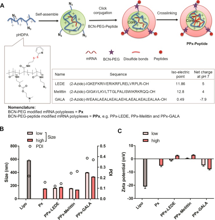
Figure 1
Preparation and characterization of peptide-modified mRNA polyplexesn(PPx). (A) Schematic illustration of the 3-step preparation methodnof PPx: (a) core self-assembly by mixing the cationic polymer an...
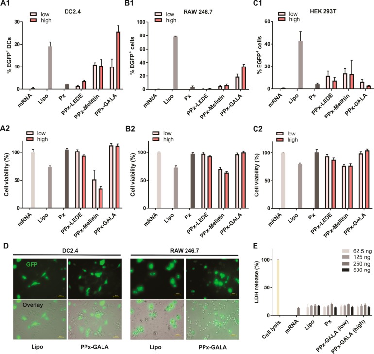
Figure 2
Evaluationnof PPx formulations for EGFP mRNA delivery. EGFP expressionn(upper panels) detected by flow cytometry and normalized cell viabilityn(lower panels) in comparison to untreated cells (100% cel...
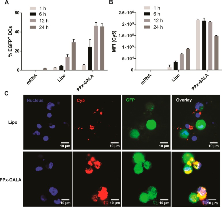
Figure 3
Transfectionn(A) and cellular uptake (B) kinetics of Cy5-EGFP mRNAnon D1 cells following incubation with the indicated formulations bynflow cytometry. (C) Confocal microscopy of D1 cells incubated wit...
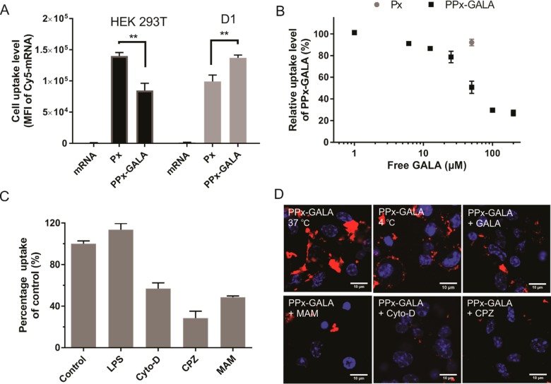
Figure 4
Cellular uptake mechanism of PPx-GALA. (A) Cellularnuptake of nakednCy5-luc_mRNA, Px and PPx-GALA by HEK293T (left) and D1 (right) cellsnafter 1 h incubation in the presence of serum. (B) Inhibition o...
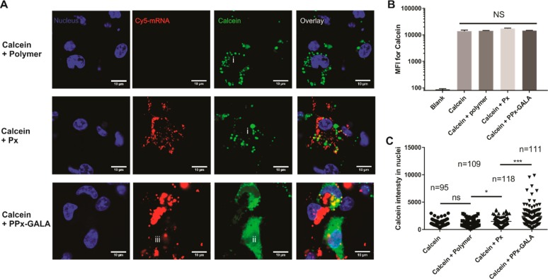
Figure 5
PPx-GALA polyplexes facilitate the delivery of the membrane-impermeablenfluorescent molecule calcein into the cytosol of dendritic cells.nConfocal microscopy (A) and flow cytometry analysis ( n = 4) (...
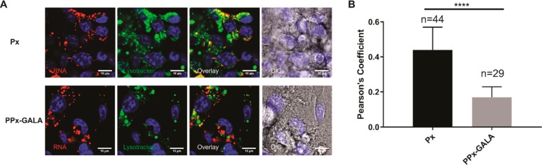
Figure 6
Lysotrackernstaining of PPx-GALA mRNA formulations in D1 cells.n(A) Confocal microscopy of D1 cells 3 h after incubation with Px ornPPx-GALA containing Cy5-luc_mRNA in the presence of serum at 37 u00b...
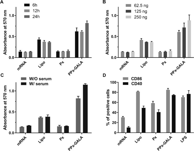
Figure 7
B3Z T cell activation by transfected D1 dendriticncells. D1 cellsnwere incubated with free mRNA and the indicated mRNA polyplexes encodingnovalbumin (OVA) for indicated lengths of time (A), or differe...
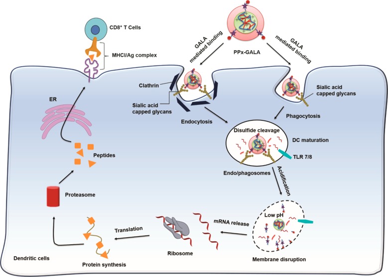
Figure 8
Proposednintracellular trafficking pathway of PPx-GALA mRNA polyplexes.nThe GALA peptide on the out-layer of polyplexes binds to the sialicnacid capped glycans on the surface of DCs, followed by inter...
Figure images are served from the NIH/NLM PubMed Central Open Access Subset or Europe PMC; copyright remains with the publishers and authors.
💬 Discussion
0 commentsNo comments yet. Be the first to start a discussion!
Leave a Comment