Abstract
AbstractLight-sheet fluorescence microscopy has emerged as a powerful platform for 3-D volumetric imaging in the life sciences. Here, we introduce an important step towards its use deep inside biological tissue. Our new technique, based on digital holography, enables delivery of the light-sheet through a multimode optical fibre – an optical element with extremely small footprint, yet permitting complex control of light transport processes within. We show that this approach supports some of the most advanced methods in light-sheet microscopy: by taking advantage of the cylindrical symmetry of the fibre, we facilitate the wavefront engineering methods for generation of both Bessel and structured Bessel beam plane illumination. Finally, we assess the quality of imaging on a sample of fluorescent beads fixed in agarose gel and we conclude with a proof-of-principle imaging of a biological sample, namely the regenerating operculum prongs of Spirobranchus lamarcki.
🔬 Techniques
🧪 Sample Preparation
🏛️ Research Organizations (ROR)
Affiliated research institutions:
📊 Figures
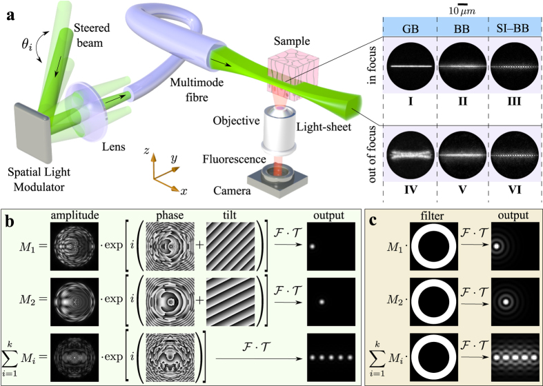
Figure 1
Light-sheet pattern generation.
( a ) A simplified scheme of the LS delivery using the MMF (detailed experimental setup is in supplementary information S4 ). The insets (Iu2013VI) show the yz -plane profiles of the GB, BB and SI-BB ...
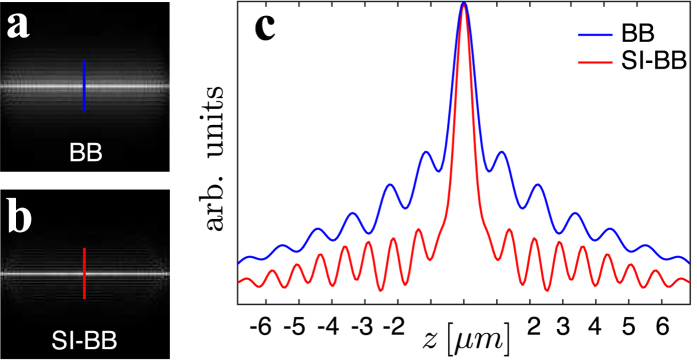
Figure 2
Comparison of the BB/SI-BB LS approach.
The simulated intensity profile of ( a ) BB LS and ( b ) SI-BB LS in the calibration (focal) plane. ( c ) One dimensional intensity profile taken from ( a ) (blue line) and ( b ) (red line) respective...
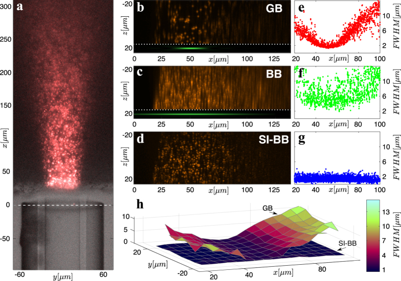
Figure 3
Axial resolution studies.
( a ) An image from the detection objective with the control illumination turned on. The MMF is at the bottom while the agarose gel with dispersed fluorescent particles is at the top. The control view...
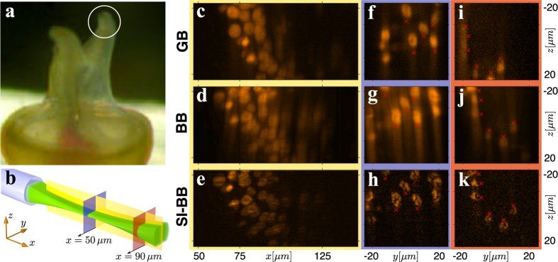
Figure 4
Imaging of biological sample.
( a ) Close-up image of operculum prongs of S. lamarcki . ( b ) The cross-sections used for the comparison of the imaging performance of GB, BB and SI-BB LS modalities. ( c , f , i ) GB LS, ( d , g , ...
Figure images are served from the NIH/NLM PubMed Central Open Access Subset or Europe PMC; copyright remains with the publishers and authors.
💬 Discussion
0 commentsNo comments yet. Be the first to start a discussion!
Leave a Comment