Abstract
In inflammation, pain is regulated by a balance of pro- and analgesic mediators. Analgesic mediators include opioid peptides which are secreted by neutrophils at the site of inflammation, leading to activation of opioid receptors on peripheral sensory neurons. In humans, local opioids and opioid peptides significantly downregulate postoperative as well as arthritic pain. In rats, inflammatory pain is induced by intraplantar injection of heat inactivated Mycobacterium butyricum, a component of complete Freund's adjuvant. We hypothesized that mycobacterially derived formyl peptide receptor (FPR) and/or toll like receptor (TLR) agonists could activate neutrophils, leading to opioid peptide release and inhibition of inflammatory pain. In complete Freund's adjuvant-induced inflammation, thermal and mechanical nociceptive thresholds of the paw were quantified (Hargreaves and Randall-Selitto methods, respectively). Withdrawal time to heat was decreased following systemic neutrophil depletion as well as local injection of opioid receptor antagonists or anti-opioid peptide (i.e. Met-enkephalin, beta-endorphin) antibodies indicating an increase in pain. In vitro, opioid peptide release from human and rat neutrophils was measured by radioimmunoassay. Met-enkephalin release was triggered by Mycobacterium butyricum and formyl peptides but not by TLR-2 or TLR-4 agonists. Mycobacterium butyricum induced a rise in intracellular calcium as determined by FURA loading and calcium imaging. Opioid peptide release was blocked by intracellular calcium chelation as well as phosphoinositol-3-kinase inhibition. The FPR antagonists Boc-FLFLF and cyclosporine H reduced opioid peptide release in vitro and increased inflammatory pain in vivo while TLR 2/4 did not appear to be involved. In summary, mycobacteria activate FPR on neutrophils, resulting in tonic secretion of opioid peptides from neutrophils and in a decrease in inflammatory pain. Future therapeutic strategies may aim at selective FPR agonists to boost endogenous analgesia.
🔬 Techniques
🔭 Microscopes
✨ Fluorophores
🧪 Sample Preparation
🔬 Cell Lines
🏭 Microscope Brands
🧪 Reagent Suppliers
📷 Detectors
🏛️ Research Organizations (ROR)
Affiliated research institutions:
📋 Methods
Antibodies and reagents Rabbit anti-Met-enkephalin or anti-rat-β-endorphin
Abs as well as purified Met-enkephalin and Boc-FLFLF were purchased from Bachem, Weil am Rhein, Germany. Naloxone, D-Phe-Cys-Tyr-D-Trp-Orn-Thr-Pen-Thr-NH 2 (CTOP), naltrindole hydrochloride (NTI) and fMLP were obtained from Sigma-Aldrich Chemie, Deisenhofen, Germany, and desiccated Mycobacterium butyricum was from BD Bioscience, Heidelberg, Germany. Complete Freund's adjuvant, LY294002, wortmannin and 1,2- bis ( o -aminophenoxy)ethane-N,N,N′,N′-tetraacetic acid acetoxymethyl ester (BAPTA/AM) were purchased from Calbiochem, San Diego, CA, USA. BAPTA/AM, LY294002, wortmannin, fMLP, and Boc-FLFLF were dissolved in dimethyl sulfoxide (maximal final concentration 1%). Anti-neutrophil serum was obtained from Accurate Chemical&Scientific Corporation, Westbury, NY, USA. Cyclosporine H was purchased from Eton Bioscience, San Diego, CA, USA. Anti-TLR-2-phycoerythrin (PE, clone TL2.1) and anti-TLR-4-PE (clone HTA125) as well as mouse IgG 2a were obtained from eBioscience, San Diego, CA, USA. Anti-TLR-2 (clone TL2.1) and anti-TLR-4 (clone HTA125) were from Alexis, Lörrach, Germany. fMLP-fluorescein isothiocyanate (FITC) was obtained from Invitrogen-Molecular Probes, Karlsruhe, Germany. CD45-CyC, RP-1-PE and ED1-PE were obtained by BD Biosciences, Heidelberg, Germany and Serotec, London, Great Britain, respectively Animals and complete Freund's adjuvant-induced inflammation Male Wistar rats weighing 180–220 g were injected intraplantarly with 150 µl complete Freund's adjuvant in the right hind paw as described [70] . Experiments were conducted at 2–24 h after inoculation. All injections were performed under brief isoflurane anesthesia. Animal protocols were approved by the animal care committee of local authorities and were in accordance with the guidelines of the International Association for the Study of Pain [71] .
Show full methods section
Antibodies and reagents Rabbit anti-Met-enkephalin or anti-rat-β-endorphin
Abs as well as purified Met-enkephalin and Boc-FLFLF were purchased from Bachem, Weil am Rhein, Germany. Naloxone, D-Phe-Cys-Tyr-D-Trp-Orn-Thr-Pen-Thr-NH 2 (CTOP), naltrindole hydrochloride (NTI) and fMLP were obtained from Sigma-Aldrich Chemie, Deisenhofen, Germany, and desiccated Mycobacterium butyricum was from BD Bioscience, Heidelberg, Germany. Complete Freund's adjuvant, LY294002, wortmannin and 1,2- bis ( o -aminophenoxy)ethane-N,N,N′,N′-tetraacetic acid acetoxymethyl ester (BAPTA/AM) were purchased from Calbiochem, San Diego, CA, USA. BAPTA/AM, LY294002, wortmannin, fMLP, and Boc-FLFLF were dissolved in dimethyl sulfoxide (maximal final concentration 1%). Anti-neutrophil serum was obtained from Accurate Chemical&Scientific Corporation, Westbury, NY, USA. Cyclosporine H was purchased from Eton Bioscience, San Diego, CA, USA. Anti-TLR-2-phycoerythrin (PE, clone TL2.1) and anti-TLR-4-PE (clone HTA125) as well as mouse IgG 2a were obtained from eBioscience, San Diego, CA, USA. Anti-TLR-2 (clone TL2.1) and anti-TLR-4 (clone HTA125) were from Alexis, Lörrach, Germany. fMLP-fluorescein isothiocyanate (FITC) was obtained from Invitrogen-Molecular Probes, Karlsruhe, Germany. CD45-CyC, RP-1-PE and ED1-PE were obtained by BD Biosciences, Heidelberg, Germany and Serotec, London, Great Britain, respectively Animals and complete Freund's adjuvant-induced inflammation Male Wistar rats weighing 180–220 g were injected intraplantarly with 150 µl complete Freund's adjuvant in the right hind paw as described [70] . Experiments were conducted at 2–24 h after inoculation. All injections were performed under brief isoflurane anesthesia. Animal protocols were approved by the animal care committee of local authorities and were in accordance with the guidelines of the International Association for the Study of Pain [71] .
Measurement of hyperalgesia and analgesia
Mechanical nociceptive thresholds were assessed using the paw pressure algesiometer (modified Randall-Selitto test; Ugo Basile) as described before [14] , [17] . The pressure required to elicit paw withdrawal using a blunt piston onto the dorsal surface of the hind paw, the paw pressure threshold, was determined. The treatments were randomized and the experimenter was blinded to the treatments. A decrease in the paw pressure threshold was interpreted as hyperalgesia (pain) whereas a rise in the paw pressure threshold was interpreted as analgesia (antinociception). Thermal nociceptive thresholds were measured by the Hargreaves test [42] . The latency (time; s) required to elicit paw withdrawal was measured with an electronic timer (IITC Inc/Life Science) after application of radiant heat to the plantar surface of a hind paw from underneath the glass floor with a high-intensity light bulb. The stimulus intensity was adjusted to give 20 s paw withdrawal latency in noninflamed paws, and the cutoff was 25 s to avoid tissue damage. The average of two measurements taken with 20 s intervals was calculated. A decrease in paw withdrawal latency was interpreted as pain (hyperalgesia) whereas a rise in paw withdrawal latency was interpreted as analgesia (antinociception). Experimental protocols fMLP-induced analgesia was evaluated in rats with complete Freund's adjuvant inflammation after intraplantar (i.pl.) injection of 0.1–3 ng fMLP dissolved in 100 µl of NaCl 0.9% or of solvent only. Paw pressure threshold or paw withdrawal latency were measured 5 min after fMLP injection. In some experiments the opioid receptor antagonist naloxone (0.56 ng i.pl.), CTOP (2 µg i.pl.), NTI (50 µg i.pl.), or an antibody against Met-enkephalin (1.25 µg i.pl.) were injected concomitantly. Optimal doses were determined in pilot experiments and in previous studies [17] , [18] . To deplete rats of neutrophils, animals were injected with 80 µl anti-neutrophil serum intravenously 18 h before complete Freund's adjuvant injection as described previously [14] , [17] . Modulation of baseline inflammatory thermal hyperalgesia was analyzed in rats with complete Freund's adjuvant (2–24 h) inflammation after i.pl. treatment with naloxone (0.56 ng), anti-Met-enkephalin (1.25 µg), anti-β-endorphin (2 µg), FPR antagonists (Boc-FLFLF: 0.3 and 3 µg or cyclosporine H: 0.9 and 9 µg) or after systemic neutrophil depletion. Monocyte isolation by MACS separation To obtain human monocytes from healthy blood donors, red blood cells were lyzed using buffer EL (Qiagen, Hilden, Germany). The remaining white blood cells were incubated with anti-CD14-coupled magnetic beads (Miltenyi Biotec, Bergisch-Gladbach, Germany) in phosphate-buffered saline (PBS) containing 0.5% bovine serum albumin and 2 mM ethylene diamine tetraacetic acid. CD14 + monocytes were isolated using a LS column (Miltenyi Biotec). Purity was confirmed by staining with anti-CD14 FITC antibody (BD Biosciences) and FACS analysis (see below). Opioid peptide release Human neutrophils from healthy blood donors were purified using dextran sedimentation, Ficoll separation and hypotonic lysis (all Amersham Biosciences). Rat peritoneal neutrophils were obtained 4 h after intraperitoneal injection of 1% oyster glycogen (Sigma-Aldrich Chemie) [17] , [72] . For determination of opioid peptide release, 5×10 7 neutrophils or 1×10 7 CD14 + monocytes were stimulated with fMLP (1–1000 nM) or Mycobacterium butyricum (0.06–0.66 mg/ml) after preincubation with cytochalasin B (5 µg/ml) for 5 min in Hank's balanced salt solution containing the proteinase inhibitors bestatin (5 µg/ml), aprotinin (40 µg/ml) and thiorphan (100 µM, all Sigma-Aldrich Chemie) [17] , [73] , [74] . Doses of fMLP and Mycobacterium butyricum were based on pilot experiments and the literature [74] , [75] . In some experiments, cells were concomitantly incubated with inhibitors as described in the results section. Doses were established in pilot experiments and according to the literature [17] , [31] , [35] . Control samples with the solvent dimethyl sulfoxide did not induce significant release. Doses for anti-TLR-2 (clone TL2.1) and anti-TLR-4 (clone HTA125) antibodies were chosen based on their blocking effects according to the literature [46] , [49] – [52] . Supernatants were obtained after 7 min stimulation and stored at −20°C until further analysis by radioimmunoassay using commercially available kits for rat or human Met-enkephalin and β-endorphin (Bachem) [17] , [76] , [77] .
Cell culture and transfection
Construction of plasmids coding for the human fMLP receptor has been described elsewhere [78] . HEK293 cells were grown at 37°C and 5% CO 2 in Dulbecco's modified Eagle's medium or minimal essential medium with Earle's salts, supplemented with 10% fetal calf serum, 2 mM glutamine, 100 µg/ml streptomycin, and 100 units/ml penicillin. HEK293 cells were transfected using Fugene 6 transfection reagent (Roche Applied Science, Mannheim, Germany) according to the manufacturer's recommendations. The amount of transfected human FPR plasmid cDNA was 250 ng per 35 mm dish and was kept constant by addition of empty expression vector (pcDNA3 up to 2 µg) where necessary. Ca 2+ imaging Fluorescence imaging was performed with a monochromator-equipped xenon lamp and a cooled CCD camera (TILL-Photonics) connected to an inverted epifluorescence microscope (Axiovert 100; Carl Zeiss). All imaging experiments were performed in a Hepes-buffered solution containing 128 mM NaCl, 6 mM KCl, 1 mM MgCl 2 , 1 mM CaCl 2 , 5.5 mM glucose, 10 mM Hepes (pH 7.4), and 0.2% (wt/vol) bovine serum albumin. For determination of [Ca 2+ ] i , neutrophils or FPR transfected HEK293 cells were placed on dishes coated with poly-L-lysine and then loaded with 1 or 2 µM Fura 2/AM (Molecular Probes-Invitrogen) for 30 min at 37°C as previously described [79] . After basal recordings, cells were stimulated by subsequent addition of 1 µM fMLP or dimethyl sulfoxide extracted Mycobacterium butyricum . Fura-2 loaded cells were alternately excited at 340 and 380 nm, and fluorescence was detected through a 505 nm filter. Calibration of [Ca 2+ ] i was performed as described [17] , [79] .
Flow cytometry
TLRs were labeled in human neutrophils after preincubation with 10% mouse serum for 10 min using PE conjugated anti-human-TLR-2, anti-human-TLR-4 or isotype control antibodies according to manufacturer's instructions. The FPR was stained using FITC-conjugated fMLP (1 µM). In selected experiments samples were pretreated for 10 min with different concentrations of Boc-FLFLF before addition of FITC-fMLP [13] , [14] , [17] , [76] . FPR staining in subcutaneous paw tissue was performed as described before [17] . Neutrophils were identified by CD45 + and RP1 + staining while macrophages were CD45 + ED1 + .
Immunofluorescence
Immunofluorescence staining was performed using human neutrophils (5 min preincubation with cytochalasin B, then addition of 0.66 mg/ml Mycobacterium butyricum for 15 min) [41] as well as neutrophils preincubated with Boc-FLFLF. After centrifugation for 10 min at 300 g, cell pellets of neutrophils were reconstituted in 5 ml PBS, and 50,000 neutrophils in suspension were then centrifuged by a Shandon Cytospin 3 (Thermo Shandon, Pittsburgh, PA) at 20 g for 3 min on glass slides. Neutrophils were fixed for 30 min and confocal analysis was carried out as previously described [80] . Briefly, neutrophil cytospins were incubated with rabbit polyclonal antibodies against Met-enkephalin (1∶1000, Peninsula Laboratories, Belmont, CA, USA) and subsequently with a Texas red-conjugated goat anti-rabbit antibody. Thereafter, cytospins were washed with PBS and mounted in vectashield. Images were acquired on a Zeiss LSM510META confocal laser scanning system (Zeiss AIM; Jena) using a 63×/1.4 Plan-Apochromat or 40×/1.3Plan-Neofluar oil immersion objective in a series of optical sections of about 1 µm thickness. Each experiment was repeated three times. To demonstrate specificity of staining, the following controls were included as mentioned in detail elsewhere [14] , [81] : (1) preabsorption of diluted antibody against Met-enkephalin with purified Met-enkephalin (Peninsula laboratories-Bachem) and (2) omission of either the primary or the secondary antibodies.
Statistical analysis
Data are presented as raw values (mean±SEM). Normally distributed data were analyzed by student's t-test or Mann-Whitney test. Not normally distributed data were analyzed by Wilcoxon Signed Rank Test. Multiple comparisons were analyzed by one-way ANOVA or by one-way ANOVA on ranks in case of not normally distributed data. If necessary repeated measures (RM) one way ANOVA was used. Posthoc comparisons were performed by Student-Newman-Keuls', Dunnett's or Duncan's method, respectively. Differences were considered significant if p
📊 Figures
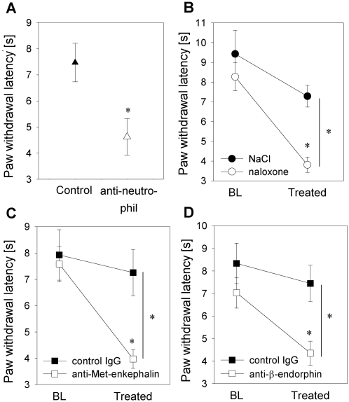
Figure 1
Neutrophils attenuate inflammatory pain by local opioid peptide release.
[A] In a rat model, inflammation was induced by intraplantar injection of heat-inactivated Mycobacterium butyricum (complete Freund's adjuvant). Prior to induction of inflammation, rats were pretreate...
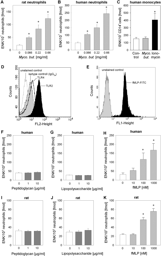
Figure 2
Opioid peptide release from neutrophils is triggered by mycobacteria and FPR agonists but not by TLR-2 or TLR-4 agonists.
[Au2013C] Rat and human neutrophils as well as CD14 + human monocytes were incubated with heat-inactivated Mycobacterium butyricum ( Myco. but. ), and Met-enkephalin (ENK) release was quantified by ra...
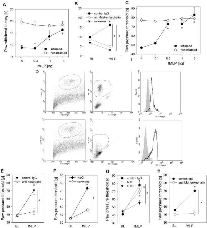
Figure 3
Formyl peptides inhibit inflammatory pain through release of opioid peptides from neutrophils.
[A, C] Rats received intraplantar injections of fMLP into inflamed paws (2 h post complete Freund's adjuvant: filled circles; noninflamed paw: open circles). Paw withdrawal latency [A] or paw pressure...
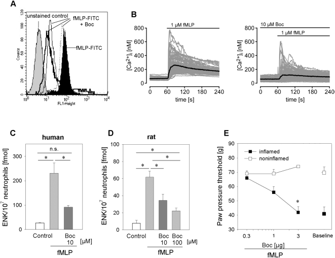
Figure 4
fMLP-induced opioid peptide release in vitro and analgesia in vivo are dependent on activation of the FPR.
[A] FPR staining of human neutrophils with fMLP-FITC (black histogram) is dose-dependently inhibited by the FPR antagonist Boc-FLFLF (Boc, dotted line: 0.1 u00b5M, thin black line: 1 u00b5M, thick bla...
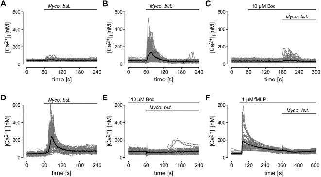
Figure 5
Mycobacterium butyricum-triggered intracellular calcium elevation is FPR dependent.
Mock [A] or human FPR [B,C] transfected HEK293 cells or human neutrophils [Du2013F] were loaded with Fura-2. Changes in [Ca 2+ ] i were analyzed after addition of Mycobacterium butyricum ( Myco. but. ...
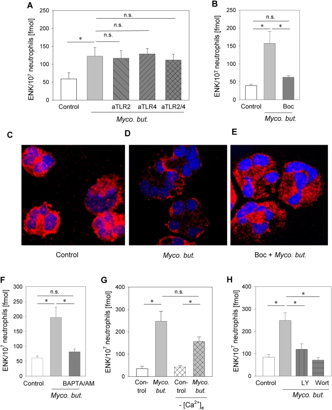
Figure 6
Mycobacterium butyricum-induced opioid peptide release and translocation of Met-enkephalin-containing granules from human neutrophils requires FPR stimulation, intracellular Ca 2+ mobilization and PI3K activation.
[A, B] Met-enkephalin release induced by Mycobacterium butyricum ( Myco. but. 0.66 mg/ml) was analyzed after preincubation with anti-TLR-2 and anti-TLR-4 ([A], anti-TLR2 or 4 both 10 u00b5g/ml, nu200a...
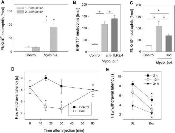
Figure 7
Tonic opioid peptide release through FPR stimulation reduces inflammatory pain.
[A] Rat neutrophils were repeatedly stimulated with Mycobacterium butyricum ( Myco. but. 0.66 mg/ml) and Met-enkephalin (ENK) content was determined in supernatants (nu200a=u200a12u201316; * p<0.05...
Figure images are served from the NIH/NLM PubMed Central Open Access Subset or Europe PMC; copyright remains with the publishers and authors.
💬 Discussion
0 commentsNo comments yet. Be the first to start a discussion!
Leave a Comment