Abstract
Abstract Expansion microscopy (ExM) enables super-resolution imaging of proteins and nucleic acids on conventional microscopes. However, imaging of details of the organization of lipid bilayers by light microscopy remains challenging. We introduce an unnatural short-chain azide- and amino-modified sphingolipid ceramide, which upon incorporation into membranes can be labeled by click chemistry and linked into hydrogels, followed by 4× to 10× expansion. Confocal and structured illumination microscopy (SIM) enable imaging of sphingolipids and their interactions with proteins in the plasma membrane and membrane of intracellular organelles with a spatial resolution of 10–20 nm. As our functionalized sphingolipids accumulate efficiently in pathogens, we use sphingolipid ExM to investigate bacterial infections of human HeLa229 cells by Neisseria gonorrhoeae, Chlamydia trachomatis and Simkania negevensis with a resolution so far only provided by electron microscopy. In particular, sphingolipid ExM allows us to visualize the inner and outer membrane of intracellular bacteria and determine their distance to 27.6 ± 7.7 nm.
🔬 Techniques
🔭 Microscopes
💻 Software
✨ Fluorophores
🧪 Sample Preparation
🏭 Microscope Brands
🧪 Reagent Suppliers
🔎 Objectives
💻 Software Details
📋 Protocols
💾 Data Repositories
🏛️ Research Organizations (ROR)
Affiliated research institutions:
📋 Methods
A step-by-step protocol describing the expansion of cellular and bacterial membranes can be found at Protocol Exchange 48 . Chemical synthesis of α -amino- ω -azido-C 6 -ceramide Starting from N -Boc-protected l -lysine ( 1 ) the introduction of the azide-functionality was accomplished via catalytic diazotransfer reaction to obtain azido-acid 2 in 85% yield (Fig. 1a ). For that, triflyl azide was prepared based on a method of Yan et al. with a reduced amount of highly toxic sodium azide and triflyl anhydride compared to previous protocols 49 . Subsequent amide coupling of 2 with sphingosine was performed in DMF under basic conditions using HATU as coupling reagent. The resulting Boc-protected azido-ceramide analog 3 was isolated in 48% yield. In the last step the amine group was deprotected by the treatment with TFA in dichloromethane. After basic workup, followed by column chromatography, the target ceramide analog 4 was successfully isolated in 39% yield (Fig. 1a ). Details on the experimental procedures can be found in the Supporting Information. All isolated compounds were characterized by a combination of HRMS, NMR and IR spectroscopy (Supplementary Figs. 1 – 13 ).
Cell lines and bacteria Human
HeLa229 cells (ATCC CCL-2.1tm) and human epithelial conjunctival cells (Chang) were cultured in 10% (v/v) heat inactivated FBS (Sigma-Aldrich) RPMI1640 + GlutaMAXtm medium (Gibcotm) and were grown in a humidified atmosphere containing 5% (v/v) CO 2 at 37 °C. HeLa229 cells were used for infection with Chlamydia trachomatis and Simkania negevensis , Chang cells for infection with Neisseria gonorrhoeae . For this study, C. trachomatis serovar L2/434/Bu (ATCC VR-902B tm ), S. negevensis and N. gonorrhoeae (strain MS11, derivative N927) were used. C. trachomatis and S. negevensis were propagated in HeLa229 cells at a multiplicity of infection (MOI) of 1 for 48 h for C. trachomatis and 72 h for S. negevensis . The cells were then detached and lysed using glass beads (3 mm, Roth). Low centrifugation supernatant (10 min at 2000 g at 4 °C for C. trachomatis and 10 minutes at 600 g at 4 °C for S. negevensis ) was transferred to high speed centrifugation (30 min at 30.000 g at 4 °C for C. trachomatis and 30 min at 20.000 g at 4 °C for S. negevensis ) to pellet the bacteria. Afterwards, the pellet was washed and resuspended in 1x SPG buffer (7.5% sucrose, 0.052% KH 2 PO 4 , 0.122% NaHPO 4 , 0.072% L-glutamate). The resuspended bacteria were then stored at −80 °C and titrated for an MOI of 1 for further experimentation. Infected cells were incubated in a humidified atmosphere with 5% (v/v) CO 2 at 35 °C. For secondary infections, Hela229 cells were infected with C. trachomatis for 48 hours and then lysed using glass beads (3 mm, Roth). Afterwards, the supernatant was diluted 1:100 to infect other cells. The cell lines as well as the Chlamydia used in this study were tested to be free of Mycoplasma via PCR. Neisseria were cultivated on gonococci (GC) agar (ThermoScientific, Waltham, USA) plates supplemented with 1% vitamin mix at 37 °C and 5% CO 2 for 16 h. On the day of infection, liquid culture was performed in protease-peptone medium (PPM) supplemented with 1% vitamin mix and 0.5% sodium bicarbonate 8.4% solution (PPM + ) at 37 °C and 120 rpm. Gonococci were grown to an OD 550 0.4 to 0.6. Before infecting the cells, the medium of the liquid culture was changed to 4-(2-Hydroxyethyl)piperazine-1-ethanesulfonic acid (HEPES buffer) medium by centrifugation with 2778 g for 5 min. After the indicated time of 4 h, the infection was stopped by washing the cells three times with Hepes medium. Western blot Western Blot lysates were collected on ice by lysing the cells in SDS sample buffer (62.5 mM Tris, pH 6.8, 2% SDS, 20% glycerol and 5% β-mercaptoethanol) and then cooked for 5 min at 95 °C. The protein samples were separated in 10% SDS-PAGE gel and then transferred to a PVDF membrane (Roche) in a semi-dry electroblotter. After transfer, the membrane was blocked for 1 h in Tris-buffer containing 0.05% Tween 20 and 5% dry milk powder and afterwards incubated in primary antibody over night at 4 °C. The primary antibodies used were: cHSP60 (Santa Cruz, sc-57840, dilution 1:1000) and β-actin (Sigma, A5441, dilution 1:10,000). Proteins were detected with secondary antibodies coupled to horseradish peroxidase (Santa Cruz Bioscience) using the ECL system (Pierce) on an Intas Chem HR 16-3200 reader.
Show full methods section
A step-by-step protocol describing the expansion of cellular and bacterial membranes can be found at Protocol Exchange 48 . Chemical synthesis of α -amino- ω -azido-C 6 -ceramide Starting from N -Boc-protected l -lysine ( 1 ) the introduction of the azide-functionality was accomplished via catalytic diazotransfer reaction to obtain azido-acid 2 in 85% yield (Fig. 1a ). For that, triflyl azide was prepared based on a method of Yan et al. with a reduced amount of highly toxic sodium azide and triflyl anhydride compared to previous protocols 49 . Subsequent amide coupling of 2 with sphingosine was performed in DMF under basic conditions using HATU as coupling reagent. The resulting Boc-protected azido-ceramide analog 3 was isolated in 48% yield. In the last step the amine group was deprotected by the treatment with TFA in dichloromethane. After basic workup, followed by column chromatography, the target ceramide analog 4 was successfully isolated in 39% yield (Fig. 1a ). Details on the experimental procedures can be found in the Supporting Information. All isolated compounds were characterized by a combination of HRMS, NMR and IR spectroscopy (Supplementary Figs. 1 – 13 ).
Cell lines and bacteria Human
HeLa229 cells (ATCC CCL-2.1tm) and human epithelial conjunctival cells (Chang) were cultured in 10% (v/v) heat inactivated FBS (Sigma-Aldrich) RPMI1640 + GlutaMAXtm medium (Gibcotm) and were grown in a humidified atmosphere containing 5% (v/v) CO 2 at 37 °C. HeLa229 cells were used for infection with Chlamydia trachomatis and Simkania negevensis , Chang cells for infection with Neisseria gonorrhoeae . For this study, C. trachomatis serovar L2/434/Bu (ATCC VR-902B tm ), S. negevensis and N. gonorrhoeae (strain MS11, derivative N927) were used. C. trachomatis and S. negevensis were propagated in HeLa229 cells at a multiplicity of infection (MOI) of 1 for 48 h for C. trachomatis and 72 h for S. negevensis . The cells were then detached and lysed using glass beads (3 mm, Roth). Low centrifugation supernatant (10 min at 2000 g at 4 °C for C. trachomatis and 10 minutes at 600 g at 4 °C for S. negevensis ) was transferred to high speed centrifugation (30 min at 30.000 g at 4 °C for C. trachomatis and 30 min at 20.000 g at 4 °C for S. negevensis ) to pellet the bacteria. Afterwards, the pellet was washed and resuspended in 1x SPG buffer (7.5% sucrose, 0.052% KH 2 PO 4 , 0.122% NaHPO 4 , 0.072% L-glutamate). The resuspended bacteria were then stored at −80 °C and titrated for an MOI of 1 for further experimentation. Infected cells were incubated in a humidified atmosphere with 5% (v/v) CO 2 at 35 °C. For secondary infections, Hela229 cells were infected with C. trachomatis for 48 hours and then lysed using glass beads (3 mm, Roth). Afterwards, the supernatant was diluted 1:100 to infect other cells. The cell lines as well as the Chlamydia used in this study were tested to be free of Mycoplasma via PCR. Neisseria were cultivated on gonococci (GC) agar (ThermoScientific, Waltham, USA) plates supplemented with 1% vitamin mix at 37 °C and 5% CO 2 for 16 h. On the day of infection, liquid culture was performed in protease-peptone medium (PPM) supplemented with 1% vitamin mix and 0.5% sodium bicarbonate 8.4% solution (PPM + ) at 37 °C and 120 rpm. Gonococci were grown to an OD 550 0.4 to 0.6. Before infecting the cells, the medium of the liquid culture was changed to 4-(2-Hydroxyethyl)piperazine-1-ethanesulfonic acid (HEPES buffer) medium by centrifugation with 2778 g for 5 min. After the indicated time of 4 h, the infection was stopped by washing the cells three times with Hepes medium. Western blot Western Blot lysates were collected on ice by lysing the cells in SDS sample buffer (62.5 mM Tris, pH 6.8, 2% SDS, 20% glycerol and 5% β-mercaptoethanol) and then cooked for 5 min at 95 °C. The protein samples were separated in 10% SDS-PAGE gel and then transferred to a PVDF membrane (Roche) in a semi-dry electroblotter. After transfer, the membrane was blocked for 1 h in Tris-buffer containing 0.05% Tween 20 and 5% dry milk powder and afterwards incubated in primary antibody over night at 4 °C. The primary antibodies used were: cHSP60 (Santa Cruz, sc-57840, dilution 1:1000) and β-actin (Sigma, A5441, dilution 1:10,000). Proteins were detected with secondary antibodies coupled to horseradish peroxidase (Santa Cruz Bioscience) using the ECL system (Pierce) on an Intas Chem HR 16-3200 reader.
LDH assay
LDH-assays were performed using the Cytotoxicity Detection KitPLUS (LDH) (Sigma). For this, Hela229 cells were treated with 10 µM C 6 -Cer, ω-N 3 -C 6 -Cer, α-NH 2 -ω-N 3 -C 6 -ceramide and the controls with 10 µl DMSO for 1 or 24 h in 12-well plates. Additionally, one control sample was treated with 20 µl Lysis Solution for 10 min at 37 °C. Afterwards, 500 µl of the cells supernatant was centrifuged at 14.000 g. 100 µl of the centrifuged supernatant was then transferred to a 96-well plate and incubated with 100 µl of a 1:45 mixture of Catalyst (Diaphorase/NAD + mixture) and Dye-solution (INT and sodium lactate). The reaction was performed for 15 min in the dark and then stopped with 50 µl of the Stop Solution. The light absorbance of the samples was then measured on a TECAN infinite M200 and compared to the DMSO treated (low control) and the DMSO and Lysis Solution treated (max control) control samples. Chemistry and immunolabeling For immunostaining, cells were seeded on 15 mm coverslips. α-amino-ω-azido-C 6 -ceramide, ω-azido-C 6 -ceramide, as well as ω-azido-sphingosine were fed with 10 µM final concentration for 1 h at 37 °C. For chlamydial infection, the cells were fed with ceramide-analogs 23 h post infection and for infection with Simkania for 72 h and for neisserial infection, the cells were fed with the sphingosine analog immediately before infection. Afterwards, the cells were fixed in 4% PFA and 0.1% GA for 15 min, washed 3x in 1xPBS and then permeabilized for 15 min in 0.2% Triton X-100 in PBS. The cells were then washed again 3x in 1xPBS and then incubated with 5 µM DBCO-488 (Jena Bioscience, CLK-1278-1) at 37 °C for 30 min or 5 µM Click-IT Alexa Fluor® 488 DIBO alkyne dye (ThermoScientific, Waltham, USA) at 37 °C for 30 min. For staining with antibodies, the cells were washed, blocked using 2% FCS in 1xPBS for 1 h and then incubated in primary antibody diluted in blocking buffer for 1 h in a humid chamber. The primary antibodies used in this study were: anti-HSP60 ms (Santa Cruz, sc-57840, dilution 1:200), anti- Neisseria gonorrhoeae primary antibody rb (US biological, dilution 1:200), anti-Prx3 (Origene, TA322470, dilution 1:100), anti-CERT (Abcam, ab72536, 1:100) and anti-LPS (BioRAD, MCA2718, dilution 1:200). After that, the cells were washed 3x in 1× PBS and then incubated in the corresponding secondary antibody diluted in blocking buffer for 1 h and then washed 3x with 1x PBS. The secondary antibodies used were: ATTO 647 N ms (Rockland, 610-156-121 S, dilution 1:200) and ATTO 647 N rb (Sigma, 40839, dilution 1:200). mCling 150 nmol mCling was incubated in 3 molar excess of ATTO 643-Maleimide (ATTO-TEC, AD 643-45) in 100 mM TCEP overnight at RT under continuous shaking. The label product was purified by HPLC (JASCO) and the concentration was determined using a UV-vis spectrophotometer (Jasco V-650). Staining with mCling was performed by the incubation of living cells in 0.5 µM mCling dissolved in media for 10 min at 37 °C.
Expansion microscopy
Stained cells were treated for 10 min with 0.25% GA at RT and gelated after three washing steps. In case of 4x expansion a monomer solution consisting of 8.625% sodium acrylate (Sigma, 408220), 2.5% acrylamide (Sigma, A9926), 0.15% N,N'-methylenbisacrylamide (Sigma, A9926), 2 M NaCl (Sigma, S5886) and 1xPBS and 0.2% freshly added ammonium persulfate (APS, Sigma, A3678) and tetramethylethylenediamine (TEMED, Sigma, T7024) was used. Here gelation was performed for 1 h at RT followed by proteinase digestion. In case of 10x expansion 1 ml of the monomer solution containing 0.267 g DMAA (Sigma, 274135) and 0.064 g sodium acrylate (Sigma, 408220) dissolved in 0.57 g ddH 2 O was degassed for 45 min on ice with nitrogen followed by the addition of 100 µl KPS (0.036 g/ml, Sigma, 379824). After another 15 min of degassing and the addition of 4 µl TEMED per ml monomer solution, gelation was performed for 30 min at RT followed by an incubation of 1.5 h at 37 °C. Hereafter the samples were digested for 3 h – overnight in digestion buffer (50 mM Tris pH 8.0, 1 mM EDTA (Sigma, ED2P), 0.5% Triton X-100 (Thermo Fisher, 28314) and 0.8 M guanidine HCl (Sigma, 50933)), supplied with 8 U/ml protease K (Thermo Fisher, AM2548) and for expansion of Neisseria additional 1 mg/ml Lysozyme according to Lim et al. 50 . Digested gels were expanded in hourly changed ddH 2 O until the expansion saturated. The expansion factor was determined by the gel size using calipers directly after gelation and by the gel size of the digested and expanded samples. We achieved experimental expansion factors of 4.1 for the 4x monomer solution and 10 for the 10x monomer solution, and the expansion factor remained constant for the used monomer solutions. Expanded and chopped gels were stored at 4 °C in ddH 2 O immobilized prior to imaging on PDL-coated glass chambers (Merck, 734-2055).
Confocal microscopy and SIM
Confocal imaging was performed on an inverted microscope (Zeiss LSM700 using software ZEN 12.0.1.362, 2012) or on a Leica TCS SP5 confocal microscope (Leica Biosystems using software LAS AF version 2.7.3.9723) and SIM-imaging on a Zeiss ELYRA S.1 SR-SIM structured illumination platform using a 63x water-immersion objective (C-Apochromat, 63×1.2 NA, Zeiss, 441777-9970). Reconstruction of SIM-images was performed using the ZEN image-processing platform with a SIM module. Z-stacks were processed using Imaris 8.4.1 and FIJI 1.51n 51 .
FRAP
HeLa229 cells were seeded in an 8-well chambered high precision coverglass (Sarstedt 8-well on coverglass II) and incubated for 24 h at 37 °C and 5% CO 2 . The cells were fed with 10 µM of the corresponding azido-ceramide analog for 30 min in cell culture media. Afterwards, the cells were washed with HBSS with magnesium and calcium and fixed with 4% formaldehyde and 0.1% glutaraldehyde in HBSS for 15 min at room temperature and washed. Ceramides were labeled by strain-promoted alkyne-azide cycloaddition (SPAAC) with 10 µM DBCO-Alexa Fluor 488 in HBSS for 30 min at 37 °C and washed. FRAP-imaging was performed at a confocal laser scanning microscope (CLSM) LSM700 (Zeiss, Germany) using the Plan-Apochromat 63×1.4 oil objective. Using the 488 nm laser line as excitation, a time series with 30 frames every 1.5 s was recorded. After three frames, a circular region of interest with diameter 1.8 µm was bleached and fluorescence recovery followed over time. Reporting summary Further information on research design is available in the Nature Research Reporting Summary linked to this article.
Supplementary information Peer Review File Reporting Summary Description of Additional Supplementary Files Supplementary Movie 1 Supplementary Information
📊 Figures
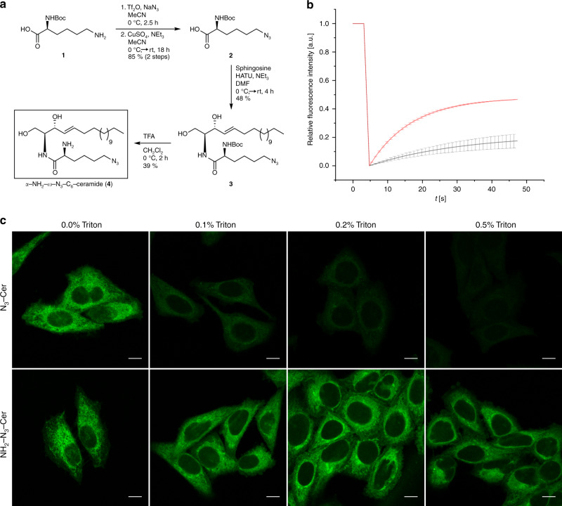
Fig. 1
Amino- and azido-functionalized sphingolipids enable fixation and fluorescence labeling of lipids.
a Schematic overview of the synthesis of u03b1-NH 2 -u03c9-N 3 -C 6 -ceramide (for synthesis details see Material and Methods and Supporting Information). To investigate the mobility of membrane-incor...
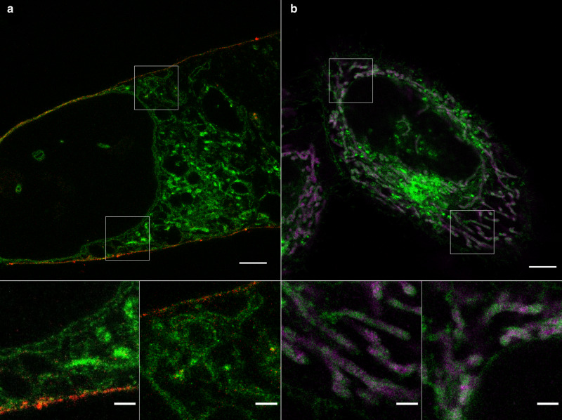
Fig. 2
Sphingolipid ExM enables super-resolution imaging of cellular membranes and protein interactions.
a Confocal fluorescence image of a 10x expanded HeLa229 cell fed with ATTO643-mCling (red) and u03b1-NH 2 -u03c9-N 3 -C 6 -ceramide clicked with DBCO-Alexa Fluor 488 (green). Scale bars, 20u2009u00b5m...
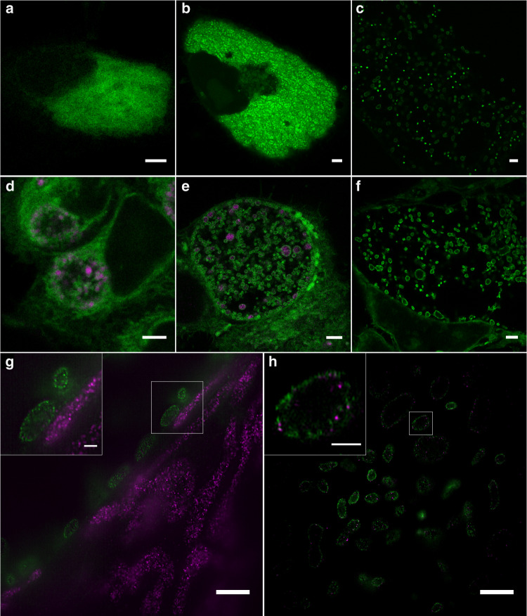
Fig. 3
Sphingolipid ExM visualizes intracellular pathogens and their interactions with mitochondrial proteins.
a u2013 c Cells were infected with Simkania negevensis for 96u2009h, fed with u03b1-NH 2 -u03c9-N 3 -C 6 -ceramide, fixed, permeabilized and stained with DBCO-Alexa Fluor 488 (green), and then imaged....
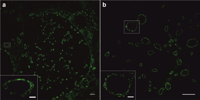
Fig. 4
10x Sphingolipid ExM in combination with SIM resolves the distance between the OM and IM of gram-negative bacteria.
HeLa229 cells infected with Chlamydia trachomatis for 24u2009h, fed with u03b1-NH 2 -u03c9-N 3 -C 6 -ceramide, fixed, permeabilized and click-labeled with DBCO-Alexa Fluor 488 (green). Confocal ( a ) ...
Figure images are served from the NIH/NLM PubMed Central Open Access Subset or Europe PMC; copyright remains with the publishers and authors.
💬 Discussion
0 commentsNo comments yet. Be the first to start a discussion!
Leave a Comment