Abstract
In many tissues, actin monomers polymerize into actin (thin) filaments of precise lengths. Although the exact mechanisms involved remain unresolved, it is proposed that "molecular rulers" dictate the lengths of the actin filaments. The giant nebulin molecule is a prime candidate for specifying thin filament lengths in striated muscle, but this idea has never been proven. To test this hypothesis, we used RNA interference technology in rat cardiac myocytes. Live cell imaging and triple staining revealed a dramatic elongation of the preexisting thin filaments from their pointed ends upon nebulin knockdown, demonstrating its role in length maintenance; the barbed ends were unaffected. When the thin filaments were depolymerized with latrunculin B, myocytes with decreased nebulin levels reassembled them to unrestricted lengths, demonstrating its importance in length specification. Finally, knockdown of nebulin in skeletal myotubes revealed its involvement in myofibrillogenesis. These data are consistent with nebulin functioning as a thin filament ruler and provide insight into mechanisms dictating macromolecular assembly.
🔬 Techniques
🧬 Organisms
✨ Fluorophores
🧪 Sample Preparation
🏭 Microscope Brands
🧪 Reagent Suppliers
📷 Detectors
💻 Software Details
💾 Data Repositories
🏛️ Research Organizations (ROR)
Affiliated research institutions:
📋 Methods
Cell culture, siRNA, and Lat B treatment Rat fetal cardiac myocytes were isolated by J. Bahl and Y. Wu (University of Arizona, Tucson, AZ) and maintained as described previously ( Bang et al., 2001 ; Kazmierski et al., 2003 ). 2 nM dsRNAs were incubated with 6 μl of Cytofectene (Bio-Rad Laboratories) in 100 μl OptiMEM for 15 min at 37°C and added to myocytes on day 3. Transfection efficiency was >80%, and >50 cultures were analyzed. Four dsRNAs to the rat nebulin sequence (19 mers + 5′ AA overhangs) were synthesized, using Ambion's website and Silencer Construction kit, and blasted to ensure no significant homology to nebulette or other known sequences in the National Center for Biotechnology Information database. Three of the four dsRNAs significantly knocked down nebulin in cardiac myocytes (target cDNA: 5′-GGAGACGGTGTACGAGGAG-3′; 5′-CCACAACTACAAGGACATC-3′; and 5′-CAAGGACATCTGACTATGA-3′), using concentrations titrated from 0.5–10 nM, with 2 nM being the lowest effective dosage. Control siRNAs included GAPDH (Ambion), a scrambled nebulin siRNA, and the three siRNAs to nebulin containing 6-bp changes in their centers. Nebulin reduction was confirmed by immunofluorescence with three independent antibodies ( Millevoi et al., 1998 ; McElhinny et al., 2001 ; Kazmierski et al., 2003 ). For live cell imaging, cells were cotransfected with GFP-tropomyosin (a gift from J.-C. Perriard, Swiss Federal Institute of Technology, Zurich, Switzerland) and imaged as described previously ( Mudry et al., 2003 ). To depolymerize thin filaments, we added 20 μM Lat B (Invitrogen) to cells for 4 h (8 h after siRNA treatment), and then washed them out and let them recover for 4 h. For skeletal myotube cultures, myoblasts were isolated from rat hind limbs. The deboned tissue was suspended in 10 ml trypsin/EDTA (Life Technologies) and incubated for 10 min at 37°C. After centrifugation, the pellets were resuspended in MEM + 15% chick embryo extract + 10% horse serum, and cells were preplated at 37°C for 1 h on 100-mm dishes. Collected cells were plated at 4 × 10 5 cells/ml in 35-mm culture dishes containing 12-mm round coverslips coated with rat-tail collagen (Sigma-Aldrich). Cells were transfected with siRNA 24 h later, except this time using 10 nM siRNA. After 3 d, cells were treated with 1 μg/ml 1-[β-D-arabinofuranosyl]-cytosine (Ara-; Sigma-Aldrich) in MEM with 2.5% chick embryo extract and 10% horse serum and fixed on days 5 and 6. RT-PCR Total RNA was harvested from cardiac myocytes within 24 h of siRNA treatment, or from skeletal myotubes within 48 h, using the RNeasy Mini kit (QIAGEN), or with Trizol (Sigma-Aldrich), and quantified using a Biophotometer (Eppendorf). cDNA was synthesized from 1–3 μg of total RNA using SuperScript II Reverse Transcriptase (Invitrogen). 0.5–3 μl of template were used per 25 μl PCR reaction. Reagents were from the TripleMaster Taq kit (Eppendorf). For nebulin amplification, at least 28–30 cycles were performed (with the threshold of detection being ∼30 cycles in cDNA from cardiac myocytes and ∼27 from skeletal myotubes), and for other transcripts, 26–30 cycles were used. Primers used included rat nebulin (forward, 5′-ACTGTCTTCCATCCCGTCAC-3′, and reverse, 5′-GCCATACATCCAGCCTTCAT-3', to amplify a 202-bp product; rat nebulette (forward, 5′-ATTGGGAAGGGCTACAGCTT-3′, and reverse, 5′-GAAGCCTCTTCCCTTCGTCT-3′ to amplify a 196-bp product); rat GAPDH (forward, 5′-CCAGTATGATTCTACCCACGGC-3′, and reverse, 5′-CGGAGATGATGACCCTTTTGGC-3′, to amplify a 227-bp product); and Tmod1 (forward, 5′-ACTGTAAGGCCATGGACAGC-3′, and reverse, 5′-GCTGCAGTTGTGTTTCAAGG-3′, to amplify a 141-bp product). All PCR products were sequenced.
Show full methods section
Cell culture, siRNA, and Lat B treatment Rat fetal cardiac myocytes were isolated by J. Bahl and Y. Wu (University of Arizona, Tucson, AZ) and maintained as described previously ( Bang et al., 2001 ; Kazmierski et al., 2003 ). 2 nM dsRNAs were incubated with 6 μl of Cytofectene (Bio-Rad Laboratories) in 100 μl OptiMEM for 15 min at 37°C and added to myocytes on day 3. Transfection efficiency was >80%, and >50 cultures were analyzed. Four dsRNAs to the rat nebulin sequence (19 mers + 5′ AA overhangs) were synthesized, using Ambion's website and Silencer Construction kit, and blasted to ensure no significant homology to nebulette or other known sequences in the National Center for Biotechnology Information database. Three of the four dsRNAs significantly knocked down nebulin in cardiac myocytes (target cDNA: 5′-GGAGACGGTGTACGAGGAG-3′; 5′-CCACAACTACAAGGACATC-3′; and 5′-CAAGGACATCTGACTATGA-3′), using concentrations titrated from 0.5–10 nM, with 2 nM being the lowest effective dosage. Control siRNAs included GAPDH (Ambion), a scrambled nebulin siRNA, and the three siRNAs to nebulin containing 6-bp changes in their centers. Nebulin reduction was confirmed by immunofluorescence with three independent antibodies ( Millevoi et al., 1998 ; McElhinny et al., 2001 ; Kazmierski et al., 2003 ). For live cell imaging, cells were cotransfected with GFP-tropomyosin (a gift from J.-C. Perriard, Swiss Federal Institute of Technology, Zurich, Switzerland) and imaged as described previously ( Mudry et al., 2003 ). To depolymerize thin filaments, we added 20 μM Lat B (Invitrogen) to cells for 4 h (8 h after siRNA treatment), and then washed them out and let them recover for 4 h. For skeletal myotube cultures, myoblasts were isolated from rat hind limbs. The deboned tissue was suspended in 10 ml trypsin/EDTA (Life Technologies) and incubated for 10 min at 37°C. After centrifugation, the pellets were resuspended in MEM + 15% chick embryo extract + 10% horse serum, and cells were preplated at 37°C for 1 h on 100-mm dishes. Collected cells were plated at 4 × 10 5 cells/ml in 35-mm culture dishes containing 12-mm round coverslips coated with rat-tail collagen (Sigma-Aldrich). Cells were transfected with siRNA 24 h later, except this time using 10 nM siRNA. After 3 d, cells were treated with 1 μg/ml 1-[β-D-arabinofuranosyl]-cytosine (Ara-; Sigma-Aldrich) in MEM with 2.5% chick embryo extract and 10% horse serum and fixed on days 5 and 6. RT-PCR Total RNA was harvested from cardiac myocytes within 24 h of siRNA treatment, or from skeletal myotubes within 48 h, using the RNeasy Mini kit (QIAGEN), or with Trizol (Sigma-Aldrich), and quantified using a Biophotometer (Eppendorf). cDNA was synthesized from 1–3 μg of total RNA using SuperScript II Reverse Transcriptase (Invitrogen). 0.5–3 μl of template were used per 25 μl PCR reaction. Reagents were from the TripleMaster Taq kit (Eppendorf). For nebulin amplification, at least 28–30 cycles were performed (with the threshold of detection being ∼30 cycles in cDNA from cardiac myocytes and ∼27 from skeletal myotubes), and for other transcripts, 26–30 cycles were used. Primers used included rat nebulin (forward, 5′-ACTGTCTTCCATCCCGTCAC-3′, and reverse, 5′-GCCATACATCCAGCCTTCAT-3', to amplify a 202-bp product; rat nebulette (forward, 5′-ATTGGGAAGGGCTACAGCTT-3′, and reverse, 5′-GAAGCCTCTTCCCTTCGTCT-3′ to amplify a 196-bp product); rat GAPDH (forward, 5′-CCAGTATGATTCTACCCACGGC-3′, and reverse, 5′-CGGAGATGATGACCCTTTTGGC-3′, to amplify a 227-bp product); and Tmod1 (forward, 5′-ACTGTAAGGCCATGGACAGC-3′, and reverse, 5′-GCTGCAGTTGTGTTTCAAGG-3′, to amplify a 141-bp product). All PCR products were sequenced.
Western blotting
Myotube lysates were solubilized in SDS sample buffer, sonicated, and incubated at 70°C for 5 min before loading onto a 4–20% gradient SDS-PAGE gel. After transfer to nitrocellulose, strips were probed with anti–COOH- and anti–NH 2 -terminal nebulin antibodies (∼1 μg/ml), followed by anti–rabbit IgG-conjugated HRP (1:25,000; Jackson ImmunoResearch Laboratories). After incubation in SuperSignal chemiluminescent substrate (Pierce Chemical Co.), the strips were exposed to BioMax MR film (Eastman Kodak Co.), and band intensity was quantified using National Institutes of Health image.
Immunofluorescence microscopy
Cells were stained as described previously ( Gregorio and Fowler, 1995 ). All cultures were double or triple stained to distinguish myocytes from fibroblasts and/or to evaluate the intensity of nebulin staining. Myocytes were treated with relaxing buffer (150 mM KCl, 5 mM MgCl 2 , 10 mM MOPS, pH 7.4, 1 mM EGTA, and 4 mM ATP) and fixed in 3% PFA for 15 min. Cells were incubated with affinity purified rabbit anti–NH 2 and anti–COOH-terminal nebulin antibodies (∼5 μg/ml), monoclonal antimyosin F59 antibodies (1:10 of culture supernatant; provided by F. Stockdale, Stanford University, Stanford, CA), rabbit antititin antibodies (A168-170 at 1:500; Z1-Z1 at 1:100; Centner et al., 2000 ), monoclonal antisarcomeric α-actinin antibodies (1:1,500; EA-53; Sigma-Aldrich), affinity-purified rabbit anti–human Tmod1 antibodies (10 μg/ml), monoclonal anticardiac actin antibodies (1:10; Ac1-20.4.2; American Research Products, Inc.), and monoclonal antitropomyosin CH1 antibodies (1 μg/ml; Developmental Studies Hybridoma Bank). The following secondary antibodies were obtained from Jackson ImmunoResearch Laboratories and Invitrogen: goat anti–mouse AlexaFluor 488 (1:1,000), goat anti–mouse Texas red (1:600), donkey anti–rabbit Texas red (1:600), goat anti–rabbit AlexaFluor 350 (1:300), and goat anti–mouse AlexaFluor 350 (1:200) IgG. AlexaFluor phalloidin 488 (Invitrogen) or Texas red phalloidin (Sigma-Aldrich) labeled thin filaments, and DAPI (5 μg/ml; Sigma-Aldrich) labeled myotube nuclei. Coverslips were mounted using Aqua Poly/Mount (Polysciences, Inc.) and analyzed on a microscope (Axiovert; Carl Zeiss MicroImaging, Inc.) using a 63 (NA 1.4) or 100× (NA 1.25) objective, and micrographs were collected as digital images on a camera (Orca-ER; Hamamatsu) using OpenLab software (Improvision). Imaging was also performed using a microscope (DeltaVision Deconvolution model D-OL; Olympus) with a 100× objective (1.3 NA) using a charge-coupled device camera (series 300; Photometrics), and on a multiphoton microscope (model 510; Carl Zeiss MicroImaging, Inc.) using a 100× objective (1.4 NA). For pixel intensity plots, images of phalloidin-stained thin filaments were oriented parallel to the long axis in Adobe Photoshop, and intensity levels along the lengths of two adjacent sarcomeres were quantified using boxes drawn to encompass 69 × 6 total pixels (SoftWorx Data Inspector). Images were processed using Adobe Photoshop 7.0, and statistical analyses were performed using Microsoft Excel.
📊 Figures
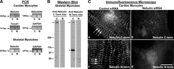
Figure 1.
Treatment of cardiac myocytes with nebulin dsRNA significantly reduces nebulin levels. (A) A specific 202-bp nebulin product was amplified by RT-PCR from rat cardiomyocyte cDNA; this product was virtu...
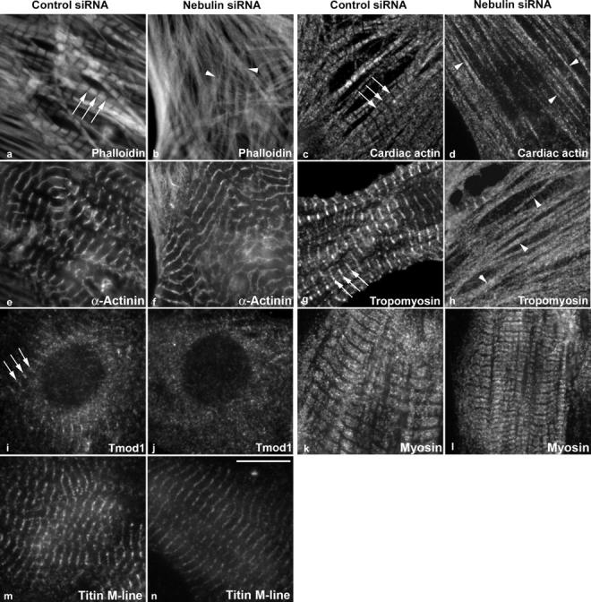
Figure 2.
Nebulin is critical for maintaining thin filament lengths from their pointed ends in cardiomyocytes. After 24 h of siRNA treatment, cardiomyocytes were stained with phalloidin to label F-actin (a and ...
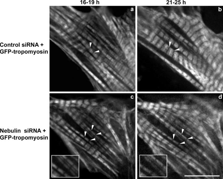
Figure 3.
Real-time imaging using GFP-tropomyosin revealed thin filament elongation after nebulin siRNA treatment. (A) Cardiomyocytes were cotransfected with siRNAs and GFP-tropomyosin and imaged in real time. ...
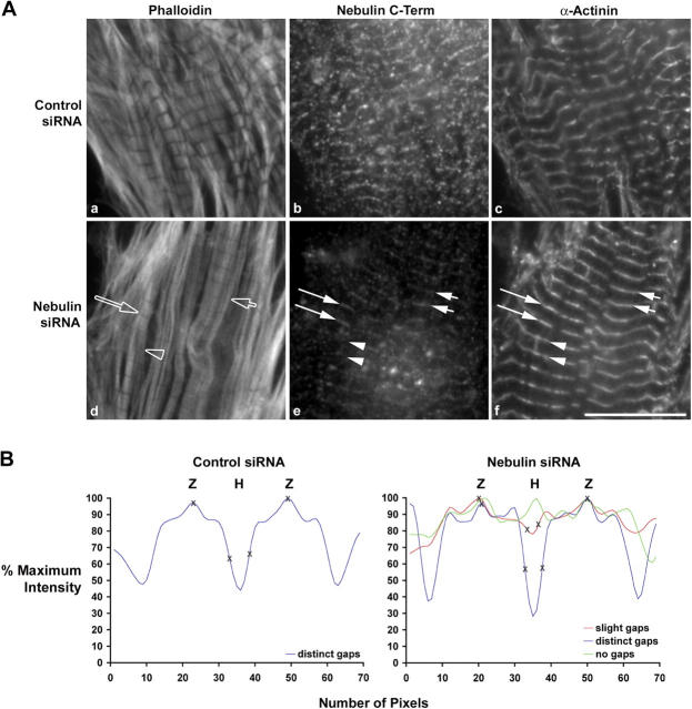
Figure 4.
Analysis at u223c 21 h after siRNA treatment revealed that thin filament elongation correlates with a reduction in nebulin staining intensity. (A) Myocytes were relaxed, fixed, and triple stained with...
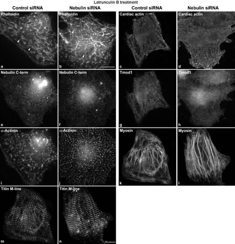
Figure 5.
Thin filaments in cardiomyocytes are specifically depolymerized by Lat B treatment. Myocytes were treated with control or nebulin siRNA for u223c18 h, followed by Lat B for 4 h. Lat B depolymerized th...
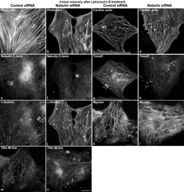
Figure 6.
Nebulin is required for proper thin filament assembly after depolymerization. After depolymerization, Lat B was washed out and the thin filaments were allowed to reassemble in the presence (control si...
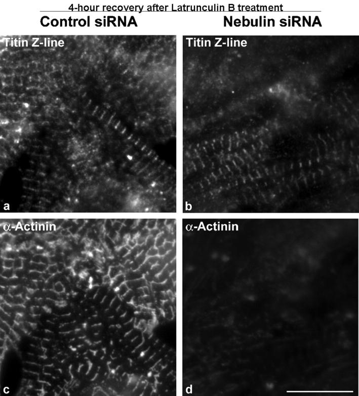
Figure 7.
The thin filament barbed ends are specifically perturbed in the absence of nebulin after Lat B treatment. Cardiac myocytes were double stained with antibodies against u03b1-actinin, a marker of the th...
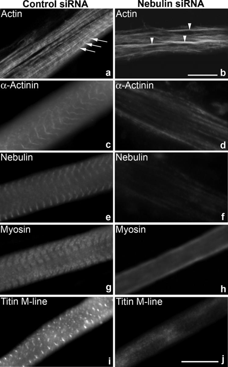
Figure 8.
Nebulin is involved in de novo myofibrillogenesis in rat skeletal myotubes. Nebulin staining was significantly reduced in nebulin siRNAu2013treated myotubes (f), compared with control myotubes (e). By...
Figure images are served from the NIH/NLM PubMed Central Open Access Subset or Europe PMC; copyright remains with the publishers and authors.
💬 Discussion
0 commentsNo comments yet. Be the first to start a discussion!
Leave a Comment