Abstract
The anterior cingulate cortex (ACC) is thought to be important for acute pain perception as well as the development of chronic pain after peripheral nerve injury. Nevertheless, how ACC neurons respond to sensory stimulation under chronic pain states is not well understood. Here, we used an in vivo two-photon imaging technique to monitor the activity of individual neurons in the ACC of awake, head restrained mice. Calcium imaging in the dorsal ACC revealed robust somatic activity in layer 5 (L5) pyramidal neurons in response to peripheral noxious stimuli, and the degree of evoked activity was correlated with the intensity of noxious stimulation. Furthermore, the activation of ACC neurons occurred bilaterally upon noxious stimulation to either contralateral or ipsilateral hind paws. Notably, with nerve injury-induced neuropathic pain in one limb, L5 pyramidal neurons in both sides of the ACC showed enhanced activity in the absence or presence of pain stimuli. These results reveal hyperactivity of L5 pyramidal neurons in the bilateral ACC during the development of neuropathic pain.
🔬 Techniques
✨ Fluorophores
🧪 Sample Preparation
🏭 Microscope Brands
🔴 Lasers
💻 Software Details
🏛️ Research Organizations (ROR)
Affiliated research institutions:
📋 Methods
Animals
All experiments were performed in accordance with the National Institutes of Health guidelines and regulations. The animal protocol was approved by the New York University School of Medicine Animal Care and Use Committee. Thy1 -GCaMP6s mice (Founder Line 3) expressing GCaMP6s in cortical pyramidal neurons were generated in the laboratory of Dr. Wen-Biao Gan. Mice were group-housed in the New York University Skirball animal facility. Two to three-month-old mice of both sexes were used in this study in order to include sex as a potential biological variable. Because the sex of the mice had no effect on pain-related neuronal responses in the ACC, data from male and female mice were grouped together. An average of four mice per group was used in Ca 2+ imaging experiments. Surgical Preparation for Imaging Awake, Head-Restrained Mice In vivo Ca 2+ imaging was performed in awake, head-restrained mice. The surgical procedure for preparing awake animal imaging has been described previously (Yang et al., 2013 ; Cichon et al., 2017 ). In short, while the animal was under deep anesthesia via an intraperitoneal injection of ketamine (100 mg/kg) and xylazine (15 mg/kg), a midline incision of the scalp was made to expose the periosteum, and the skull above the bilateral ACC was located based on stereotactic coordinates (+0.5–1.0 mm anterior of bregma and 0.3–0.8 mm lateral to midline; Allen Mouse Brain Atlas) and marked with ink. A head holder composed of two parallel micro-metal bars was attached to the animal’s skull with glue (Loctite 495) to help restrain the head and reduce motion-induced artifacts during imaging. The head holder was further fortified with dental acrylic cement. After the cement was completely dry, the marked skull region was thinned with a dental drill to a thickness less than 30 μm and then peeled off with the surgical forceps. Dura over the exposed cortex was partially removed. Precaution was taken to avoid tearing the dura on top of the superior sagittal sinus and bridging veins. A glass coverslip (approximately the same size as the bone being removed) was glued to the skull, over the exposed cortex. The surrounding area of the glass window was fortified with dental cement. Mice were given at least 24 h to recover from the surgery-related anesthesia. Before imaging, mice with head mounts were habituated to the imaging apparatus three times (10 min each) to minimize potential stress effects of head restraining and imaging. No obvious distress was observed in habituated animals during imaging experiments. In Vivo Ca 2+ Imaging To image neuronal Ca 2+ activity in the cortex of awake mice, the head holder was screwed to two metal cubes attached to a solid metal plate. Together with the metal plate, the head-restrained animal was placed on the stage of a two-photon microscope. The in vivo Ca 2+ imaging experiments were performed using a Bruker two-photon laser scanning system equipped with a Ti:sapphire laser (Mai Tai DeepSee; Spectra Physics) tuned to 890 nm. To identify L5 of the ACC, an image stack of neurons within a depth of 900 μm from the pial surface was collected to generate a 3-dimensional map and compared with the sagittal atlas. Ca 2+ images were collected from the dorsal part of the ACC (AP: +0.5–1.0 mm, ML: 0.3–0.8 mm, DV: 0.75–0.9 mm) while the animal was under a quiet resting condition, as well as when a mechanical stimulation was applied to the contralateral or ipsilateral hind paw. The average laser power on the brain sample was ~20–30 mW. All experiments were performed using a 25× objective immersed in an artificial cerebral spinal fluid (ACSF) solution and with a 1× digital zoom. Images were collected at a resolution of 512 × 512 pixels and a frame rate of 2 Hz. Image acquisition was performed using Bruker PrairieView v.5.4. software. After Ca 2+ imaging, a red dye was intracerebrally injected to the imaged area and the animal was fixed with 4% paraformaldehyde. The brain was then sectioned at 200 μm thickness and imaged using confocal microscopy to confirm that the dye-labeled region was within the ACC.
Show full methods section
Animals
All experiments were performed in accordance with the National Institutes of Health guidelines and regulations. The animal protocol was approved by the New York University School of Medicine Animal Care and Use Committee. Thy1 -GCaMP6s mice (Founder Line 3) expressing GCaMP6s in cortical pyramidal neurons were generated in the laboratory of Dr. Wen-Biao Gan. Mice were group-housed in the New York University Skirball animal facility. Two to three-month-old mice of both sexes were used in this study in order to include sex as a potential biological variable. Because the sex of the mice had no effect on pain-related neuronal responses in the ACC, data from male and female mice were grouped together. An average of four mice per group was used in Ca 2+ imaging experiments. Surgical Preparation for Imaging Awake, Head-Restrained Mice In vivo Ca 2+ imaging was performed in awake, head-restrained mice. The surgical procedure for preparing awake animal imaging has been described previously (Yang et al., 2013 ; Cichon et al., 2017 ). In short, while the animal was under deep anesthesia via an intraperitoneal injection of ketamine (100 mg/kg) and xylazine (15 mg/kg), a midline incision of the scalp was made to expose the periosteum, and the skull above the bilateral ACC was located based on stereotactic coordinates (+0.5–1.0 mm anterior of bregma and 0.3–0.8 mm lateral to midline; Allen Mouse Brain Atlas) and marked with ink. A head holder composed of two parallel micro-metal bars was attached to the animal’s skull with glue (Loctite 495) to help restrain the head and reduce motion-induced artifacts during imaging. The head holder was further fortified with dental acrylic cement. After the cement was completely dry, the marked skull region was thinned with a dental drill to a thickness less than 30 μm and then peeled off with the surgical forceps. Dura over the exposed cortex was partially removed. Precaution was taken to avoid tearing the dura on top of the superior sagittal sinus and bridging veins. A glass coverslip (approximately the same size as the bone being removed) was glued to the skull, over the exposed cortex. The surrounding area of the glass window was fortified with dental cement. Mice were given at least 24 h to recover from the surgery-related anesthesia. Before imaging, mice with head mounts were habituated to the imaging apparatus three times (10 min each) to minimize potential stress effects of head restraining and imaging. No obvious distress was observed in habituated animals during imaging experiments. In Vivo Ca 2+ Imaging To image neuronal Ca 2+ activity in the cortex of awake mice, the head holder was screwed to two metal cubes attached to a solid metal plate. Together with the metal plate, the head-restrained animal was placed on the stage of a two-photon microscope. The in vivo Ca 2+ imaging experiments were performed using a Bruker two-photon laser scanning system equipped with a Ti:sapphire laser (Mai Tai DeepSee; Spectra Physics) tuned to 890 nm. To identify L5 of the ACC, an image stack of neurons within a depth of 900 μm from the pial surface was collected to generate a 3-dimensional map and compared with the sagittal atlas. Ca 2+ images were collected from the dorsal part of the ACC (AP: +0.5–1.0 mm, ML: 0.3–0.8 mm, DV: 0.75–0.9 mm) while the animal was under a quiet resting condition, as well as when a mechanical stimulation was applied to the contralateral or ipsilateral hind paw. The average laser power on the brain sample was ~20–30 mW. All experiments were performed using a 25× objective immersed in an artificial cerebral spinal fluid (ACSF) solution and with a 1× digital zoom. Images were collected at a resolution of 512 × 512 pixels and a frame rate of 2 Hz. Image acquisition was performed using Bruker PrairieView v.5.4. software. After Ca 2+ imaging, a red dye was intracerebrally injected to the imaged area and the animal was fixed with 4% paraformaldehyde. The brain was then sectioned at 200 μm thickness and imaged using confocal microscopy to confirm that the dye-labeled region was within the ACC.
Analysis of Imaging Data
Imaging data were analyzed post hoc using NIH ImageJ software. When the animal was in a quiet resting state, motion-related artifacts due to the animal’s respiration and heart beat were typically less than 2 μm as detected in our cortical measurements. Vertical movements were infrequent and minimized by habituation and the use of head holder attached to the animal’s skull by dental acrylic. If the animal struggled on the stage of the microscope, imaging time points from those segments were excluded from quantification. Δ F / F 0 was calculated as ( F – F 0 )/ F 0 , where F 0 is the baseline fluorescence signal averaged over a 2-s period corresponding to the lowest fluorescence signal over the recording period. GCaMP6 fluorescence has a long decay time constant (Chen et al., 2013 ), therefore it is difficult to report firing rates based on calcium responses. To compare neuronal activity under various behavioral states (with or without peripheral mechanical stimuli) and among different cells, we performed an integrated measurement of a cell’s output activity over 40-s recording, termed total integrated calcium activity. The total integrated calcium activity was the average of Δ F / F 0 over 10 s per trial (a total of four trials). Neurons responsive to peripheral stimulation were defined as the cells in which evoked somatic activity was beyond three standard deviations of the mean baseline activity. Spared Nerve Injury Spared nerve injury (SNI) of the sciatic nerve or sham operation was performed under strict sterile conditions (Decosterd and Woolf, 2000 ; Bourquin et al., 2006 ; Cichon et al., 2018 ). In short, adult mice (2–3 months old) were deeply anesthetized with an intraperitoneal injection of ketamine (100 mg/kg) and xylazine (15 mg/kg). A small incision was made in the left or right thigh to expose the sciatic nerve and its peripheral branches (common peroneal, tibial and sural nerves). The 8/0 nylon thread (S&T, Fine Science Tools, CA, USA) was slipped under the common peroneal and the tibial nerves to make a ligation and cut, leaving the sural nerve intact. Great care was taken to avoid any contact with or stretching of the intact sural nerve. Muscle and skin were closed in two layers. For sham surgery, the sciatic nerve was exposed but not ligated or cut.
Withdrawal Threshold Test
Mechanical nociceptive threshold was assessed using von Frey (vF) filaments. Mice were individually placed into a plexiglass chamber over a mesh table and habituated for 20 min before testing. Beginning with 0.008 g, vF filaments in a set with logarithmically incremental stiffness (0.02, 0.04, 0.07, 0.16, 0.4, 0.6, 1.0, 1.4, 2.0, 4.0 and 10.0 g) were applied to the hind paws. The 50% withdrawal threshold was calculated using the up-down method as described previously (Chaplan et al., 1994 ). In some experiments, muscimol (2 μg/μl; 0.5 μl) or ACSF was unilaterally injected into the ACC (AP: +1.0 mm, ML: 0.5 mm, DV: 0.9 mm) under brief isoflurane anesthesia. Mechanical thresholds in the contralateral paws were measured before and 30 min after muscimol injection.
Statistics
Prism software (GraphPad 7.0, La Jolla, CA, USA) was used to conduct the statistical analysis. Data were presented as mean ± SEM. Tests for differences between two populations were performed using Mann-Whitney test or Wilcoxon signed-rank test as specified in the text. Multiple group comparison was performed with one-way ANOVA followed by a Tukey’s post hoc test. Significant levels were set at P < 0.05.
Supplementary Material The Supplementary Material for this article can be found online at: https://www.frontiersin.org/articles/10.3389/fncel.2018.00107/full#supplementary-material Click here for additional data file.
📊 Figures
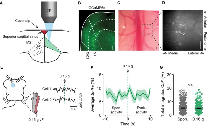
Figure 1
In vivo Ca 2+ imaging of pyramidal neurons in L5 of the anterior cingulate cortex (ACC). (A) Schematic diagram showing in vivo two-photon imaging in the dorsal ACC (dACC) through a glass window. M2, s...
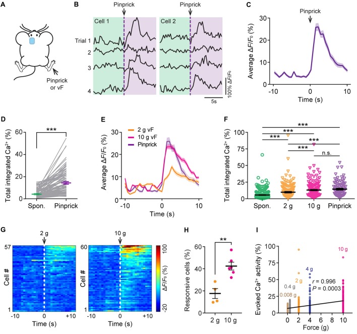
Figure 2
Evoked Ca 2+ activity of ACC neurons in response to peripheral noxious stimuli. (A) Schematic showing in vivo Ca 2+ imaging in the ACC in response to noxious stimulation (pinprick or vF) applied to th...
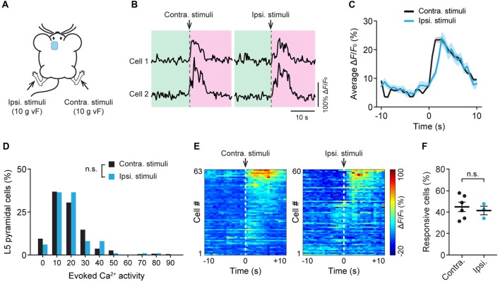
Figure 3
Bilateral responses of ACC neurons to mechanical stimulation to the hind paw. (A) Schematic showing in vivo Ca 2+ imaging in the ACC while mechanical stimulation was applied to the contralateral (cont...
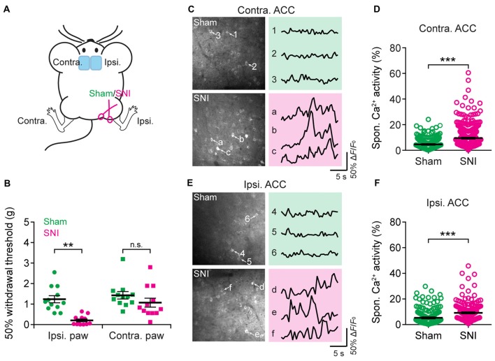
Figure 4
Elevated spontaneous neuronal activity in the ACC of neuropathic pain mice. (A) Schematic showing in vivo Ca 2+ imaging in the ACC of sham or spared nerve injury (SNI) mice. (B) Mechanical paw withdra...
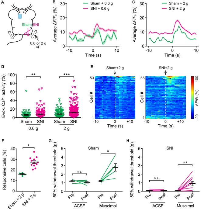
Figure 5
Increased evoked neuronal activity in the ACC of neuropathic pain mice after stimulating the injured limb. (A) Schematic showing in vivo Ca 2+ imaging in the ACC of sham or SNI mice 2 weeks after surg...
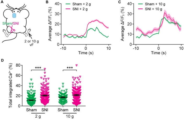
Figure 6
The ACC of neuropathic pain mice shows altered representation of acute pain in the uninjured site. (A) Schematic showing in vivo Ca 2+ imaging in the ACC of sham or SNI mice 2 weeks after surgery. VF ...
Figure images are served from the NIH/NLM PubMed Central Open Access Subset or Europe PMC; copyright remains with the publishers and authors.
💬 Discussion
0 commentsNo comments yet. Be the first to start a discussion!
Leave a Comment