Abstract
At their margins, tumors often contain neutrophils, dendritic cells, and activated macrophages, which express class II MHC and CD11b products. The interplay between stromal cells, tumor cells, and migratory cells such as lymphocytes creates opportunities for noninvasive imaging of immune responses. We developed alpaca-derived antibody fragments specific for mouse class II MHC and CD11b products, expressed on the surface of a variety of myeloid cells. We validated these reagents by flow cytometry and two-photon microscopy to obtain images at cellular resolution. To enable noninvasive imaging of the targeted cell populations, we developed a method to site-specifically label VHHs [the variable domain (VH) of a camelid heavy-chain only antibody] with (18)F or (64)Cu. Radiolabeled VHHs rapidly cleared the circulation (t1/2 ≈ 20 min) and clearly visualized lymphoid organs. We used VHHs to explore the possibility of imaging inflammation in both xenogeneic and syngeneic tumor models, which resulted in detection of tumors with remarkable specificity. We also imaged the infiltration of myeloid cells upon injection of complete Freund's adjuvant. Both anti-class II MHC and anti-CD11b VHHs detected inflammation with excellent specificity. Given the ease of manufacture and labeling of VHHs, we believe that this method could transform the manner in which antitumor responses and/or infectious events may be tracked.
🔬 Techniques
✨ Fluorophores
🧪 Sample Preparation
🔬 Cell Lines
💾 Data Repositories
🏛️ Research Organizations (ROR)
Affiliated research institutions:
📋 Methods
Synthesis of (Gly) 3 -Tetrazine. The tetrapeptide GGGC was synthesized by standard solid-phase peptide synthesis. Maleimide-tetrazine (ClickChemistryTools) was dissolved in 0.1 M phosphate buffer (PB), pH 7. The tetrapeptide GGGC was added and left to stir at room temperature for 3 h until TLC (1:1 Hex:EtOAc vol/vol) indicated near-complete conversion to the product. The solution was filtered and purified by reverse-phase HPLC with a semipreparative column (C 18 column, Gemini, 5 µm, 10 × 250 mm; Phenomenex) at a flow rate of 5.0 mL/min: solvent A, 0.1% TFA in H 2 O; solvent B, 0.1% TFA in CH 3 CN. (G) 3 -Tetrazine eluted at 30–35% (vol/vol) solvent B. Fractions containing pure product were collected and lyophilized. LC-MS calculated for C 37 H 54 N 11 O 13 S[M+H] + was 892.362, found 892.370. Synthesis of (Gly) 3 -NOTA. Maleimide-NOTA (Macromolecules) was dissolved in 0.1 M PB, pH 7. The tetrapeptide GGGC was added at room temperature for 3 h until TLC (1:1 Hex:EtOAc vol/vol) indicated almost complete conversion to the product. The solution was purified by RP-HPLC on a semipreparative column (C 18 column, Gemini, 5 µm, 10 × 250 mm; Phenomenex) at a flow rate of 5.0 mL/min: solvent A, 0.1% TFA in H 2 O; solvent B, 0.1% TFA in CH 3 CN. The desired product eluted from 15% to 20% (vol/vol) solvent B. Fractions containing pure product were collected and lyophilized. LC-MS calculated for C 27 H 45 N 10 O 11 S [M+H] + was 717.298, found 717.305. Enzymatic Incorporation of Substrates into Proteins Using Sortase. The penta-mutant sortase A, with an improved k cat , was used ( 35 ). Reaction mixtures (1 mL) contained Tris · HCl (50 mM, pH 7.5), CaCl 2 (10 mM), NaCl (150 mM), triglycine-containing probe (500 µM), LPETG-containing probe (100 µM), and sortase (5 µM) ( 11 , 36 ). After incubation at 4 °C with agitation for 2 h, reaction products were analyzed by LC-MS, with yields generally >90%. When the yield was below 90%, the reaction was allowed to proceed for an additional 2 h, with addition of sortase to 10 µM and triglycine-containing probe to 750 µM. Ni-NTA beads were added to the reaction mixture with agitation for 5 min at 25 °C, followed by centrifugation to remove sortase and any remaining unreacted His-tagged substrate. The final product—either the tetrazine-labeled protein, NOTA-labeled protein, or fluorophore-labeled protein—was purified by size-exclusion chromatography in PBS or Tris · HCl (50 mM, pH 7.5). The labeled protein was stored at −20 °C with 5% (vol/vol) glycerol and was stable for up to 3 mo. Synthesis and Characterization of 18 F-VHHs. In a typical reaction, a 1.5-mL centrifuge tube was loaded with VHH7-Tz in 1× PBS (40 μL, 150 μM), 1× PBS (300 μL), and 18 F-TCO in DMSO [4.0 mCi (148.0 MBq), 100 μL]. The tube was sealed and shaken at room temperature for 20 min. The mixture was analyzed by radio-TLC [instant thin layer chromatography (ITLC), 100% MeCN, R f 18 F-TCO = 0.9, R f 18 F-VHH7 = 0.0] showing 90% conversion to 18 F-VHH7. The reaction mixture was loaded onto a PD-10 size-exclusion cartridge (GE Healthcare), and elution with 1× PBS provided 2.3 mCi (85.1 MBq) of 18 F-VHH7 in 75.8% decay-corrected radiochemical yield. Starting with 5.3 mCi (196.1 MBq) 18 F-TCO, 18 F-VHHDC13 was prepared following the same procedure as described for 18 F-VHH7 to give 2.8 mCi (103.6 MBq) after size-exclusion chromatography, a 69.7% decay-corrected radiochemical yield. Synthesis and Characterization of 64 Cu-VHHs. In a typical reaction, a 1.5-mL centrifuge tube was loaded with VHH7-NOTA [400 μL, 20 μM in 200 mM NH 4 OAc buffer (pH 6.5)] and 64 CuCl 2 (5.7 mCi, 210.8 MBq) in 200 mM NH 4 OAc buffer (75 μL, pH 6.5). The tube was sealed and shaken at 37 °C for 20 min. The mixture was analyzed by radio-TLC (ITLC, 50 mM EDTA, pH 7, R f 64 Cu/EDTA = 1.0, R f 64 Cu-VHH7 = 0.0) showing 98% conversion to 64 Cu-VHH7. At this time, the mixture was loaded onto a PD-10 size-exclusion cartridge, and elution with 1× PBS provided 5.2 mCi (192.4 MBq) of 64 Cu-VHH7 in 94.2% decay-corrected radiochemical yield. Starting with 3.5 mCi (129.5 MBq) 64 CuCl 2 , 64 Cu-VHHDC13 was prepared following the same procedure as described for 64 Cu-VHH7 to give 3.1 mCi (114.7 MBq) after size-exclusion chromatography, a 92.3% decay-corrected radiochemical yield. Before injection, both 64 Cu-VHH7 and 64 Cu-VHHDC13 were analyzed by radio-TLC (ITLC, 50 mM EDTA, pH 7, R f 64 Cu/EDTA = 1.0, R f 64 Cu-VHH7 and 64 Cu-VHHDC13 = 0.0) and were found to have 99.6% and 99.8% radiochemical purity, respectively. PET-CT Imaging. All procedures and animal protocols were approved by the Massachusetts General Hospital subcommittee on research animal care.
Show full methods section
Synthesis of (Gly) 3 -Tetrazine. The tetrapeptide GGGC was synthesized by standard solid-phase peptide synthesis. Maleimide-tetrazine (ClickChemistryTools) was dissolved in 0.1 M phosphate buffer (PB), pH 7. The tetrapeptide GGGC was added and left to stir at room temperature for 3 h until TLC (1:1 Hex:EtOAc vol/vol) indicated near-complete conversion to the product. The solution was filtered and purified by reverse-phase HPLC with a semipreparative column (C 18 column, Gemini, 5 µm, 10 × 250 mm; Phenomenex) at a flow rate of 5.0 mL/min: solvent A, 0.1% TFA in H 2 O; solvent B, 0.1% TFA in CH 3 CN. (G) 3 -Tetrazine eluted at 30–35% (vol/vol) solvent B. Fractions containing pure product were collected and lyophilized. LC-MS calculated for C 37 H 54 N 11 O 13 S[M+H] + was 892.362, found 892.370. Synthesis of (Gly) 3 -NOTA. Maleimide-NOTA (Macromolecules) was dissolved in 0.1 M PB, pH 7. The tetrapeptide GGGC was added at room temperature for 3 h until TLC (1:1 Hex:EtOAc vol/vol) indicated almost complete conversion to the product. The solution was purified by RP-HPLC on a semipreparative column (C 18 column, Gemini, 5 µm, 10 × 250 mm; Phenomenex) at a flow rate of 5.0 mL/min: solvent A, 0.1% TFA in H 2 O; solvent B, 0.1% TFA in CH 3 CN. The desired product eluted from 15% to 20% (vol/vol) solvent B. Fractions containing pure product were collected and lyophilized. LC-MS calculated for C 27 H 45 N 10 O 11 S [M+H] + was 717.298, found 717.305. Enzymatic Incorporation of Substrates into Proteins Using Sortase. The penta-mutant sortase A, with an improved k cat , was used ( 35 ). Reaction mixtures (1 mL) contained Tris · HCl (50 mM, pH 7.5), CaCl 2 (10 mM), NaCl (150 mM), triglycine-containing probe (500 µM), LPETG-containing probe (100 µM), and sortase (5 µM) ( 11 , 36 ). After incubation at 4 °C with agitation for 2 h, reaction products were analyzed by LC-MS, with yields generally >90%. When the yield was below 90%, the reaction was allowed to proceed for an additional 2 h, with addition of sortase to 10 µM and triglycine-containing probe to 750 µM. Ni-NTA beads were added to the reaction mixture with agitation for 5 min at 25 °C, followed by centrifugation to remove sortase and any remaining unreacted His-tagged substrate. The final product—either the tetrazine-labeled protein, NOTA-labeled protein, or fluorophore-labeled protein—was purified by size-exclusion chromatography in PBS or Tris · HCl (50 mM, pH 7.5). The labeled protein was stored at −20 °C with 5% (vol/vol) glycerol and was stable for up to 3 mo. Synthesis and Characterization of 18 F-VHHs. In a typical reaction, a 1.5-mL centrifuge tube was loaded with VHH7-Tz in 1× PBS (40 μL, 150 μM), 1× PBS (300 μL), and 18 F-TCO in DMSO [4.0 mCi (148.0 MBq), 100 μL]. The tube was sealed and shaken at room temperature for 20 min. The mixture was analyzed by radio-TLC [instant thin layer chromatography (ITLC), 100% MeCN, R f 18 F-TCO = 0.9, R f 18 F-VHH7 = 0.0] showing 90% conversion to 18 F-VHH7. The reaction mixture was loaded onto a PD-10 size-exclusion cartridge (GE Healthcare), and elution with 1× PBS provided 2.3 mCi (85.1 MBq) of 18 F-VHH7 in 75.8% decay-corrected radiochemical yield. Starting with 5.3 mCi (196.1 MBq) 18 F-TCO, 18 F-VHHDC13 was prepared following the same procedure as described for 18 F-VHH7 to give 2.8 mCi (103.6 MBq) after size-exclusion chromatography, a 69.7% decay-corrected radiochemical yield. Synthesis and Characterization of 64 Cu-VHHs. In a typical reaction, a 1.5-mL centrifuge tube was loaded with VHH7-NOTA [400 μL, 20 μM in 200 mM NH 4 OAc buffer (pH 6.5)] and 64 CuCl 2 (5.7 mCi, 210.8 MBq) in 200 mM NH 4 OAc buffer (75 μL, pH 6.5). The tube was sealed and shaken at 37 °C for 20 min. The mixture was analyzed by radio-TLC (ITLC, 50 mM EDTA, pH 7, R f 64 Cu/EDTA = 1.0, R f 64 Cu-VHH7 = 0.0) showing 98% conversion to 64 Cu-VHH7. At this time, the mixture was loaded onto a PD-10 size-exclusion cartridge, and elution with 1× PBS provided 5.2 mCi (192.4 MBq) of 64 Cu-VHH7 in 94.2% decay-corrected radiochemical yield. Starting with 3.5 mCi (129.5 MBq) 64 CuCl 2 , 64 Cu-VHHDC13 was prepared following the same procedure as described for 64 Cu-VHH7 to give 3.1 mCi (114.7 MBq) after size-exclusion chromatography, a 92.3% decay-corrected radiochemical yield. Before injection, both 64 Cu-VHH7 and 64 Cu-VHHDC13 were analyzed by radio-TLC (ITLC, 50 mM EDTA, pH 7, R f 64 Cu/EDTA = 1.0, R f 64 Cu-VHH7 and 64 Cu-VHHDC13 = 0.0) and were found to have 99.6% and 99.8% radiochemical purity, respectively. PET-CT Imaging. All procedures and animal protocols were approved by the Massachusetts General Hospital subcommittee on research animal care.
Supplementary Material Supplementary File Supplementary File Supplementary File Supplementary File Supplementary File Supplementary File Supplementary File Supplementary File Supplementary File Supplementary File Supplementary File Supplementary File Supplementary File Supplementary File Supplementary File Supplementary File Supplementary File Supplementary File Supplementary File Supplementary File Supplementary File Supplementary File Supplementary File Supplementary File Supplementary File
📊 Figures
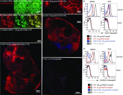
Fig. 1.
VHH7 (anti-mouse class II MHC) and VHHDC13 (anti-mouse CD11b) stain secondary lymphoid organs. VHHs were site-specifically labeled with Texas Red or Alexa 647 via sortagging. In A u2013 F , images wer...
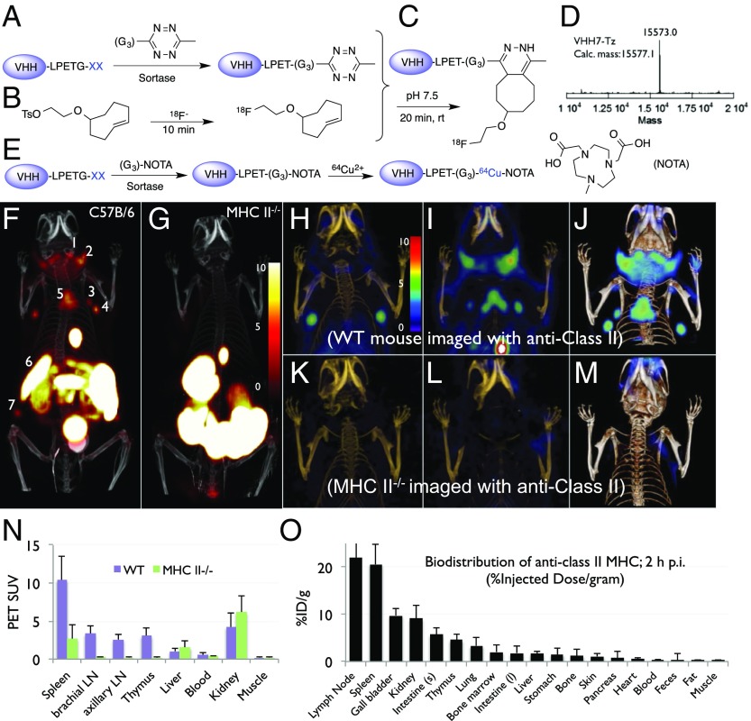
Fig. 2.
( A u2013 E ) Site-specific 18 F or 64 Cu labeling of single-domain antibodies (VHHs) using sortase. ( A ) A single-domain antibody fragment (VHH), equipped at its C terminus with the LPXTG sortase re...
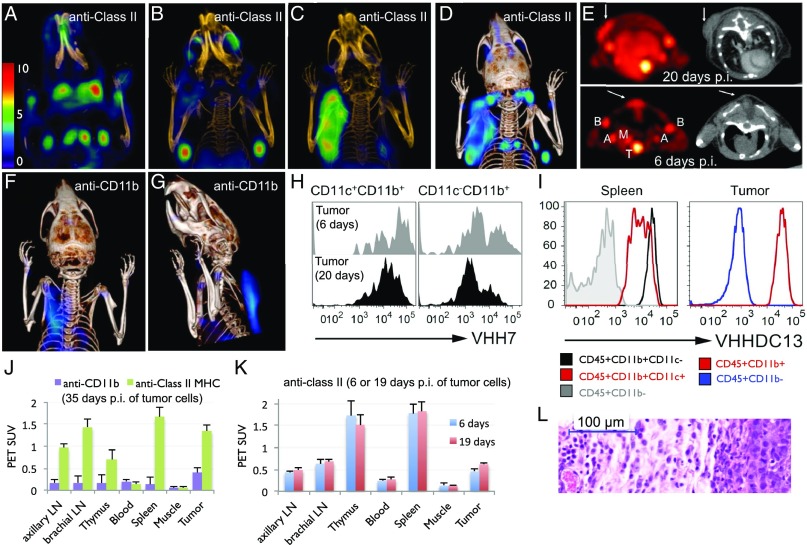
Fig. 3.
18 F-VHH7 (anti-mouse class II MHC) and 18 F-VHHDC13 (anti-mouse CD11b) detects inflammation. Tumor-associated class II MHC + cells were visualized using 18 F-VHH7. A NOD-SCID mouse was inoculated sub...
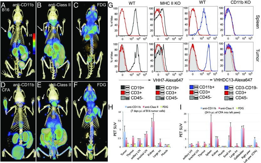
Fig. 4.
18 F-VHH7 (anti-mouse class II MHC) and 18 F-VHHDC13 (anti-mouse CD11b) detects inflammation. ( A u2013 C ) Tumor-associated CD11b + and class II MHC + cells were visualized using 18 F-VHHDC13 and VHH...
Figure images are served from the NIH/NLM PubMed Central Open Access Subset or Europe PMC; copyright remains with the publishers and authors.
💬 Discussion
0 commentsNo comments yet. Be the first to start a discussion!
Leave a Comment