Abstract
Abstract Oocytes form before birth and remain viable for several decades before fertilization 1 . Although poor oocyte quality accounts for most female fertility problems, little is known about how oocytes maintain cellular fitness, or why their quality eventually declines with age 2 . Reactive oxygen species (ROS) produced as by-products of mitochondrial activity are associated with lower rates of fertilization and embryo survival 3–5 . Yet, how healthy oocytes balance essential mitochondrial activity with the production of ROS is unknown. Here we show that oocytes evade ROS by remodelling the mitochondrial electron transport chain through elimination of complex I. Combining live-cell imaging and proteomics in human and Xenopus oocytes, we find that early oocytes exhibit greatly reduced levels of complex I. This is accompanied by a highly active mitochondrial unfolded protein response, which is indicative of an imbalanced electron transport chain. Biochemical and functional assays confirm that complex I is neither assembled nor active in early oocytes. Thus, we report a physiological cell type without complex I in animals. Our findings also clarify why patients with complex-I-related hereditary mitochondrial diseases do not experience subfertility. Complex I suppression represents an evolutionarily conserved strategy that allows longevity while maintaining biological activity in long-lived oocytes.
🔬 Techniques
🔭 Microscopes
🧬 Organisms
💻 Software
✨ Fluorophores
🧪 Sample Preparation
🔬 Cell Lines
🏭 Microscope Brands
🧪 Reagent Suppliers
💻 Software Details
💾 Data Repositories
🏛️ Research Organizations (ROR)
Affiliated research institutions:
📋 Methods
Ethics
Ethical committee permission to work with primordial oocytes from human ovary samples was obtained from the Comité Étic d’Investigació Clínica CEIC-Parc de Salut MAR (Barcelona) and Comité Ético de Investigación Clínica–Hospital Clínic de Barcelona with approval number HCB/2018/0497. Written informed consent was obtained from all participants before their inclusion in the study. Animals used in this study were housed in the Barcelona Biomedical Research Park, accredited by the International Association for Assessment and Accreditation of Laboratory Animal Care. Animal euthanasia was performed by personnel certified by the competent authority (Generalitat de Catalunya) and conformed to the guidelines from the European Community Directive 2010/63 EU, transposed into Spanish legislation on RD 53/2013 for the experimental use of animals.
Animal models
Xenopus laevis adult females of between 2 and 4 years old were purchased from Nasco and maintained in water tanks in the following controlled conditions: 18–21 °C, pH 6.8–7.5, O 2 4–20 ppm, conductivity 500–1,500 µs, ammonia
📊 Figures
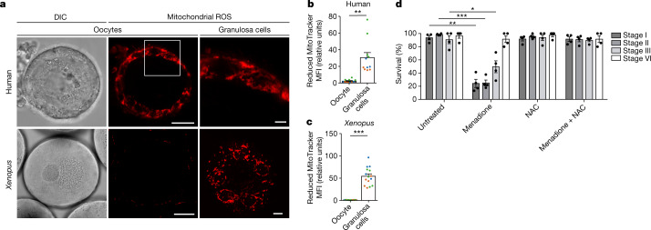
Fig. 1
Early oocytes have undetectable levels of ROS.
a , Live-cell imaging of human and Xenopus early oocytes, both with attached granulosa cells. The ROS level was measured using MitoTracker Red CM-H 2 XRos (H2X), a reduced mitochondrial dye that does ...
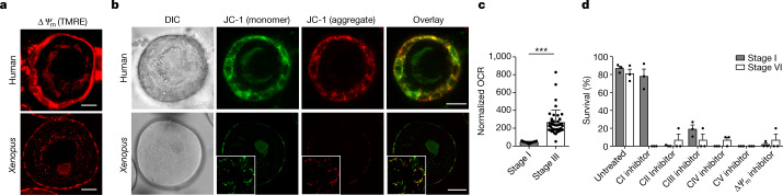
Fig. 2
OXPHOS is low, but essential, in early oocytes.
a , b , Live-cell imaging of human and Xenopus early oocytes with attached granulosa cells labelled with tetramethylrhodamine ethyl ester perchlorate (TMRE) to detect mitochondrial membrane potential ...
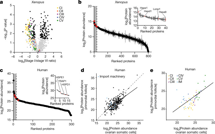
Fig. 3
The mitochondrial proteomes of Xenopus and human oocytes.
a , A volcano plot showing P values versus fold changes of mitochondrial proteins between early (stage I) and late (stage VI) oocytes. The subunits of the mitochondrial OXPHOS machinery are indicated ...
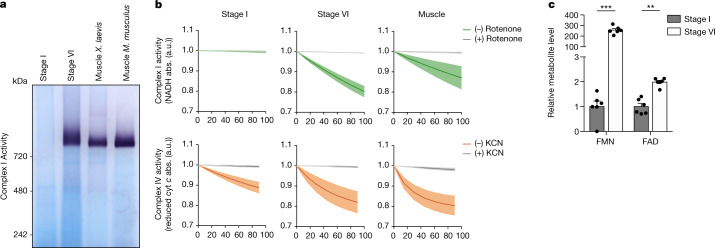
Fig. 4
Complex I is not assembled in early oocytes.
a , Mitochondrial fractions solubilized in n -dodecyl-u03b2- d -maltosideu00a0(DDM) were resolved by BN-PAGE and complex I activity was assayed by reduction of nitro blue tetrazolium chlorideu00a0(NBT...
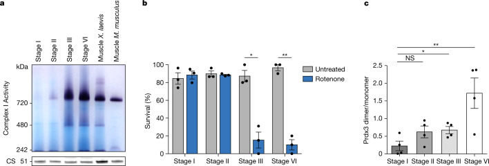
Fig. 5
Complex I and ROS levels correlate throughout oogenesis.
a , Mitochondrial fractions from early (stage I), maturing (stage II and stage III) and late-stage (stage VI) Xenopus oocytes and muscle solubilized in n -dodecyl-u03b2- d -maltosideu00a0(DDM) were re...
Extended Data Fig. 1
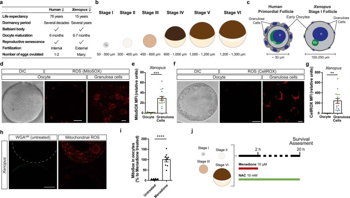
Undetectable levels of ROS in the early oocytes.
a , A comparison between select reproductive traits of humans and Xenopus laevis , which live on average 76 years 59 and 15 years in captivity 60 , respectively. Xenopus oocytes have long been used in...
Extended Data Fig. 2
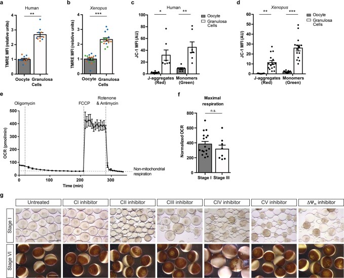
Oxidative phosphorylation is essential in early oocytes.
a , b , Quantification of the mean fluorescence intensity (MFI) of TMRE in the oocyte and in the population of granulosa cells surrounding the equatorial plane of the oocyte for human ( a ) and Xenopu...
Extended Data Fig. 3
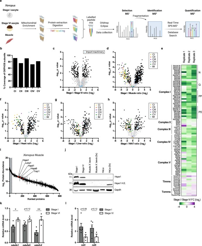
OXPHOS machinery is reduced in Xenopus early oocytes.
a , A schematic representation of isobaric-tag-based quantification of the mitochondrial proteomes of early (stage I), late (stage VI) oocytes, and muscle. The image was created with BioRender.com. b ...
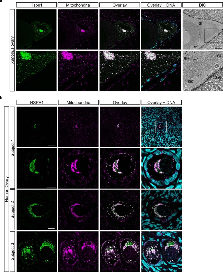
Extended Data Fig. 4
UPR mt is constitutively active in early oocytes of human and Xenopus .
a , b , Immunofluorescence of paraffin embedded sections of Xenopus ( a ) and human ( b ) ovaries with antibodies against HSPE1 (to monitor UPR mt ) and ATP5A1 (to mark mitochondria). Hoechst was used...
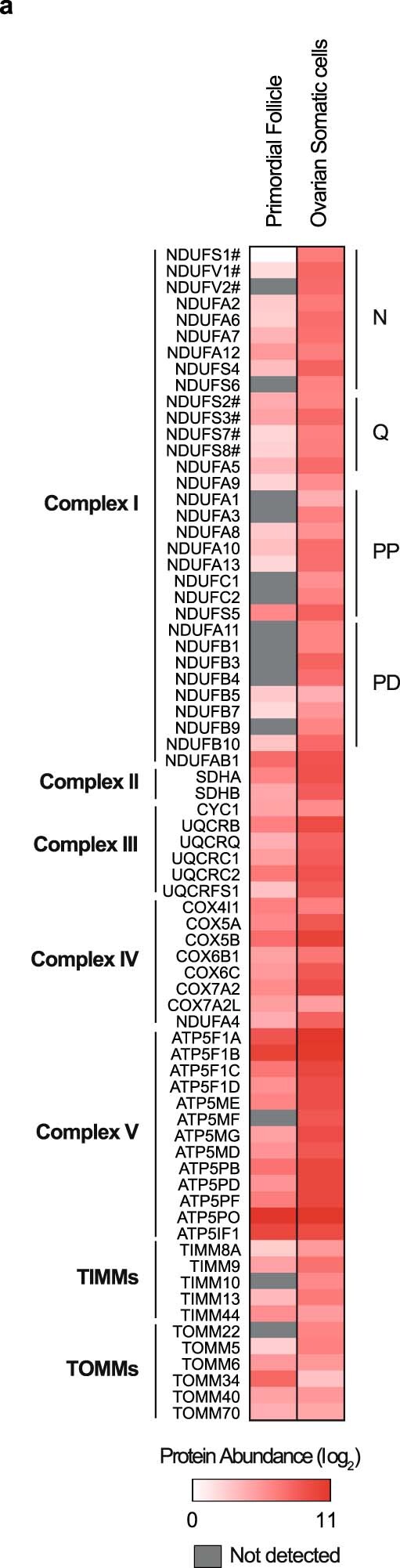
Extended Data Fig. 5
OXPHOS machinery is reduced in human early oocytes.
a , Heatmap of normalized mitochondrial protein abundances in human primordial follicles and ovarian somatic cells organized by subunits of OXPHOS and mitochondrial import machinery. # marks core subu...
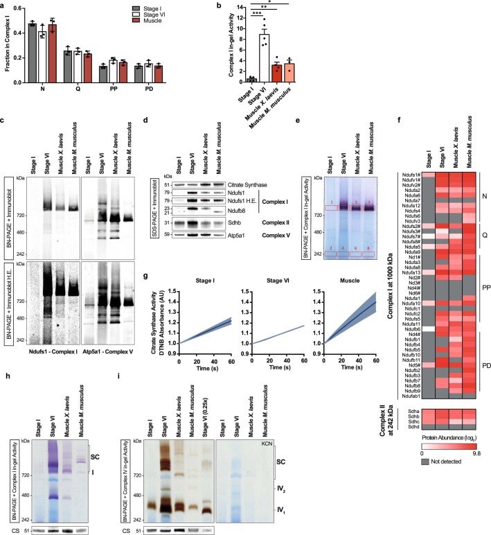
Extended Data Fig. 6
Complex I is not assembled in early oocytes.
a , Fraction of the mean abundance of the indicated modules among all complex I modules in Xenopus oocytes and muscle tissue. Note that this analysis inevitably misses undetected subunits. The data re...
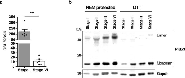
Extended Data Fig. 7
Complex I and ROS levels throughout oogenesis.
a , GSH/GSSG ratio in early (stage I) and late-stage (stage VI) oocytes. The data represent the mean u00b1 s.e.m., n=6. ** P =u20091.24 u00d7 10 u22126 using two-sided Studentu2019s t -test. b , Immun...
Figure images are served from the NIH/NLM PubMed Central Open Access Subset or Europe PMC; copyright remains with the publishers and authors.
💬 Discussion
0 commentsNo comments yet. Be the first to start a discussion!
Leave a Comment