Abstract
Aging is accompanied by impaired glucose homeostasis and an increased risk of type 2 diabetes, culminating in the failure of insulin secretion from pancreatic β-cells. To investigate the effects of age on β-cell metabolism, we established a novel assay to directly image islet metabolism with NAD(P)H fluorescence lifetime imaging (FLIM). We determined that impaired mitochondrial activity underlies an age-dependent loss of insulin secretion in human islets. NAD(P)H FLIM revealed a comparable decline in mitochondrial function in the pancreatic islets of aged mice (≥24 months), the result of 52% and 57% defects in flux through complex I and II, respectively, of the electron transport chain. However, insulin secretion and glucose tolerance are preserved in aged mouse islets by the heightened metabolic sensitivity of the β-cell triggering pathway, an adaptation clearly encoded in the metabolic and Ca2+ oscillations that trigger insulin release (Ca2+ plateau fraction: young 0.211 ± 0.006, aged 0.380 ± 0.007, P < 0.0001). This enhanced sensitivity is driven by a reduction in KATP channel conductance (diazoxide: young 5.1 ± 0.2 nS; aged 3.5 ± 0.5 nS, P < 0.01), resulting in an ∼2.8 mmol/L left shift in the β-cell glucose threshold. The results demonstrate how mice but not humans are able to successfully compensate for age-associated metabolic dysfunction by adjusting β-cell glucose sensitivity and highlight an essential mechanism for ensuring the maintenance of insulin secretion.
🔬 Techniques
💻 Software
✨ Fluorophores
🧪 Sample Preparation
🏭 Microscope Brands
🧪 Reagent Suppliers
🔴 Lasers
📷 Detectors
🔎 Objectives
🎨 Filters
💻 Software Details
🏛️ Research Organizations (ROR)
Affiliated research institutions:
📋 Methods
Research Design and Methods Human Islets An exemption was granted for all protocols by the institutional review board at the University of Wisconsin-Madison under protocol number 2015-0356. Human islets were obtained from the Integrated Islet Distribution Program, including the center at the University of Wisconsin. The Integrated Islet Distribution Program isolation protocols and insulin secretion quality control assays can be found at https://iidp.coh.org/investigator_sops.aspx . After shipment, the islets were cultured overnight in RPMI 1640/10% FBS and used within 24 h for insulin secretion measurements.
Mice
All animal studies were approved by the Institutional Animal Care and Use Committees of the University of Wisconsin-Madison and the William S. Middleton Memorial Veterans Hospital. Aged (18-, 24-, and 30-month-old) and young (4- to 6-month-old) C57BL/6J male mice used for the insulin secretion and mitochondrial assays were from the National Institute on Aging (NIA) Aged Rodent Colony. Additional 4- to 6-month-old C57BL/6J male mice used for the calcium and electrophysiology experiments were purchased from The Jackson Laboratory. The median life expectancy of C57BL/6J male mice from these sources is ∼28 months ( 20 ) ( http://phenome.jax.org ). In Vivo Studies Glucose tolerance tests were performed by fasting the mice overnight for 16 h and then injecting 1 g/kg glucose intraperitoneally; glucose measurements were then performed at the indicated times by using a Bayer Contour blood glucose meter and test strips. In vivo GSIS and fasting insulin levels were determined by fasting the mice overnight for 16 h, measuring blood glucose, and collecting tail blood (using Sarstedt heparinized tubes) immediately before and 15 min after injecting 1 g/kg glucose. Insulin levels in heparinized plasma were determined using Mouse Insulin ELISA kits (Crystal Chem).
Show full methods section
Research Design and Methods Human Islets An exemption was granted for all protocols by the institutional review board at the University of Wisconsin-Madison under protocol number 2015-0356. Human islets were obtained from the Integrated Islet Distribution Program, including the center at the University of Wisconsin. The Integrated Islet Distribution Program isolation protocols and insulin secretion quality control assays can be found at https://iidp.coh.org/investigator_sops.aspx . After shipment, the islets were cultured overnight in RPMI 1640/10% FBS and used within 24 h for insulin secretion measurements.
Mice
All animal studies were approved by the Institutional Animal Care and Use Committees of the University of Wisconsin-Madison and the William S. Middleton Memorial Veterans Hospital. Aged (18-, 24-, and 30-month-old) and young (4- to 6-month-old) C57BL/6J male mice used for the insulin secretion and mitochondrial assays were from the National Institute on Aging (NIA) Aged Rodent Colony. Additional 4- to 6-month-old C57BL/6J male mice used for the calcium and electrophysiology experiments were purchased from The Jackson Laboratory. The median life expectancy of C57BL/6J male mice from these sources is ∼28 months ( 20 ) ( http://phenome.jax.org ). In Vivo Studies Glucose tolerance tests were performed by fasting the mice overnight for 16 h and then injecting 1 g/kg glucose intraperitoneally; glucose measurements were then performed at the indicated times by using a Bayer Contour blood glucose meter and test strips. In vivo GSIS and fasting insulin levels were determined by fasting the mice overnight for 16 h, measuring blood glucose, and collecting tail blood (using Sarstedt heparinized tubes) immediately before and 15 min after injecting 1 g/kg glucose. Insulin levels in heparinized plasma were determined using Mouse Insulin ELISA kits (Crystal Chem).
Islet Isolation and Adenoviral Infection
The mouse pancreas was inflated through the common bile duct with 3–5 mL of 0.5 mg/mL collagenase (C5138; Sigma) and 0.2 mg/mL BSA (A8806; Sigma) in Hanks’ balanced salt solution (HBSS) (Invitrogen), excised, and incubated in a glass vial at 37°C in 5 mL HBSS/BSA/collagenase solution for 5 min on an orbital shaking water bath (SHKA7000; Thermo Fisher Scientific) at 250 rpm. Beginning 6 min postincubation, the digest was agitated for 20 s at 375 rpm every 2 min until minute 28, washed three times with 30 mL ice-cold HBSS/BSA solution, and pelleted at 50 g for 2 min. The pellets were resuspended by vortex at medium speed in 1–2 mL of solution and hand-picked from 60 mL of ice-cold HBSS/BSA solution. After isolation, the islets were maintained in RPMI 1640 supplemented with 10% FBS (volume for volume), 100 units/mL penicillin, and 100 μg/mL streptomycin (Invitrogen). Adenoviruses were used to express PercevalHR ATP/ADP sensors (49082; Addgene) in islet β-cells under control of the rat insulin promoter as in Merrins et al. ( 21 ). Groups of 25 freshly isolated islets were immediately infected with 2,000 multiplicity of infection of each adenoviral construct for 2 h in a 95%/5% air/CO 2 incubator at 37°C followed by removal to fresh culture media lacking virus. Islets were imaged after 1–3 days in culture. We confirmed that the presence of the PercevalHR sensors, which were expressed in only a small fraction of β-cells, had no effect on downstream Ca 2+ oscillations. Respirometry After overnight incubation, 150 islets/condition/mouse (six mice/condition) were transferred to a 35-mm Petri dish containing 2 mL of MiR05 (in mmol/L: 0.5 EGTA, 3 MgCl 2 , 60 lactobionic acid, 20 taurine, 10 KH 2 PO 4 , 20 HEPES, 110 sucrose, 1% BSA; pH 7.1, adjusted with 5 mol/L KOH) ( 22 ) and 5 mg/mL saponin. Preliminary experiments showed that permeabilized islets demonstrated higher coupled respiration rates. Islets were gently rocked in the permeabilization medium for 20 min. After 20 min, the islets were rinsed with fresh MiR05 in the absence of saponin, and then pipetted into an Oxygraph-2k chamber (OROBOROS) containing 2 mL of MiR05 at 37°C. Oxygen electrodes were calibrated to air-saturated MiR05 buffer at 37°C using published oxygen solubilities ( 23 ) corrected for local atmospheric pressure. Oxygen concentration and oxygen flux were recorded using DatLab software (OROBOROS). A modified substrate-uncoupler-inhibitor titration protocol based on Votion et al. ( 24 ) was applied. Respiratory flux through complex I was measured by adding 5 mmol/L glutamate and 1 mmol/L malate followed by 1.25 mmol/L ADP to induce oxidative phosphorylation. To assess the integrity of the outer mitochondrial membrane, cytochrome c was added to the chamber (8 μmol/L) ( 25 ), and no significant increases in respiration were observed. Damage to the outer mitochondrial membrane during permeabilization would result in a loss of cytochrome c and a large increase in respiration upon the addition of exogenous cytochrome c to the respiration chamber. Convergent electron flow through complexes I and II was measured with the addition of 10 mmol/L succinate, allowing the electron transport system to achieve maximal coupled respiration. Next, a stepwise addition of carbonyl cyanide p-trifluoromethoxyphenylhydrazone was used to completely uncouple mitochondria (0.250 μmol/L initial + 0.250 μmol/L additions). To assess oxygen flux through complex II, 0.5 μmol/L of rotenone was added to inhibit complex I. Finally, electron transfer was blocked at complex III with the addition of 5 μmol/L of antimycin A and allowed to run long enough to obtain a residual oxygen consumption. Oxygen flux was expressed per milligram of total protein (Pierce 660-nm protein assay; Thermo Fisher Scientific). FLIM of NAD(P)H Islets were imaged in glass-bottom dishes on a custom-built multiphoton laser scanning system based around a Nikon TE-300 inverted microscope equipped with a Plan Apo 60×/1.4 NA oil immersion objective (Nikon Instruments) in a standard external solution containing (in mmol/L) 135 NaCl, 4.8 KCl, 5 CaCl 2 , 1.2 MgCl 2 , 20 HEPES, and 10 glucose (pH 7.35). Temperature was maintained at 35°C by using a LiveCell incubator (Pathology Devices). NAD(P)H was excited with a Mai Tai DeepSee Ti:Sapphire laser (Spectra-Physics) at 740 nm with a 450/70m bandpass emission filter (Chroma) before being collected by a Hamamatsu H7422P-40 GaAsP photomultiplier tube. FLIM images were collected at 256 × 256 resolution with 120 s (1/s) collection using SPC-830 Photon Counting Electronics (Becker & Hickl GmbH). In each experiment, urea crystals were used to define the instrument response function with a 370/10 bandpass emission filter (Chroma), and coumarin was used as a reference for lifetime (2.5 ns) by using a 450/70m bandpass emission filter (Chroma). For analysis, raw SDT files were imported into MATLAB (MathWorks), and a custom script was used to generate a phasor histogram for each treatment by using the equations in Digman et al. ( 26 ). To avoid contamination from lipofuscin fluorescence in the aged cells ( 27 ), which forms a short lifetime tail in the phasor plot, all data were reported as the phasor histogram peak ( 1-g max ,s max ). Time-lapse Imaging of NAD(P)H, ATP/ADP, and Ca 2+ For measurements of cytosolic Ca 2+ , islets were preincubated in 2.5 μmol/L Fura Red (Molecular Probes, Eugene, OR) for 45 min at 37°C. Islets were then placed in an RC-24N glass-bottom chamber (54 μL volume) (Warner Instruments) on a Nikon Eclipse Ti-E inverted microscope equipped with Super Fluor 10×/0.5 NA and 20×/0.75 NA objectives (Nikon Instruments). The chamber was perfused with standard external solution (as previously described). The flow rate was 0.3 mL/min, and temperature was maintained at 33°C using inline solution and chamber heaters (Warner Instruments). Excitation was provided by a SOLA SE II 365 (lumencor) set to 10% output. Excitation (x) or emission (m) filters (ET type; Chroma Technology) were used in combination with an FF444/521/608-Di01 dichroic beamsplitter (Semrock) as follows: Fura Red 430/20x and 500/20x, 630/70m (R430/500); NAD(P)H 365/20x, 470/24m; and PercevalHR 430/20x and 500/20x, 535/35m (R500/430). Fluorescence emission was collected with a Hamamatsu ORCA-Flash4.0 V2 Digital CMOS camera at 0.125–0.2 Hz. A single region of interest was used to quantify the average response of each islet using Nikon NIS-Elements and MathWorks MATLAB software.
Electrophysiology β-Cell membrane potential and K
ATP conductance were measured as in Ren et al. ( 28 ). Briefly, a Sutter MP-225 micromanipulator was used together with a HEKA EPC 10 patch clamp amplifier in the perforated patch clamp configuration to record membrane potential from intact islets perfused with standard external solution (as previously described). Pipette tips were filled with an internal solution (in mmol/L: 28.4 K 2 SO 4 , 63.7 KCl, 11.8 NaCl, 1 MgCl 2 , 20.8 HEPES, 0.5 EGTA, 40 sucrose; pH 7.2) containing 0.36 mg/mL of amphotericin B. Islet β-cells were identified by the presence of slow oscillations in 10 mmol/L glucose. The amplifier was then switched into voltage clamp mode. Conductance changes were determined from the current-voltage relation using 2-s voltage ramps from −120 to −50 mV every 20 s during the silent phase of bursting and following application of 200 μm diazoxide. Assuming that diazoxide acts by maximizing steady-state K ATP open probability, changes in conductance primarily reflect changes in the number of open K ATP channels. Ex Vivo Insulin Release The GSIS assay was performed on human and mouse islets by using a high-throughput multiple-well plate technique ( 29 ) in which islets were individually incubated in a tissue culture–treated 96-well V-bottom plate, allowed to adhere for 24 h, and incubated with Krebs-Ringer bicarbonate buffer at 1.7 mmol/L glucose (preincubation) followed by a stimulatory 16.7 mmol/L glucose incubation for 45 min each. Secretory medium was then collected, and the islets were lysed with a cell lysis buffer (9803; Cell Signaling Technology). Insulin secretion as a percentage of total islet insulin content was measured by ELISA.
Statistics
Data are expressed as mean ± SE. Statistical significance was determined using one- or two-way ANOVA with Sidak multiple comparisons test post hoc or Student t test as appropriate. Differences were considered to be statistically significant at P < 0.05. Statistical calculations were performed with GraphPad Prism software.
📊 Figures
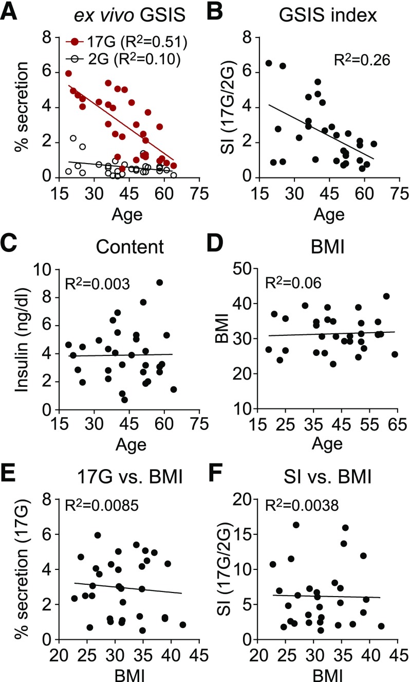
Figure 1
Insulin secretion declines with age in human islets. Insulin secretion (2 mmol/L glucose [2G] and 17 mmol/L glucose [17G]) ( A ), stimulation index (SI) (i.e., stimulated/basal secretion) ( B ), insul...
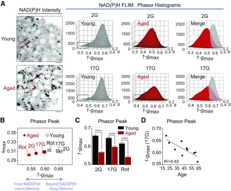
Figure 2
NAD(P)H FLIM of human islet metabolism. A : Multiphoton FLIM of NAD(P)H in human islets from representative young (24-year-old, ACJ3270) and aged (58-year-old, ACKH054A) donors. Phasor histograms show...
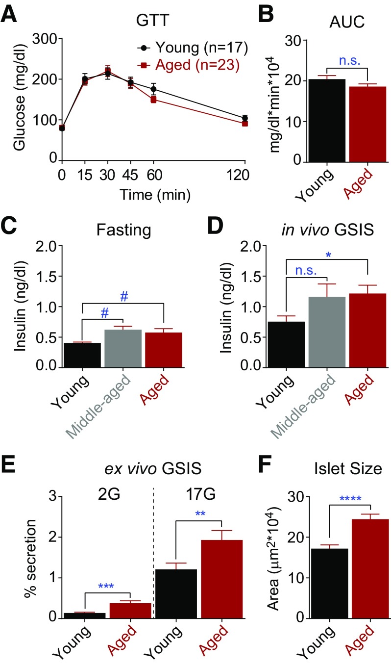
Figure 3
Insulin secretion is increased in aged mice. In vivo glucose tolerance ( A and B ) in young (6-month-old) and aged (u226524-month-old) C57BL/6J mice. C and D : Plasma insulin levels in young (6-month-...
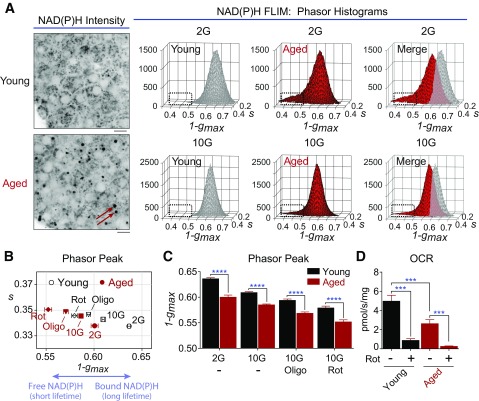
Figure 4
NAD(P)H FLIM of mouse islet metabolism. A : Multiphoton FLIM of NAD(P)H in islets isolated from young (6-month-old) and aged (30-month-old) C57BL/6J mice. Arrows in the NAD(P)H intensity images indica...
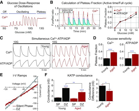
Figure 5
Loss of K ATP channel conductance underlies a left shift in the glucose threshold of aged mouse u03b2-cells. A : Recording of islet Ca 2+ in response to increased glucose levels. B : Glucose dependenc...
Figure images are served from the NIH/NLM PubMed Central Open Access Subset or Europe PMC; copyright remains with the publishers and authors.
💬 Discussion
0 commentsNo comments yet. Be the first to start a discussion!
Leave a Comment