Abstract
Abstract Aims/hypothesis Pancreatic beta cells are subjected to exogenous damaging factors such as proinflammatory cytokines or excess glucose that can cause accumulation of damage-inducing reactive oxygen species during the pathogenesis of diabetes. We and others have shown that beta cell autophagy can reduce reactive oxygen species to protect against apoptosis. While impaired islet autophagy has been demonstrated in human type 2 diabetes, it is unknown if islet autophagy is perturbed in the pathogenesis of type 1 diabetes. We hypothesised that beta cell autophagy is dysfunctional in type 1 diabetes, and that there is a progressive loss during early diabetes development. Methods Pancreases were collected from chloroquine-injected and non-injected non-obese diabetes-resistant (NOR) and non-obese diabetic (NOD) mice. Age- and BMI-matched pancreas tissue sections from human organ donors ( N = 34) were obtained from the Network for Pancreatic Organ Donors with Diabetes (nPOD). Tissue sections were stained with antibodies against proinsulin or insulin (beta cell markers), microtubule-associated protein 1 light chain 3 A/B (LC3A/B; autophagosome marker), lysosomal-associated membrane protein 1 (LAMP1; lysosome marker) and p62 (autophagy adaptor). Images collected on a scanning laser confocal microscope were analysed with CellProfiler and ImageJ. Secondary lysosomes and telolysosomes were assessed in electron micrographs of human pancreatic tissue sections ( n = 12), and energy dispersive x-ray analysis was performed to assess distribution of elements ( n = 5). Results We observed increased autophagosome numbers in islets of diabetic NOD mice ( p = 0.008) and increased p62 in islets of both non-diabetic and diabetic NOD mice ( p < 0.001) vs NOR mice. There was also a reduction in LC3–LAMP1 colocalisation in islets of diabetic NOD mice compared with both non-diabetic NOD ( p < 0.001) and NOR mice ( p < 0.001). Chloroquine elicited accumulation of autophagosomes in the islets of NOR ( p = 0.003) and non-diabetic NOD mice ( p < 0.001), but not in islets of diabetic NOD mice; and stimulated accumulation of p62 in NOR ( p < 0.001), but not in NOD mice. We observed reduced LC3–LAMP1 colocalisation ( p < 0.001) in residual beta cells of human donors with type 1 diabetes vs non-diabetic participants. We also observed reduced colocalisation of proinsulin with LAMP1 in donors with type 1 diabetes ( p < 0.001). Electron microscopy also revealed accumulation of telolysosomes with nitrogen-dense rings in beta cells of autoantibody-positive donors ( p = 0.002). Conclusions/interpretation We provide evidence of islet macroautophagy/crinophagy impairment in human type 1 diabetes. We also document accumulation of telolysosomes with peripheral nitrogen in beta cells of autoantibody-positive donors, demonstrating altered lysosome content that may be associated with lysosome dysfunction before clinical hyperglycaemia. Similar macroautophagy impairments are present in the NOD mouse model of type 1 diabetes. Graphical abstract
🔬 Techniques
🔭 Microscopes
💻 Software
✨ Fluorophores
🧪 Sample Preparation
🏭 Microscope Brands
🧪 Reagent Suppliers
📷 Detectors
💻 Software Details
💾 Data Repositories
🏷️ Research Resource Identifiers (RRIDs)
Verified research resources used in this paper:
🏛️ Research Organizations (ROR)
Affiliated research institutions:
📋 Methods
Pancreases were collected from chloroquine-injected and non-injected non-obese diabetes-resistant (NOR) and non-obese diabetic (NOD) mice. Age- and BMI-matched pancreas tissue sections from human organ donors ( N = 34) were obtained from the Network for Pancreatic Organ Donors with Diabetes (nPOD). Tissue sections were stained with antibodies against proinsulin or insulin (beta cell markers), microtubule-associated protein 1 light chain 3 A/B (LC3A/B; autophagosome marker), lysosomal-associated membrane protein 1 (LAMP1; lysosome marker) and p62 (autophagy adaptor). Images collected on a scanning laser confocal microscope were analysed with CellProfiler and ImageJ. Secondary lysosomes and telolysosomes were assessed in electron micrographs of human pancreatic tissue sections ( n = 12), and energy dispersive x-ray analysis was performed to assess distribution of elements ( n = 5).
Methods Mice
Non-obese diabetes-resistant (NOR) and non-obese diabetic (NOD) mice were purchased from The Jackson Laboratory (ME, USA) at ~7 weeks of age. Mice were housed in a temperature-controlled facility with a 12 h light/12 h dark cycle and were given free access to food and water. All experiments were approved by the Indiana University School of Medicine Institutional Animal Care and Use Committee. Random-fed blood glucose for NOR mice and NOD mice was monitored bi-weekly with an AlphaTrak2 glucometer (Zoetis, NJ, USA), and NOD mice were characterised as diabetic after 2 consecutive days of blood glucose readings >13.9 mmol/l. Mice were euthanised by cervical dislocation and pancreases were collected. Harvested tissues were fixed in 3.7% paraformaldehyde (vol./vol.) for 4–5 h at room temperature with gentle agitation and then transferred to 70% ethanol. Pancreases were then paraffin embedded and sectioned in the Histology Core of the Indiana Center for Musculoskeletal Health, Indiana University School of Medicine. For all experiments, female mice aged 11–26 weeks were used.
Show full methods section
Pancreases were collected from chloroquine-injected and non-injected non-obese diabetes-resistant (NOR) and non-obese diabetic (NOD) mice. Age- and BMI-matched pancreas tissue sections from human organ donors ( N = 34) were obtained from the Network for Pancreatic Organ Donors with Diabetes (nPOD). Tissue sections were stained with antibodies against proinsulin or insulin (beta cell markers), microtubule-associated protein 1 light chain 3 A/B (LC3A/B; autophagosome marker), lysosomal-associated membrane protein 1 (LAMP1; lysosome marker) and p62 (autophagy adaptor). Images collected on a scanning laser confocal microscope were analysed with CellProfiler and ImageJ. Secondary lysosomes and telolysosomes were assessed in electron micrographs of human pancreatic tissue sections ( n = 12), and energy dispersive x-ray analysis was performed to assess distribution of elements ( n = 5).
Methods Mice
Non-obese diabetes-resistant (NOR) and non-obese diabetic (NOD) mice were purchased from The Jackson Laboratory (ME, USA) at ~7 weeks of age. Mice were housed in a temperature-controlled facility with a 12 h light/12 h dark cycle and were given free access to food and water. All experiments were approved by the Indiana University School of Medicine Institutional Animal Care and Use Committee. Random-fed blood glucose for NOR mice and NOD mice was monitored bi-weekly with an AlphaTrak2 glucometer (Zoetis, NJ, USA), and NOD mice were characterised as diabetic after 2 consecutive days of blood glucose readings >13.9 mmol/l. Mice were euthanised by cervical dislocation and pancreases were collected. Harvested tissues were fixed in 3.7% paraformaldehyde (vol./vol.) for 4–5 h at room temperature with gentle agitation and then transferred to 70% ethanol. Pancreases were then paraffin embedded and sectioned in the Histology Core of the Indiana Center for Musculoskeletal Health, Indiana University School of Medicine. For all experiments, female mice aged 11–26 weeks were used.
Chloroquine injections
To analyse dynamic autophagic flux, a subset of NOR mice (11 weeks; n = 5), non-diabetic NOD mice (14 weeks; n = 5) and diabetic NOD mice (14–26 weeks; n = 3) were intra-peritoneally injected with 50 mg/kg of chloroquine diphosphate (Tocris Bioscience #4109, Bristol, UK). At 2 h post injection, pancreases were collected, paraffin embedded and sectioned. For non-injected controls, a subset of NOR mice ( n = 5), non-diabetic NOD mice ( n = 6) and diabetic NOD mice ( n = 7) were used.
Human organ donor characteristics
We obtained deidentified pancreatic tissue sections from 34 human organ donors through the JDRF Network for Pancreatic Organ Donors with Diabetes (nPOD; Table 1 ). Because samples were deidentified, they were exempt from institutional review board oversight. Samples included sections from 12 non-diabetic control donors (six male and six female donors), 12 autoantibody-positive donors (six male, six female) and ten donors with type 1 diabetes (five male, five female) that had residual insulin-positive islets. Age- (26.15 ± 2 years) and BMI- (26.55 ± 1 kg/m 2 ) matched donor samples were used. A breakdown of age and BMI in each group is shown in electronic supplementary material (ESM) Fig. 1 and in the Human Islet Checklist . The duration of diabetes ranged from
📊 Figures
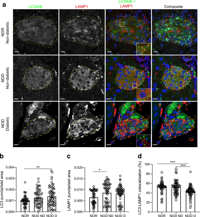
Fig. 1
Reduced autophagy in the pancreatic islets of diabetic NOD mice. ( a ) Representative images showing immunofluorescent staining of islets in pancreas tissue sections from non-diabetic NOR, non-diabeti...
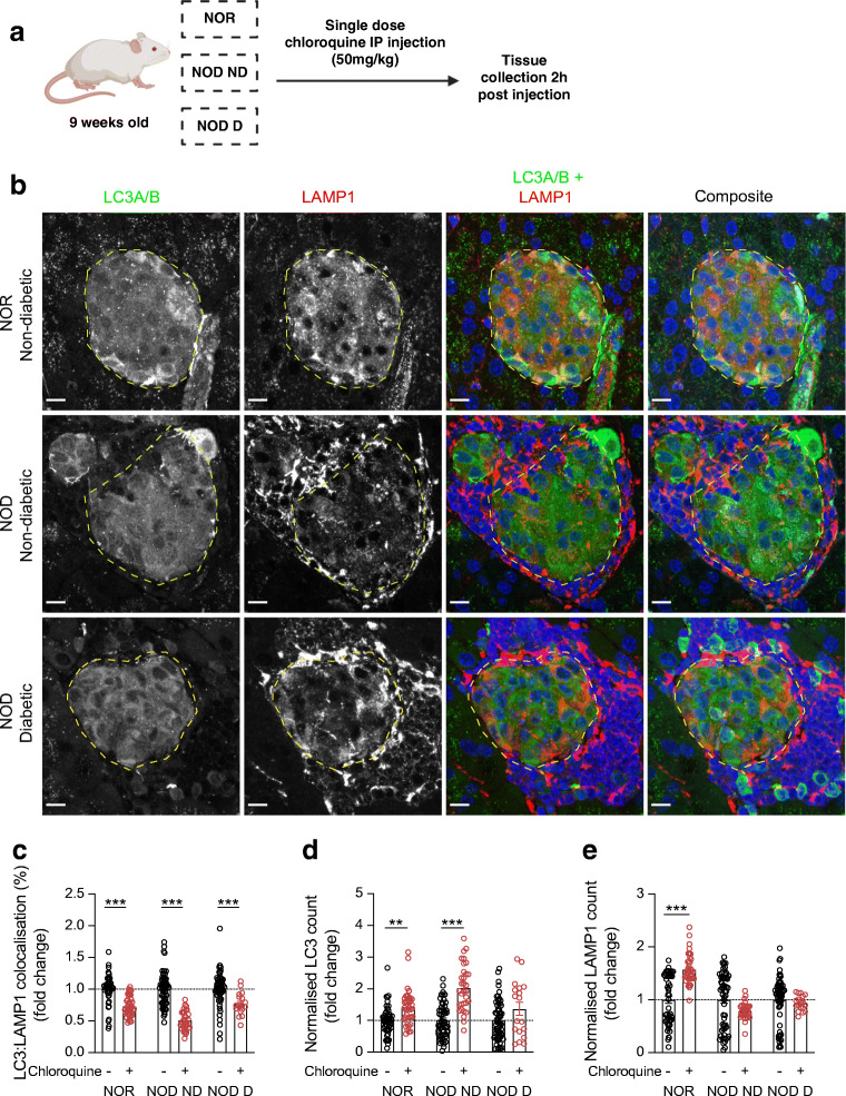
Fig. 2
Impaired autophagic flux in the islets of diabetic NOD mice . ( a ) Schematic of autophagic flux assessment experiment, created in BioRender.com . ( b ) Representative images showing immunofluorescent...
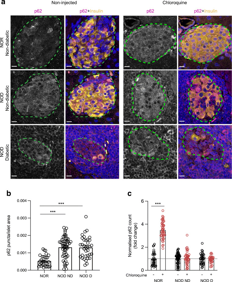
Fig. 3
Impaired islet p62 degradation in non-diabetic and diabetic NOD mice. ( a ) Representative images showing immunofluorescent staining of islets of chloroquine-injected NOR and NOD mouse pancreatic tiss...
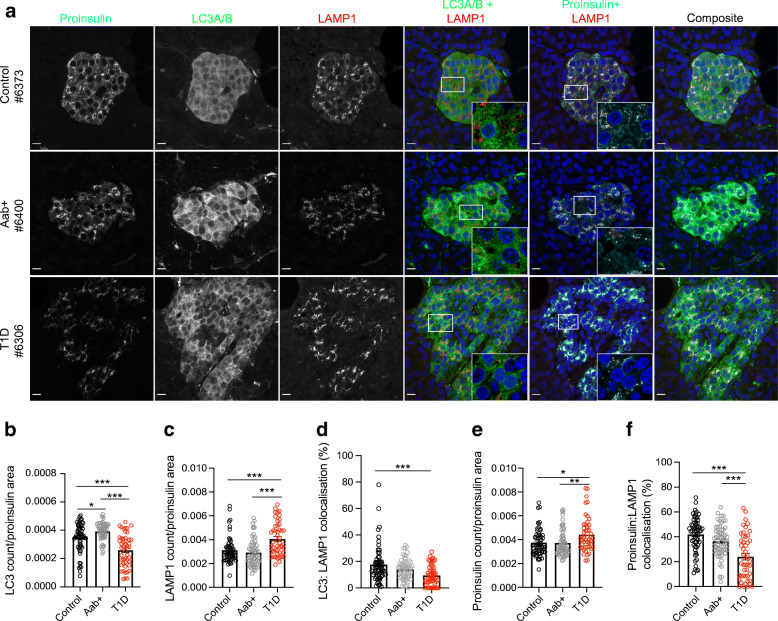
Fig. 4
Reduced autophagy and crinophagy in the pancreatic beta cells of human donors with type 1 diabetes. ( a ) Representative images showing immunofluorescent staining of islets in proinsulin-positive cell...
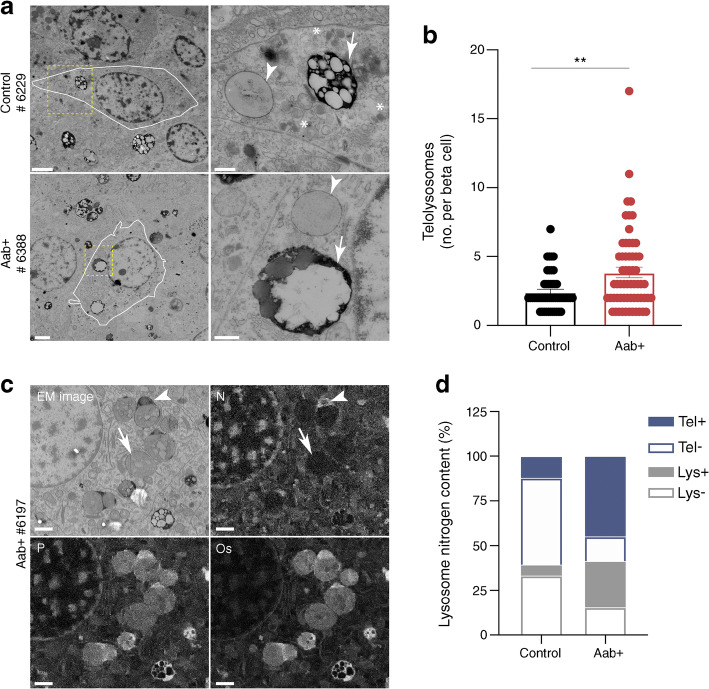
Fig. 5
Increased presence of telolysosomes in beta cells of autoantibody-positive human donors. ( a ) Representative images showing electron microscopic images of beta cells of non-diabetic and autoantibody-...
Figure images are served from the NIH/NLM PubMed Central Open Access Subset or Europe PMC; copyright remains with the publishers and authors.
💬 Discussion
0 commentsNo comments yet. Be the first to start a discussion!
Leave a Comment