Abstract
Particles of sub-micron size possess significant capacity to adsorb organic molecules from aqueous media. Semiconductor photocatalysts in particle form could potentially be utilized for dye removal through either physical adsorption or photo-induced chemical process. The photocatalytic and adsorption capabilities of Cu₂O particles with various exposed crystal facets have been studied through separate adsorption capacity test and photocatalytic degradation test. These crystals display unique cubic, octahedral, rhombic dodecahedral, and truncated polyhedral shapes due to specifically exposed crystal facet(s). For comparison, Cu₂O particles with no clear exposed facets were also prepared. The current work confirms that the surface charge critically affects the adsorption performance of the synthesized Cu₂O particles. The octahedral shaped Cu₂O particles, with exposed {111} facets, possess the best adsorption capability of methyl orange (MO) dye due to the strongest positive surface charge among the different types of particles. In addition, we also found that the adsorption of MO follows the Langmuir monolayer mechanism. The octahedral particles also performed the best in photocatalytic dye degradation of MO under visible light irradiation because of the assistance from dye absorption. On top of the photocatalytic study, the stability of these Cu₂O particles during the photocatalytic processes was also investigated. Cu(OH)₂ and CuO are the likely corrosion products found on the particle surface after the photocorrosion in MO solution. By adding hole scavengers in the solution, the photocorrosion of Cu₂O was greatly reduced. This observation confirms that the photocatalytically generated holes were responsible for the photocorrosion of Cu₂O.
🔬 Techniques
🧪 Sample Preparation
🔬 Cell Lines
🏭 Microscope Brands
🧪 Reagent Suppliers
🏛️ Research Organizations (ROR)
Affiliated research institutions:
📋 Methods
3.1. Synthesis of Cu 2 O Particles Copper(II) acetate monohydrate (C 4 H 6 CuO 4 ·H 2 O, 99.0%) was purchased from Fluka (Singapore). Copper(II) sulfate pentahydrate (CuSO 4 ·5H 2 O, 98.0%), l -ascorbic acid (C 6 H 8 O 6 , reagent grade), hydrazine hydrate (H 4 N 2 ·xH 2 O, reagent grade 50–60%), oleic acid (C 18 H 34 O 2 , technical grade 90.0%), and d -(+)-Glucose (C 6 H 12 O 6 , 99.5%) were obtained from Sigma Aldrich (Singapore). Sodium hydroxide pellets (NaOH, 99.0%) were acquired from Schedelco (Singapore). Absolute ethanol (C 2 H 5 OH, 99.0%) was purchased from Merck (Singapore). Absolute methanol (CH 3 OH, analytical reagent grade) was purchased from Fisher Scientific (Singapore). Hexane (C 6 H 14 , reagent grade) was obtained from Riverbank Chemical Pte Ltd (Singapore). All chemicals were used as received without further purification. 3.1.1. Spherical Cu 2 O Particles Copper(II) acetate (Cu(OAc) 2 ) (0.01 mol) was dissolved into 20 mL of deionized water under constant stirring at 500 rpm. Forty milliliters of 0.01 M NaOH was then added to the solution followed by 0.01 M of ascorbic acid (20 mL). After stirring for 30 min, the solution was centrifuged at 6000 rpm for 3 min and then washed with deionized water and ethanol solution. The powder collected was then dried in a vacuum oven at 60 °C for 6 h. 3.1.2. Cubic Cu 2 O Particles Copper(II) acetate (Cu(OAc) 2 ) (0.01 mol) was dissolved in 20 mL of deionized water under constant stirring at 500 rpm. Forty milliliters NaOH solution (0.03 M) was then added, followed by 20 mL of 0.01 M ascorbic acid. After stirring for 30 min, the solution was centrifuged at 6000 rpm for 3 min followed by washing with deionized water and ethanol solution. The powder collected was then dried in a vacuum oven at 60 °C for 6 h. 3.1.3. Octahedral Cu 2 O Particles Copper(II) acetate (Cu(OAc) 2 ) (0.01 mol) was dissolved in 20 mL of deionized water under constant stirring at 500 rpm. Forty milliliters NaOH solution (0.03 M) was then added to the solution followed by addition of 40 mL hydrazine hydrate solution (0.05 M). After 45 min of stirring, the solution was centrifuged at 6000 rpm for 3 min and washed with deionized water and ethanol solution. The powder collected was dried in a vacuum oven at 60 °C for 6 h. 3.1.4. Rhombic Dodecahedral Cu 2 O Particles Copper(II) sulfate (CuSO 4 ·5H 2 O) (0.25 g) was dissolved in 40 mL deionized water. When the powder was fully dissolved, 5 mL oleic acid and 20 mL absolute ethanol were added to the solution. The solution was vigorously stirred at 700 rpm for 30 min before being heated at 90 °C. Twenty milliliters NaOH solution (0.08 M) was then added to the CuSO 4 solution under constant stirring for 5 min at 90 °C. Afterwards, 3.42 g of glucose, which was dissolved in 30 mL of heated deionized water, was added to the solution. The final solution was left to stir at 90 °C for 45 min. After that, the solution was centrifuged and washed with hexane (10 times) and ethanol (3 times). The powder collected was then dried in a vacuum oven at 60 °C for 6 h. 3.1.5. Truncated Polyhedral Cu 2 O Particles Copper(II) acetate (Cu(OAc) 2 ) (0.015 mol) was dissolved in 40 mL deionized water at 70 °C. After continuous stirring at 500 rpm for 5 min, 10 mL of 9.0 M NaOH and 0.6 g glucose were added into the solution. The solution continued to be stirred at 70 °C for about 60 min before being cooled down to room temperature. The powder was washed and centrifuged with ethanol (6 times) and deionized water (3 times). Finally, the powder was dried in a vacuum oven at 60 °C for 6 h. 3.2.
Show full methods section
3.1. Synthesis of Cu 2 O Particles Copper(II) acetate monohydrate (C 4 H 6 CuO 4 ·H 2 O, 99.0%) was purchased from Fluka (Singapore). Copper(II) sulfate pentahydrate (CuSO 4 ·5H 2 O, 98.0%), l -ascorbic acid (C 6 H 8 O 6 , reagent grade), hydrazine hydrate (H 4 N 2 ·xH 2 O, reagent grade 50–60%), oleic acid (C 18 H 34 O 2 , technical grade 90.0%), and d -(+)-Glucose (C 6 H 12 O 6 , 99.5%) were obtained from Sigma Aldrich (Singapore). Sodium hydroxide pellets (NaOH, 99.0%) were acquired from Schedelco (Singapore). Absolute ethanol (C 2 H 5 OH, 99.0%) was purchased from Merck (Singapore). Absolute methanol (CH 3 OH, analytical reagent grade) was purchased from Fisher Scientific (Singapore). Hexane (C 6 H 14 , reagent grade) was obtained from Riverbank Chemical Pte Ltd (Singapore). All chemicals were used as received without further purification. 3.1.1. Spherical Cu 2 O Particles Copper(II) acetate (Cu(OAc) 2 ) (0.01 mol) was dissolved into 20 mL of deionized water under constant stirring at 500 rpm. Forty milliliters of 0.01 M NaOH was then added to the solution followed by 0.01 M of ascorbic acid (20 mL). After stirring for 30 min, the solution was centrifuged at 6000 rpm for 3 min and then washed with deionized water and ethanol solution. The powder collected was then dried in a vacuum oven at 60 °C for 6 h. 3.1.2. Cubic Cu 2 O Particles Copper(II) acetate (Cu(OAc) 2 ) (0.01 mol) was dissolved in 20 mL of deionized water under constant stirring at 500 rpm. Forty milliliters NaOH solution (0.03 M) was then added, followed by 20 mL of 0.01 M ascorbic acid. After stirring for 30 min, the solution was centrifuged at 6000 rpm for 3 min followed by washing with deionized water and ethanol solution. The powder collected was then dried in a vacuum oven at 60 °C for 6 h. 3.1.3. Octahedral Cu 2 O Particles Copper(II) acetate (Cu(OAc) 2 ) (0.01 mol) was dissolved in 20 mL of deionized water under constant stirring at 500 rpm. Forty milliliters NaOH solution (0.03 M) was then added to the solution followed by addition of 40 mL hydrazine hydrate solution (0.05 M). After 45 min of stirring, the solution was centrifuged at 6000 rpm for 3 min and washed with deionized water and ethanol solution. The powder collected was dried in a vacuum oven at 60 °C for 6 h. 3.1.4. Rhombic Dodecahedral Cu 2 O Particles Copper(II) sulfate (CuSO 4 ·5H 2 O) (0.25 g) was dissolved in 40 mL deionized water. When the powder was fully dissolved, 5 mL oleic acid and 20 mL absolute ethanol were added to the solution. The solution was vigorously stirred at 700 rpm for 30 min before being heated at 90 °C. Twenty milliliters NaOH solution (0.08 M) was then added to the CuSO 4 solution under constant stirring for 5 min at 90 °C. Afterwards, 3.42 g of glucose, which was dissolved in 30 mL of heated deionized water, was added to the solution. The final solution was left to stir at 90 °C for 45 min. After that, the solution was centrifuged and washed with hexane (10 times) and ethanol (3 times). The powder collected was then dried in a vacuum oven at 60 °C for 6 h. 3.1.5. Truncated Polyhedral Cu 2 O Particles Copper(II) acetate (Cu(OAc) 2 ) (0.015 mol) was dissolved in 40 mL deionized water at 70 °C. After continuous stirring at 500 rpm for 5 min, 10 mL of 9.0 M NaOH and 0.6 g glucose were added into the solution. The solution continued to be stirred at 70 °C for about 60 min before being cooled down to room temperature. The powder was washed and centrifuged with ethanol (6 times) and deionized water (3 times). Finally, the powder was dried in a vacuum oven at 60 °C for 6 h. 3.2.
Materials Characterization
Crystal structure was identified by X-ray diffraction (XRD) using a Shimadzu LabX-6000 diffractometer (Shimadzu Corporation, Tokyo, Japan) with Cu Kα radiation (λ = 1.54178 Å). A step size of 0.02° over 2θ ranging from 10° to 80° was used with a scanning rate at 2.33° per minute. The accelerating voltage and emission current were 40 kV and 30 mA, respectively. The morphology of the samples was examined by field emission scanning electron microscopy (FESEM, JEOL JSM-7600F, JEOL Ltd., Tokyo, Japan) and transmission electron microscopy (TEM, JEOL JEM-2010, JEOL Ltd., Tokyo, Japan). The specific surface areas were evaluated using a Micromeritics ASAP 2020 (Micromeritics Instrument Corporation, Norcross, GA, USA) surface analyzer based on the BET theory. The samples were outgassed under vacuum and heated to 100 °C before the test. The surface chemical analysis was carried out by X-ray photoelectron spectroscopy (XPS) using a VG ESCALab 220i-XL system (Thermo Scientific, Waltham, MA, USA). Mg Kα X-ray (hν = 1253.6 eV) from twin anode X-ray gun was employed using a large area lens mode for analysis with photoelectron takeoff angle of 90° with respect to surface plane. The maximum analysis depth is in the range of 4–8 nm. Survey spectra were acquired for elemental identification while high-resolution spectra were acquired for chemical state identification and surface composition calculation. For chemical state analysis, a spectral deconvolution was performed by a curve-fitting procedure based on Lorentzians broadened by Gaussian using the manufacturer’s standard software. The error of binding energy is estimated to be within 0.2 eV. FTIR was carried out in a Perkin Elmer Instruments Spectrum GX FTIR spectrometer (PerkinElmer, Waltham, MA, USA). Synthesized particles were mixed with standard KBr particles and pressed into thin pellets. The spectral range was 400–4000 cm −1 and a total of 40 scans were recorded at a resolution of 4 cm −1 averaging each spectrum. Zeta potential was measured by a Mavern Nanosizer system (Malvern Instruments Ltd, Malvern, UK). Particles (5–10 mg) were dispersed in deionized water (~pH 7) and sonicated for 5 min. The equilibrium time was set at 120 s and each sample was run for 10 times to obtain the average value of the surface charge. Optical absorption of bulk powders was measured on a Perkin Elmer Lambda 900 UV-Visible spectrometer (PerkinElmer, Waltham, MA, USA) in the diffuse reflectance spectroscopy mode over the spectra ranging from 250 to 800 nm. The optical diffuse reflectance spectrum of Cu 2 O was measured on a Shimadzu 2550 UV-vis-NIR spectrometer (Shimadzu Corporation, Tokyo, Japan) using BaSO 4 as a reference standard. The bandgap of Cu 2 O was calculated using the Kubelka–Munk function. The flat band potentials were measured by impedance spectroscopy based on the Mott–Schottky plots. To prepare for the test samples, 15 mg of the as-synthesized Cu 2 O powder was sonicated in 1 mL of ethanol to obtain a homogeneous mixture. The Cu 2 O suspension was then drop-casted on a conductive fluorine-tin oxide (FTO) glass substrate with adhesive tapes acting as spacers attached on the four edges. The substrate was then dried at 80 °C, and the adhesive tape attached on the top side of the substrate was removed. Electrical contact was formed by first applying silver paint on the top uncoated area of FTO, and then sticking a conductive copper tape onto the dried silver paint. Three electrodes were used for the impedance measurements which include the working electrode (the Cu 2 O film), a Pt counter electrode, and a reference electrode (Ag/AgCl, saturated in KCl). A 0.1 M Na 2 SO 4 solution was used as the electrolyte. The measurements were carried out by a Gamry electrochemical impedance spectrometer, and the potential was systemically varied between +0 V and +2.0 V with frequency of 50 Hz. 3.3. Photocatalytic and Adsorption Experiments Methyl orange (MO) was chosen as a representative dye to test the photocatalytic degradation activity and the adsorption capacity of the prepared Cu 2 O samples. Solution of the dye was prepared by dissolving the dye in deionized water at 20 ppm concentration, and the solution pH was around 6.7. One hundred mg of the powder samples were dispersed in 50 mL MO solution for the dye degradation test under visible light irradiation. A 100 mL glass beaker, wrapped with aluminum foil on its side wall, was used as the reactor with light shone from the top. The irradiation source comes from a solar simulator equipped with a 300 W Xe-lamp (HAL-320, Asahi Spectra Co., Ltd., Kita-ku, Japan). Super cold filter (YSC0750) was used to provide visible light ranging from 420 to 700 nm. The light intensity was around 50 mW/cm 2 . The amount of photocatalyst used was chosen through a few trial runs based on their degradation speed. An adsorption isotherm test was carried out in the dark to prevent the potential photocatalytic degradation of MO under light. The equilibrium adsorption isotherm was determined using various concentrations of MO (10, 20, 30, 50 and 80 ppm). For each test, 100 mg of adsorbent was added to 50 mL MO solution. After 48 h, the equilibrium concentration was measured. To determine the adsorption capacity, 100 mg of synthesized powder was added to 50 mL MO solution of various concentrations (10, 20, 30, 50 and 80 ppm). Stirring was applied throughout the duration of the test (1440 min) at a speed of 400 rpm in dark. At different intervals, the sample was collected, centrifuged and measured using the UV-visible spectrometer to determine the dye concentration. 3.4. Photocatalytic Stability Study Two separate tests were carried out to measure the photocatalytic stability of Cu 2 O particles and to determine the possible root cause of the instability. In the first experiment, 50 mg powder was dispersed in 50 mL MO solution with 10 ppm concentration. For the second experiment, 50 mg of the powder was dispersed in 40 mL of MO solution (10 ppm) mixed with 10 mL of absolute methanol (purity > 99%). Both were constantly stirred under a solar simulator equipped with a 300 W Xe-lamp (HAL-320, Asahi Spectra Co., Ltd., Kita-ku, Japan) for 12 h under the intensity of 100 mW/cm 2 . An AM 1.5 G filter (400 to 1100 nm) was used.
3.2.
Materials Characterization
Crystal structure was identified by X-ray diffraction (XRD) using a Shimadzu LabX-6000 diffractometer (Shimadzu Corporation, Tokyo, Japan) with Cu Kα radiation (λ = 1.54178 Å). A step size of 0.02° over 2θ ranging from 10° to 80° was used with a scanning rate at 2.33° per minute. The accelerating voltage and emission current were 40 kV and 30 mA, respectively. The morphology of the samples was examined by field emission scanning electron microscopy (FESEM, JEOL JSM-7600F, JEOL Ltd., Tokyo, Japan) and transmission electron microscopy (TEM, JEOL JEM-2010, JEOL Ltd., Tokyo, Japan). The specific surface areas were evaluated using a Micromeritics ASAP 2020 (Micromeritics Instrument Corporation, Norcross, GA, USA) surface analyzer based on the BET theory. The samples were outgassed under vacuum and heated to 100 °C before the test. The surface chemical analysis was carried out by X-ray photoelectron spectroscopy (XPS) using a VG ESCALab 220i-XL system (Thermo Scientific, Waltham, MA, USA). Mg Kα X-ray (hν = 1253.6 eV) from twin anode X-ray gun was employed using a large area lens mode for analysis with photoelectron takeoff angle of 90° with respect to surface plane. The maximum analysis depth is in the range of 4–8 nm. Survey spectra were acquired for elemental identification while high-resolution spectra were acquired for chemical state identification and surface composition calculation. For chemical state analysis, a spectral deconvolution was performed by a curve-fitting procedure based on Lorentzians broadened by Gaussian using the manufacturer’s standard software. The error of binding energy is estimated to be within 0.2 eV. FTIR was carried out in a Perkin Elmer Instruments Spectrum GX FTIR spectrometer (PerkinElmer, Waltham, MA, USA). Synthesized particles were mixed with standard KBr particles and pressed into thin pellets. The spectral range was 400–4000 cm −1 and a total of 40 scans were recorded at a resolution of 4 cm −1 averaging each spectrum. Zeta potential was measured by a Mavern Nanosizer system (Malvern Instruments Ltd, Malvern, UK). Particles (5–10 mg) were dispersed in deionized water (~pH 7) and sonicated for 5 min. The equilibrium time was set at 120 s and each sample was run for 10 times to obtain the average value of the surface charge. Optical absorption of bulk powders was measured on a Perkin Elmer Lambda 900 UV-Visible spectrometer (PerkinElmer, Waltham, MA, USA) in the diffuse reflectance spectroscopy mode over the spectra ranging from 250 to 800 nm. The optical diffuse reflectance spectrum of Cu 2 O was measured on a Shimadzu 2550 UV-vis-NIR spectrometer (Shimadzu Corporation, Tokyo, Japan) using BaSO 4 as a reference standard. The bandgap of Cu 2 O was calculated using the Kubelka–Munk function. The flat band potentials were measured by impedance spectroscopy based on the Mott–Schottky plots. To prepare for the test samples, 15 mg of the as-synthesized Cu 2 O powder was sonicated in 1 mL of ethanol to obtain a homogeneous mixture. The Cu 2 O suspension was then drop-casted on a conductive fluorine-tin oxide (FTO) glass substrate with adhesive tapes acting as spacers attached on the four edges. The substrate was then dried at 80 °C, and the adhesive tape attached on the top side of the substrate was removed. Electrical contact was formed by first applying silver paint on the top uncoated area of FTO, and then sticking a conductive copper tape onto the dried silver paint. Three electrodes were used for the impedance measurements which include the working electrode (the Cu 2 O film), a Pt counter electrode, and a reference electrode (Ag/AgCl, saturated in KCl). A 0.1 M Na 2 SO 4 solution was used as the electrolyte. The measurements were carried out by a Gamry electrochemical impedance spectrometer, and the potential was systemically varied between +0 V and +2.0 V with frequency of 50 Hz.
📊 Figures
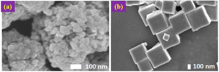
Figure 1
Field Emission Scanning Electron Microscopy (FESEM) images of Cu 2 O particles: ( a ) Cu 2 O of agglomerated spherical shape with no clear exposed facets; and ( b ) Cu 2 O of cubic shape with exposed ...
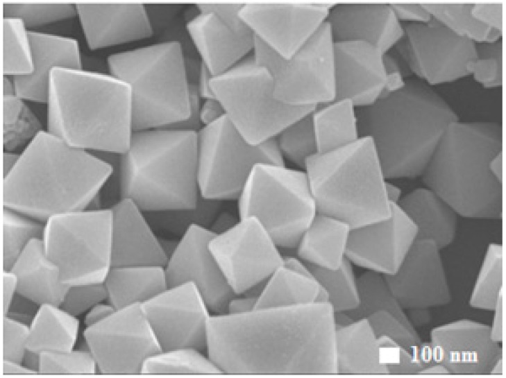
Figure 2
FESEM images of octahedral shaped Cu 2 O particles exposing the {111} facets.
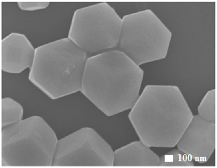
Figure 3
FESEM images of particles in rhombic dodecahedral shape with exposed {110} facets.
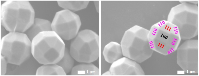
Figure 4
FESEM images of the truncated polyhedral Cu 2 O particles, exposing eight {111} facets, six {100} facets and 12 {110} facets.
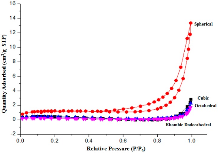
Figure 5
Isotherm N 2 adsorption of Cu 2 O particles. STP: Standard Temperature and Pressure.
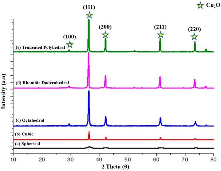
Figure 6
The X-ray diffraction (XRD) patterns of Cu 2 O particles: ( a ) spherical; ( b ) cubic; ( c ) octahedral; ( d ) rhombic dodecahedral; and ( e ) truncated polyhedral.
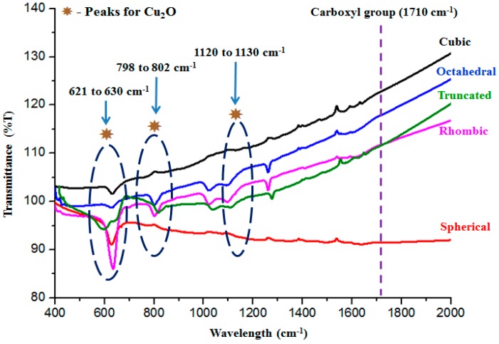
Figure 7
The Fourier Transform Infrared (FTIR) spectra of the faceted Cu 2 O particles.
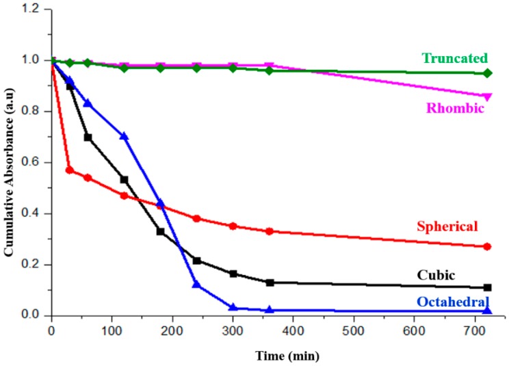
Figure 8
Adsorption performance of the Cu 2 O (spherical, cubic, octahedral, rhombic dodecahedral and truncated polyhedral) particles in 20 ppm methyl orange (MO) solution.
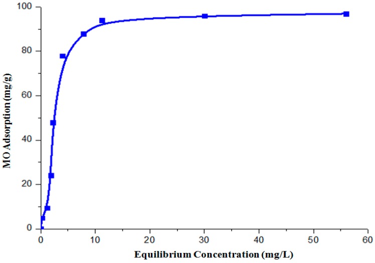
Figure 9
The adsorption isotherms of the octahedral shaped Cu 2 O.
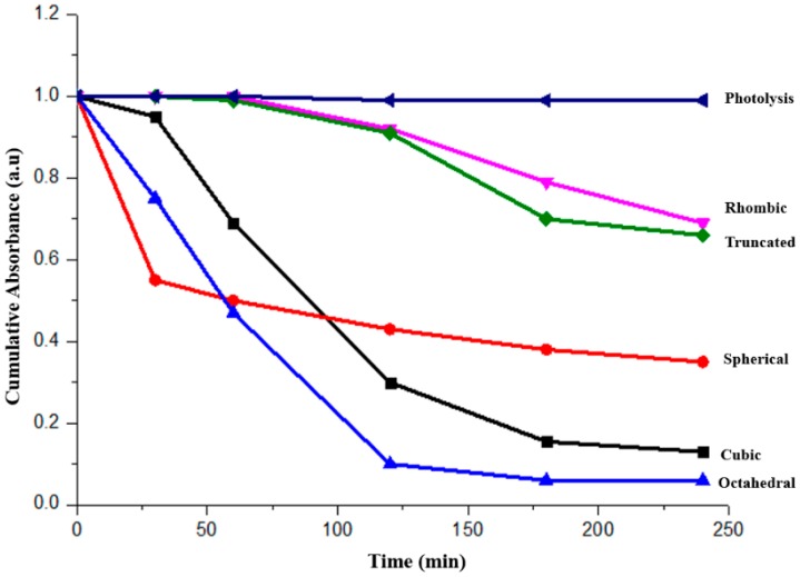
Figure 10
The photocatalytic performance of the faceted Cu 2 O (spherical, cubic, octahedral, rhombic dodecahedral and truncated polyhedral) particles under visible light irradiation.
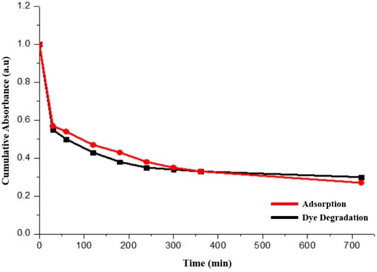
Figure 11
Comparison of adsorption (red line) and dye degradation (black line) performances of the spherical Cu 2 O particles.
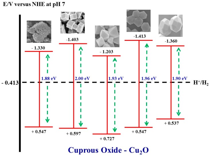
Figure 12
The band edge potential positions of the faceted Cu 2 O particles.
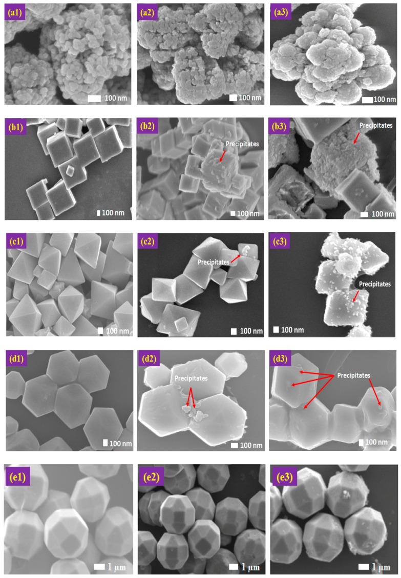
Figure 13
Column 1u2014FESEM images of as-synthesized Cu 2 O: ( a1 ) spherical; ( b1 ) cubic; ( c1 ) octahedral; ( d1 ) rhombic dodecahedral; and ( e1 ) truncated polyhedral. Column 2u2014FESEM images of Cu 2 O...
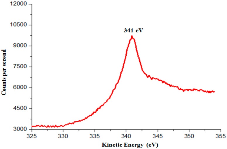
Figure 14
The kinetic energy (Auger LMM) of the photocorroded Cu 2 O particles.
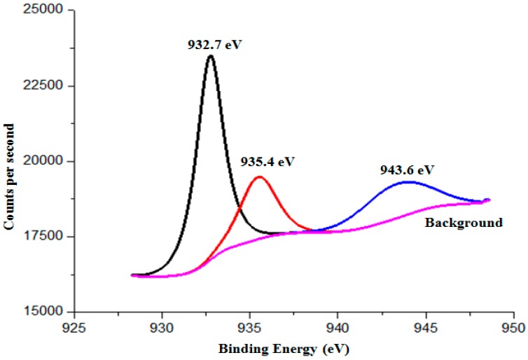
Figure 15
The binding energy of the photocorroded Cu 2 O particles.
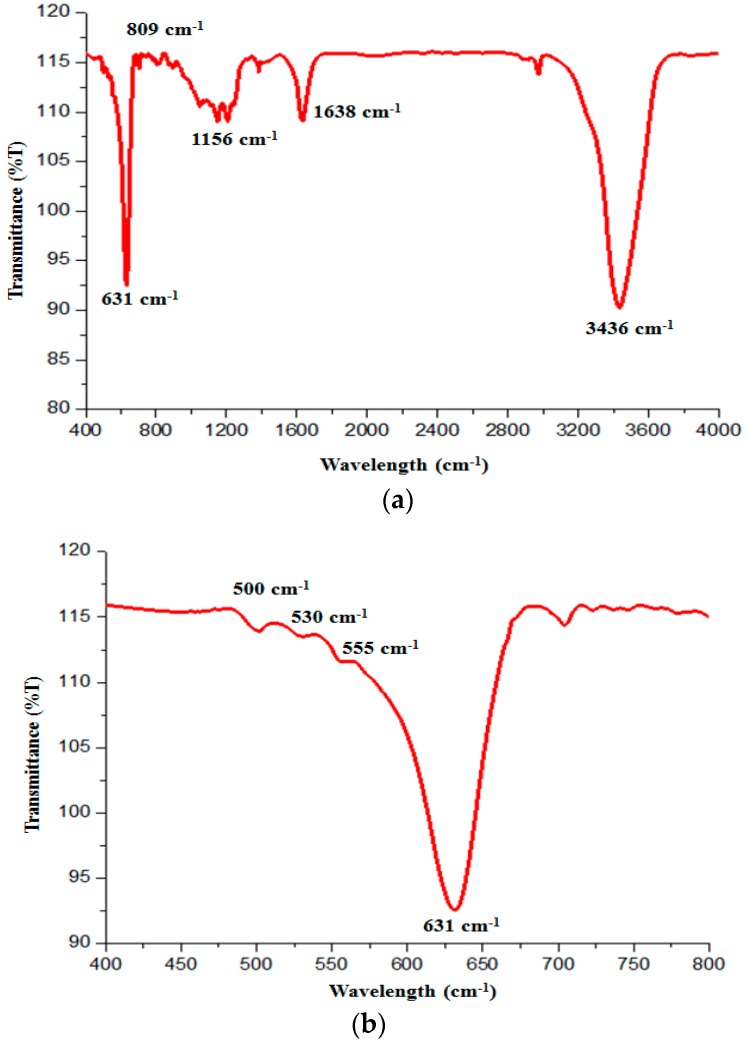
Figure 16
( a ) FTIR spectrum of the photocorroded Cu 2 O particles; and ( b ) zoom-in FTIR showing the existence of CuO.
Figure images are served from the NIH/NLM PubMed Central Open Access Subset or Europe PMC; copyright remains with the publishers and authors.
💬 Discussion
0 commentsNo comments yet. Be the first to start a discussion!
Leave a Comment