Abstract
Abstract Background Cultured epidermal stem cells (Epi-SCs) and skin-derived precursors (SKPs) were capable of reconstituting functional hair follicles after implantation, while the signaling pathways that regulate neogenic hair follicle formation are poorly investigated. In this study, we aimed to understand the interactions between Epi-SCs and SKPs during skin organoid formation and to uncover key signal pathways crucial for de novo hair follicle regeneration. Methods To track their fate after transplantation, Epi-SCs derived from neonatal C57BL/6 mice were labeled with tdTomato, and SKPs were isolated from neonatal C57BL/6/GFP mice. A mixture of Epi-SCs-tdTomato and SKPs-EGFP in Matrigel was observed under two-photon microscope in culture and after implantation into excisional wounds in nude mice, to observe dynamic migrations of the cells during hair follicle morphogenesis. Signaling communications between the two cell populations were examined by RNA-Seq analysis. Potential signaling pathways revealed by the analysis were validated by targeting the pathways using specific inhibitors to observe a functional loss in de novo hair follicle formation. Results Two-photon microscopy analysis indicated that when Epi-SCs and SKPs were mixed in Matrigel and cultured, they underwent dynamic migrations resulting in the formation of a bilayer skin-like structure (skin organoid), where Epi-SCs positioned themselves in the outer layer; when the mixture of Epi-SCs and SKPs was grafted into excisional wounds in nude mice, a bilayer structure resembling the epidermis and the dermis formed at the 5th day, and de novo hair follicles generated subsequently. RNA-Seq analysis of the two cell types after incubation in mixture revealed dramatic alterations in gene transcriptome, where PI3K-Akt signaling pathway in Epi-SCs was significantly upregulated; meanwhile, elevated expressions of several growth factors and cytokine potentially activating PI3K were found in SKPs, suggesting active reciprocal communications between them. In addition, inhibition of PI3K or Akt by specific inhibitors markedly suppressed the hair follicle regeneration mediated by Epi-SCs and SKPs. Conclusions Our data indicate that the PI3K-Akt signaling pathway plays a crucial role in de novo hair follicle regeneration, and the finding may suggest potential therapeutic applications in enhancing hair regeneration.
🔬 Techniques
🔭 Microscopes
✨ Fluorophores
🧪 Sample Preparation
🔬 Cell Lines
🏭 Microscope Brands
🧪 Reagent Suppliers
🏛️ Research Organizations (ROR)
Affiliated research institutions:
📋 Methods
To track their fate after transplantation, Epi-SCs derived from neonatal C57BL/6 mice were labeled with tdTomato, and SKPs were isolated from neonatal C57BL/6/GFP mice. A mixture of Epi-SCs-tdTomato and SKPs-EGFP in Matrigel was observed under two-photon microscope in culture and after implantation into excisional wounds in nude mice, to observe dynamic migrations of the cells during hair follicle morphogenesis. Signaling communications between the two cell populations were examined by RNA-Seq analysis. Potential signaling pathways revealed by the analysis were validated by targeting the pathways using specific inhibitors to observe a functional loss in de novo hair follicle formation.
Materials and methods Mice
Five- and six-week old BALB/c nu/nu mice and 7-week-old C57BL/6 mice were purchased from the Guangdong Medical Laboratory Animal Center, Guangzhou, China. Six-week-old C57BL/6/GFP mice were derived from the Cyagen BioSciences, Guangzhou, China. These mice were maintained in a temperature-controlled environment (20 ± 1 °C). All animal procedures were performed with the approval of the Animal Ethics Committee of Tsinghua Shenzhen International Graduate School.
Isolation and culture of Epi-SCs and SKPs
Full-thickness dorsal skin tissue was collected from C57BL/6 or C57BL/GFP mice 0~72 h after birth. The tissue was washed 3 times in PBS, cut into 2~3 mm 2 pieces, and digested with 0.35% Dispase II (sigma) for 40 min at 37 °C. The epithelial layer was removed manually. Epi-SCs were isolated on account of their high-adhesive property as described previously [ 8 , 14 ]. Briefly, the epidermis was cut into pieces, which were treated with 0.035% collagenase I (Sigma) at 37 °C for 1 h with shaking gently and filtered with a 40-μm cell strainer. The cells were seeded in tissue culture dishes and cultured in CnT-07 PCT Epidermal Keratinocyte Medium (CELLnTEC Advanced Cell Systems). The non-adherent cells were removed and the adherent cells (Epi-SCs) were maintained. When reaching 70–80% confluence, the cells were digested with accutase (Sigma) and subcultured. SKPs were prepared as previously described [ 8 , 15 ]. Briefly, single dermal cells derived from the dermal tissue were incubated in a 10-cm non-treated dish in 10 ml Dulbecco’s modified Eagle’s medium (DMEM)/F12, 3:1 (Gibco) containing B27 (Gibco), 20 ng/ml epidermal growth factor (EGF, Peprotech), and 40 ng/ml basal fibroblast growth factor (bFGF, Peprotech) and incubated in a 37 °C, 5% CO 2 tissue culture incubator. Cell labeling and in vitro cell tracing Epi-SCs were transduced with tdTomato by retroviruses (Epi-SCs-tdTomato). 10 6 Epi-SCs seeded in a 10-cm tissue culture dish were infected with tdTomato by retroviruses (MOI = 10) in the presence of 5 μg/mL polybrene (Sigma) in the culture medium. The medium was replaced by regular growth medium after 12 h. The cells were examined for tdTomato expression at 48 h and subjected to transplantation. 1 × 10 6 Epi-SCs-tdTomato and 2 × 10 6 SKPs-EGFP were mixed in Matrigel, seeded in a 33-mm confocal dish, incubated at 37 °C for 15 min, and then added with 2 ml DMEM/F12 3:1. Live-cell images were recorded for 36 h at 37 °C and 5% CO 2 on Leica DMI6000 confocal live-cell imaging system.
Show full methods section
To track their fate after transplantation, Epi-SCs derived from neonatal C57BL/6 mice were labeled with tdTomato, and SKPs were isolated from neonatal C57BL/6/GFP mice. A mixture of Epi-SCs-tdTomato and SKPs-EGFP in Matrigel was observed under two-photon microscope in culture and after implantation into excisional wounds in nude mice, to observe dynamic migrations of the cells during hair follicle morphogenesis. Signaling communications between the two cell populations were examined by RNA-Seq analysis. Potential signaling pathways revealed by the analysis were validated by targeting the pathways using specific inhibitors to observe a functional loss in de novo hair follicle formation.
Materials and methods Mice
Five- and six-week old BALB/c nu/nu mice and 7-week-old C57BL/6 mice were purchased from the Guangdong Medical Laboratory Animal Center, Guangzhou, China. Six-week-old C57BL/6/GFP mice were derived from the Cyagen BioSciences, Guangzhou, China. These mice were maintained in a temperature-controlled environment (20 ± 1 °C). All animal procedures were performed with the approval of the Animal Ethics Committee of Tsinghua Shenzhen International Graduate School.
Isolation and culture of Epi-SCs and SKPs
Full-thickness dorsal skin tissue was collected from C57BL/6 or C57BL/GFP mice 0~72 h after birth. The tissue was washed 3 times in PBS, cut into 2~3 mm 2 pieces, and digested with 0.35% Dispase II (sigma) for 40 min at 37 °C. The epithelial layer was removed manually. Epi-SCs were isolated on account of their high-adhesive property as described previously [ 8 , 14 ]. Briefly, the epidermis was cut into pieces, which were treated with 0.035% collagenase I (Sigma) at 37 °C for 1 h with shaking gently and filtered with a 40-μm cell strainer. The cells were seeded in tissue culture dishes and cultured in CnT-07 PCT Epidermal Keratinocyte Medium (CELLnTEC Advanced Cell Systems). The non-adherent cells were removed and the adherent cells (Epi-SCs) were maintained. When reaching 70–80% confluence, the cells were digested with accutase (Sigma) and subcultured. SKPs were prepared as previously described [ 8 , 15 ]. Briefly, single dermal cells derived from the dermal tissue were incubated in a 10-cm non-treated dish in 10 ml Dulbecco’s modified Eagle’s medium (DMEM)/F12, 3:1 (Gibco) containing B27 (Gibco), 20 ng/ml epidermal growth factor (EGF, Peprotech), and 40 ng/ml basal fibroblast growth factor (bFGF, Peprotech) and incubated in a 37 °C, 5% CO 2 tissue culture incubator. Cell labeling and in vitro cell tracing Epi-SCs were transduced with tdTomato by retroviruses (Epi-SCs-tdTomato). 10 6 Epi-SCs seeded in a 10-cm tissue culture dish were infected with tdTomato by retroviruses (MOI = 10) in the presence of 5 μg/mL polybrene (Sigma) in the culture medium. The medium was replaced by regular growth medium after 12 h. The cells were examined for tdTomato expression at 48 h and subjected to transplantation. 1 × 10 6 Epi-SCs-tdTomato and 2 × 10 6 SKPs-EGFP were mixed in Matrigel, seeded in a 33-mm confocal dish, incubated at 37 °C for 15 min, and then added with 2 ml DMEM/F12 3:1. Live-cell images were recorded for 36 h at 37 °C and 5% CO 2 on Leica DMI6000 confocal live-cell imaging system.
Cell sorting and RNA-Seq
Epi-SCs-tdTomato and SKPs-EGFP were cultured in Matrigel individually or in mixture for 24 h. Then, cells were recovered from the matrix. Epi-SCs-tdTomato and SKPs-EGFP were separated through cell sorting by a flow cytometer (Becton Dickinson). Total RNA was extracted from the Epi-SCs and SKPs using Trizol (TAKARA) according to the manufacturer’s instructions, and libraries were constructed using VAHTS mRNA-seq V3 Library Prep Kit for Illumina® (Vazyme). The qualified libraries were used for sequencing using Illumina HiSeqTM 2500 by Gene Denovo. All raw data of RNA sequencing were corresponded to the mouse genome using TopHat V2.0.3 and Bowtie2. Gene expression was measured by FPKM (fragment per kilobase of transcript per million mapped reads), which computed with Cufflinks V2.1.1. Enricher was used to analyze gene ontology enrichment. And heatmaps were carried out by GO analysis, coupled with the KEGG pathways database. In addition, differentially expressed genes (DEGs) were identified by edgeR analysis with FDR < 0.05.
Cell transplantation and hair follicle regeneration
BALB/c nu/nu mice (4–5 weeks old) were anesthetized with sodium pentobarbital (50 mg/kg). Symmetrical full-thickness skin wounds were created on the back with a 2-mm-diameter skin biopsy punch as previously described [ 16 ]. 1 × 10 6 Epi-SCs-tdTomato were mixed with 2 × 10 6 SKPs-EGFP and encapsulated in 20 μl Matrigel (BD BioSciences). The cell-Matrigel was incubated at 37 °C for 30 min and implanted into an excisional wound. The wound was then covered with Tegaderm (3M) transparent dressing and self-adhering elastic bandage successively. Three weeks later, mice were sacrificed and wound tissue samples were obtained for histological analysis. In PI3K/Akt inhibitor treatment, Perifosine (Akt inhibitor) and LY294002 (PI3K inhibitor) were added into the cell mixture, respectively, after dilution with Matrigel, resulting in a final concentration of 2 mM Perifosine or 500 μM LY294002 in the graft. The mixture was incubated in a tissue culture incubator for 30 min and then implanted into wounds in mice.
Two-photon microscopy
Movement of Epi-SCs-tdTomato and SKPs-EGFP after implantation into skin wounds were tracked by two-photon microscopy according to a method previously described [ 17 ]. Mice were anesthetized with isoflurane and fixed on the platform of the microscope. A custom tweezer was placed to fix the skin wound. Images were recorded with a FV300 Olympus two-photon microscopy. Laser beam was 940 nm for GFP and 1040 nm for tdTomato. Serial optical sections were obtained in 5 μm steps to image a total depth of about 300 μm of tissue.
Immunofluorescence staining
Dorsal skin tissues of nu/nu mice were harvested and fixed with 4% paraformaldehyde (PFA, Sigma), washed with PBS, and dehydrated with 30% sucrose successively. Tissues were embedded in OCT and sectioned (10-μm thickness). Samples were washed with PBS and blocked with 3% BSA/PBS containing 0.2% Triton X-100 (Sigma) at 37 °C for 1 h. Samples were incubated with primary antibodies in 1% BSA/PBS at appropriate concentrations at 4 °C overnight: CD49f-biotin (BioLegend, 1:150), K14 (BioLegend, 1:100), K1 (BioLegend, 1:100), and nestin (Santa Cruz, 1:100). Samples were washed with PBS and detected with fluorescence-conjugated secondary antibodies. Nuclei were stained with DAPI. Samples were examined under confocal laser scanning microscope (FV1000, Olympus, Japan).
📊 Figures
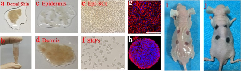
Fig. 1
A schematic diagram of Epi-SCs and SKP transplantation for hair follicle regeneration. Dorsal skin tissue in full thickness was collected from neonatal C57 mice ( a ), which was cut into pieces ( b )....
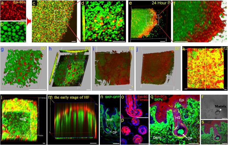
Fig. 2
Fates of Epi-SCs and SKPs in mixture in vitro and in vivo. a u2013 f Epi-SCs and SKPs in skin organoid formation . Epi-SCs labeled with tdTomato (red) were cultured in monolayer ( a ) and SKPs derived...
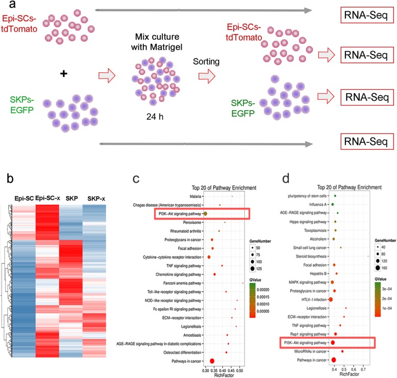
Fig. 3
RNA-Seq of Epi-SC and SKPs. a A schematic diagram depicting sample preparation for RNA-Seq. Total RNA was extracted from Epi-SCs-tdTomato and SKPs-EGFP which were cultured separately or in mixture in ...
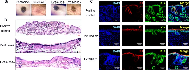
Fig. 4
Analysis of PI3K/Akt inhibition on hair follicle regeneration. a Hair follicle reconstitution assay showed that addition of Periforsine or LY294002 to Epi-SCs and SKPs mixtures before transplantation ...
Figure images are served from the NIH/NLM PubMed Central Open Access Subset or Europe PMC; copyright remains with the publishers and authors.
💬 Discussion
0 commentsNo comments yet. Be the first to start a discussion!
Leave a Comment