Abstract
AbstractEfficient differentiation of human pluripotent stem cells (hPSCs) into neurons is paramount for disease modeling, drug screening, and cell transplantation therapy in regenerative medicine. In this manuscript, we report the capability of five transcription factors (TFs) toward this aim: NEUROG1, NEUROG2, NEUROG3, NEUROD1, and NEUROD2. In contrast to previous methods that have shortcomings in their speed and efficiency, a cocktail of these TFs as synthetic mRNAs can differentiate hPSCs into neurons in 7 days, judged by calcium imaging and electrophysiology. They exhibit motor neuron phenotypes based on immunostaining. These results indicate the establishment of a novel method for rapid, efficient, and footprint-free differentiation of functional neurons from hPSCs.
🔬 Techniques
🔭 Microscopes
💻 Software
✨ Fluorophores
🧪 Sample Preparation
🔬 Cell Lines
🏭 Microscope Brands
🧪 Reagent Suppliers
📷 Detectors
💻 Software Details
📋 Protocols
🏛️ Research Organizations (ROR)
Affiliated research institutions:
📋 Methods
Cells and culture conditions
SEES3 human ES cells 20 were obtained from Dr. Hidenori Akutsu, TkDA3-4 human dermal fibroblast-derived iPS cells 29 were obtained from Dr. Koji Eto. 201B7 human iPS cells were obtained from RIKEN Cell Bank. SEES3 human ES, TkDA3-4, and 201B7 human iPS cells were cultured under feeder-free conditions in StemFit AK03 medium (Ajinomoto, Tokyo, Japan) on laminin511 (iMatrix-511: Nippi, Tokyo, Japan)-coated dishes. The chemicals used and their suppliers Recombinant Laminin 511-E8b fragments (iMatrix-511: Nippi, Tokyo, Japan). Dorsomorphin (EMD chemicals, Darmstadt, Germany). SB431542, Forskolin, Poly-L-Ornithine, Alpha-Bungarotoxin-tetramethyl rhodamine (Sigma-Aldrich, St. Louis, MO, USA). BDNF (PeproTech, Rocky Hill, NJ, USA). GDNF and NT3 (R&D systems, Minneapolis, MN, USA). DMEM/HAM F-12, Neurobasal medium, OPTIMEM, N2, and B27 supplements (Life Technologies, Carlsbad, CA, USA). Recombinant B18R protein (eBioscience-Affymetrix, San Diego, CA, USA). Modified nucleotides 5-methylcytidine-5′-triphospahte and pseudouridine-5′-triphosphate were obtained from Trilink biotechnologies (San Diego, CA, USA). KAPA HiFi Hot Start ready mix (Kapa Biosystems, Boston, MA, USA). MEGAscript T7 kit, MEGAclear kit, and RNA Millennium markers (Ambion, Thermo Fisher Scientific, Waltham, MA, USA). Anti-reverse cap analog 3′-O-Me-m7-G(5′)ppp(5′)G and Antarctic Phosphatase (New England Biolabs, Ipswich, MA, USA). 4% PFA-PBS (WAKO Pure Chemical Industries, Osaka, Japan), glass coverslips (13/15/18 mm) (Matsunami Glass IND, Osaka, Japan). 35 and 60 mm cell culture dishes (AGC techno glass, Shizuoka, Japan), Four-well plates (NUNC, Thermo Fisher Scientific, Waltham, MA, USA). Twenty-four-well plates (Costar, Sigma-Aldrich, St. Louis, MO, USA). PCR cleanup kit (Macherey-NAGEL, Duren, Germany). Trizol (Life Technologies, Carlsbad, CA, USA). Direct-zol RNA mini prep kit (Zymo Research). Total RNA from Trizol was extracted using the Direct-zol RNA mini prep kit (Zymo Research Corp., Carlsbad, CA, USA). cDNA was synthesized using the ReverTra Ace qPCR RT PCR Master mix (TOYOBO, Osaka, Japan). Reverse transcription PCR was performed using the Ex-Taq enzyme (Takara-Bio, Shiga, Japan).
Show full methods section
Cells and culture conditions
SEES3 human ES cells 20 were obtained from Dr. Hidenori Akutsu, TkDA3-4 human dermal fibroblast-derived iPS cells 29 were obtained from Dr. Koji Eto. 201B7 human iPS cells were obtained from RIKEN Cell Bank. SEES3 human ES, TkDA3-4, and 201B7 human iPS cells were cultured under feeder-free conditions in StemFit AK03 medium (Ajinomoto, Tokyo, Japan) on laminin511 (iMatrix-511: Nippi, Tokyo, Japan)-coated dishes. The chemicals used and their suppliers Recombinant Laminin 511-E8b fragments (iMatrix-511: Nippi, Tokyo, Japan). Dorsomorphin (EMD chemicals, Darmstadt, Germany). SB431542, Forskolin, Poly-L-Ornithine, Alpha-Bungarotoxin-tetramethyl rhodamine (Sigma-Aldrich, St. Louis, MO, USA). BDNF (PeproTech, Rocky Hill, NJ, USA). GDNF and NT3 (R&D systems, Minneapolis, MN, USA). DMEM/HAM F-12, Neurobasal medium, OPTIMEM, N2, and B27 supplements (Life Technologies, Carlsbad, CA, USA). Recombinant B18R protein (eBioscience-Affymetrix, San Diego, CA, USA). Modified nucleotides 5-methylcytidine-5′-triphospahte and pseudouridine-5′-triphosphate were obtained from Trilink biotechnologies (San Diego, CA, USA). KAPA HiFi Hot Start ready mix (Kapa Biosystems, Boston, MA, USA). MEGAscript T7 kit, MEGAclear kit, and RNA Millennium markers (Ambion, Thermo Fisher Scientific, Waltham, MA, USA). Anti-reverse cap analog 3′-O-Me-m7-G(5′)ppp(5′)G and Antarctic Phosphatase (New England Biolabs, Ipswich, MA, USA). 4% PFA-PBS (WAKO Pure Chemical Industries, Osaka, Japan), glass coverslips (13/15/18 mm) (Matsunami Glass IND, Osaka, Japan). 35 and 60 mm cell culture dishes (AGC techno glass, Shizuoka, Japan), Four-well plates (NUNC, Thermo Fisher Scientific, Waltham, MA, USA). Twenty-four-well plates (Costar, Sigma-Aldrich, St. Louis, MO, USA). PCR cleanup kit (Macherey-NAGEL, Duren, Germany). Trizol (Life Technologies, Carlsbad, CA, USA). Direct-zol RNA mini prep kit (Zymo Research). Total RNA from Trizol was extracted using the Direct-zol RNA mini prep kit (Zymo Research Corp., Carlsbad, CA, USA). cDNA was synthesized using the ReverTra Ace qPCR RT PCR Master mix (TOYOBO, Osaka, Japan). Reverse transcription PCR was performed using the Ex-Taq enzyme (Takara-Bio, Shiga, Japan).
RT-PCR and quantitative
RT-PCR primers used are listed in the Supplementary Table S2 . Primary and secondary antibodies used are listed the Supplementary Table S3 and were obtained from the following suppliers: Cell Signal Technologies (CST, Danvers, MA, USA); Roche Diagnostics (Mannheim, Germany); DSHB (University of Iowa, Iowa City, USA); Abcam (Cambridge, UK); Sigma-Aldrich; Merck Millipore (Darmstadt, Germany); and Santa Cruz BioTechnologies (SCBT, Dallas, TX, USA).
Generation of synthetic messenger RNAs
Using the LR reaction protocol (Thermo Fisher Scientific, USA), we cloned open reading frames (ORFs) of desired TFs into the PCR2-UTR-R1R2 vector. PCR2-UTR-B1B2 vector was linearized with a restriction enzyme that cuts outside the ORF. The linearized vector was used as the template in a tail PCR reaction which utilized a 5′ primer with a T7 polymerase promoter sequence and a 3′UTR directed primer with a long poly T tail. The PCR product was gel purified and was used as a template in an in vitro transcription (IVT) protocol essentially as described by Mandal and Rossi 11 . The IVT reaction included an anti-reverse cap analog (ARCA) and modified nucleotides, 5-methyl cytidine-5′-triphosphate and pseudouridine-5′-triphosphate, to reduce cytotoxicity due to the activation of innate immune responses. The purified synthetic mRNA obtained was size verified and stored as aliquots at −80 °C until use.
Differentiation protocol
Pluripotent stem cells were seeded and cultured overnight in four-well plates at a density of 50,000–70,000 cells per well in StemFit AK03 medium containing Y27632. The next day, before mRNA transfection, the medium was replaced with StemFit AK03 containing B18R (200 ng/ml final concentration), a recombinant receptor that binds and neutralizes the type 1 interferons to prevent toxicity during mRNA transfection. The mRNA was transfected using Lipofectamine Messenger Max reagent according to manufacturer’s instruction. Briefly, 1 μg mRNA cocktail in OptiMEM was mixed with 2 μl messenger max in OptiMEM reduced Serum Media and incubated at room temperature for five minutes for complex formation. Complexes were then added dropwise to wells. Medium was replaced three hours after transfection with StemFit AK03 containing B18R. After a two-hour recovery period, the cells were transfected again as above for a second time. In some experiments, cells were transfected two more times the next day (total of four times). After overnight incubation after the final transfection, the medium was replaced with neural differentiation medium (1:1 mixture of DMEM/F12 HAM and Neurobasal medium with N2 and B27 supplements) containing small molecule cocktail dorsomorphin, SB431542, and forskolin all at a final concentration of 3.3 μM to enhance neuronal generation 18 . Two days after the start of transfection, the cells were passaged and cultured on ornithine/LM coated glass coverslips in the differentiation medium. The medium was replaced every day for five days. After that, the medium was replaced with N2B27 containing BDNF, GDNF, and NT-3 (all at 10 ng/ml). Half the medium was replaced every other day. In all the experiments (except one experiment shown in Fig. 4f , where cells were transfected four times with syn-NGN3), the cells were transfected twice with syn-5TFs mRNA cocktail. In our hand, four-time transfections slightly enhanced the efficiency but also increased cytotoxicity. We, therefore, routinely carried out transfection twice.
Immunocytochemistry
The medium was removed from wells, and cells were rinsed with PBS once and fixed with 4% paraformaldehyde (Wako) at RT for 20 min. This was followed by permeabilization with 0.2% Triton-X-100 (in 5% BSA) for 12 min. After washing three times with PBS, the cells were blocked with either 5% BSA or 5% goat serum for 30 min at room temperature. The cells were then incubated with the primary antibodies overnight at 4 °C. After washing off the unbound primary antibodies, the cells were incubated for 45 to 60 min at room temperature with Alexa Fluor conjugated secondary antibodies. Cells were washed three times with PBS, and DAPI was added to stain the nucleus. Fluorescent labeling was verified, and the stained cells were photographed by Olympus microscope (IX73). After the staining process, the coverslips were mounted in VECTASHIELD Mounting Medium (Vector Laboratories). In some experiments, cells were photographed using a DeltaVision Elite microscope (GE Healthcare).
Electrophysiology
Neurons derived from iPSCs transfected with syn-5TFs mRNAs were used for electrophysiological analyses on Day 7 and 10 as described previously 30 . Briefly, whole-cell current-clamp or voltage-clamp recordings were performed using Axopatch 200B (Axon Instruments, USA) at room temperature. Cells were continuously perfused with the extracellular solution composed of 117 mM NaCl, 2.5 mM KCl, 2 mM CaCl 2 , 2 mM MgCl 2 , 15 mM d -glucose and 20 mM HEPES (pH 7.4 adjusted with NaOH, 304 mOsm). Patch pipettes had a resistance of 5–6 MΩ filled with the intracellular solution containing 130 mM K-gluconate, 1 mM CaCl 2 , 1 mM MgCl 2 , 10 mM EGTA, 10 mM sucrose and 20 mM HEPES (pH adjusted with KOH, 305 mOsm). In voltage-clamp recordings, differentiated cells were held at −80 mV and voltage pulses (10 mV/step, 30 msec) were applied to elicit voltage-activated currents. Action potentials were evoked by injecting currents (20 pA/step, 300 msec) at −80 mV in a current-clamp mode. Data were digitized at 10 kHz with a 2 kHz low-pass filter. Liquid junction potential was corrected.
Calcium measurement assays
Neurons derived from iPSCs transfected with syn-5TFs mRNAs were grown on glass cover slips and used for calcium imaging at Day7. Cells were loaded with 1.5 μM Fluo-4 AM (Thermo Fisher Scientific, Waltham, MA) in a buffer consisting of 117 mM NaCl, 2.5 mM KCl, 2 mM CaCl 2 , 2 mM MgSO 4 , 25 mM HEPES (pH 7.4) and 30 mM d -(+)-glucose for 20 min at 37 °C. After washing the dye with the buffer for 30 min, the coverslips were placed in a chamber equipped with electrodes for field stimulation. Cells were perfused with the buffer at room temperature with or without 0.5 μM tetrodotoxin (TTX, Alomone Labs, Israel). Images were captured at 2 Hz (exposure time of 500 ms) with a Nikon Eclipse microscope using a 20x objective equipped with a CCD camera (Andor iXon, DU897). For extracellular electric field stimulation, 40 Hz of 500 μs pulses were applied for 5 s. Images were analyzed using ImageJ software (Rasband, W.S., ImageJ, U. S. National Institutes of Health, Bethesda, Maryland, USA, http://imagej.nih.gov/ij/ , 1997–2015.)
RNA seq analysis SEES3 human
ES cells were transfected for a total of four times over two days with either Emerald (two times on Day 1) or mCherry (two times on Day 2) and Neurogenin3 synthetic mRNA (total of four times, two times in a day). Forty-eight hours after the start of the first transfection, the cells were harvested and lysed with Trizol reagent, and total RNA was extracted and was analyzed for RNA seq analysis.
Scoring marker-positive cells and statistical analysis
To assess the efficiency of cell differentiation, photographs obtained by the immunocytochemistry with various differentiation markers were visually inspected and the number of marker-positive cells were counted (e.g., Figs 3 a,c,d and 6 a). For each experiment, a total number of marker-positive cells were obtained by summing the number of marker-positive cells from 3 to 5 photographs captured from different fields of immunocytochemistry. For statistical analyses, a total number of marker-positive cells obtained from at least two independent differentiation experiments were used to calculate the mean and standard deviation (SD). Quantitative PCR analysis Quantitative RT-PCR (qPCR) was carried out using cDNAs obtained from undifferentiated or syn-5TFs-treated TkDA3-4 human iPS cells at various time points. qPCR was performed using the SYBR Premix ExTaq II (Takara Clonetech) and TAKARA CyclerDice Real Time System II (Takara, Kusatsu, Shiga, Japan). GAPDH was used as a reference gene and the relative gene expression levels were obtained by normalizing with GAPDH expression.
Differentiation protocol
Pluripotent stem cells were seeded and cultured overnight in four-well plates at a density of 50,000–70,000 cells per well in StemFit AK03 medium containing Y27632. The next day, before mRNA transfection, the medium was replaced with StemFit AK03 containing B18R (200 ng/ml final concentration), a recombinant receptor that binds and neutralizes the type 1 interferons to prevent toxicity during mRNA transfection. The mRNA was transfected using Lipofectamine Messenger Max reagent according to manufacturer’s instruction. Briefly, 1 μg mRNA cocktail in OptiMEM was mixed with 2 μl messenger max in OptiMEM reduced Serum Media and incubated at room temperature for five minutes for complex formation. Complexes were then added dropwise to wells. Medium was replaced three hours after transfection with StemFit AK03 containing B18R. After a two-hour recovery period, the cells were transfected again as above for a second time. In some experiments, cells were transfected two more times the next day (total of four times). After overnight incubation after the final transfection, the medium was replaced with neural differentiation medium (1:1 mixture of DMEM/F12 HAM and Neurobasal medium with N2 and B27 supplements) containing small molecule cocktail dorsomorphin, SB431542, and forskolin all at a final concentration of 3.3 μM to enhance neuronal generation 18 . Two days after the start of transfection, the cells were passaged and cultured on ornithine/LM coated glass coverslips in the differentiation medium. The medium was replaced every day for five days. After that, the medium was replaced with N2B27 containing BDNF, GDNF, and NT-3 (all at 10 ng/ml). Half the medium was replaced every other day. In all the experiments (except one experiment shown in Fig. 4f , where cells were transfected four times with syn-NGN3), the cells were transfected twice with syn-5TFs mRNA cocktail. In our hand, four-time transfections slightly enhanced the efficiency but also increased cytotoxicity. We, therefore, routinely carried out transfection twice.
📊 Figures
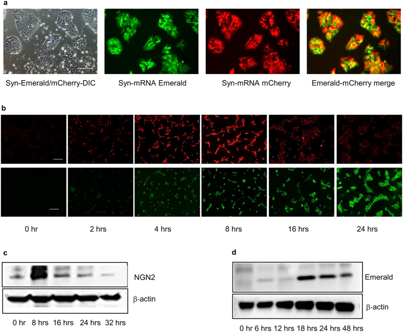
Figure 1
Delivery of syn-mRNAs into hPSCs.
( a ) Expression of synthetic messenger RNAs for fluorescent proteins Emerald and mCherry in TkDA3-4 human iPS cells 24u2009hours after transfection. Scale bar is 200u2009u03bcm. ( b ) Kinetics of NGN...
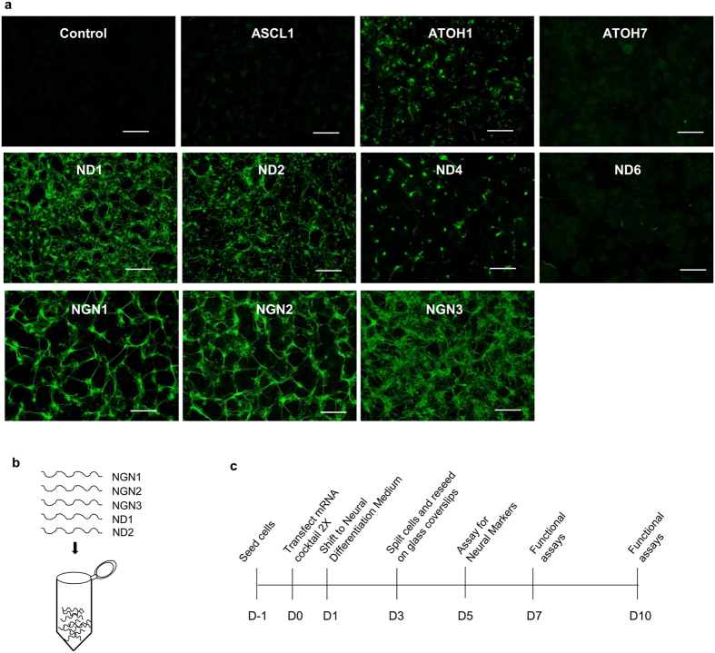
Figure 2
Induction of neurogenesis by syn-TFs mRNAs of Neurogenin and NeuroD families.
( a ) Neuronal differentiation induced by synthetic mRNAs for different Neurogenins and NeuroD TFs in TkDA3-4 human iPS cells (assayed at Day 5). For control, syn-Emerald and syn-mCherry were transfec...
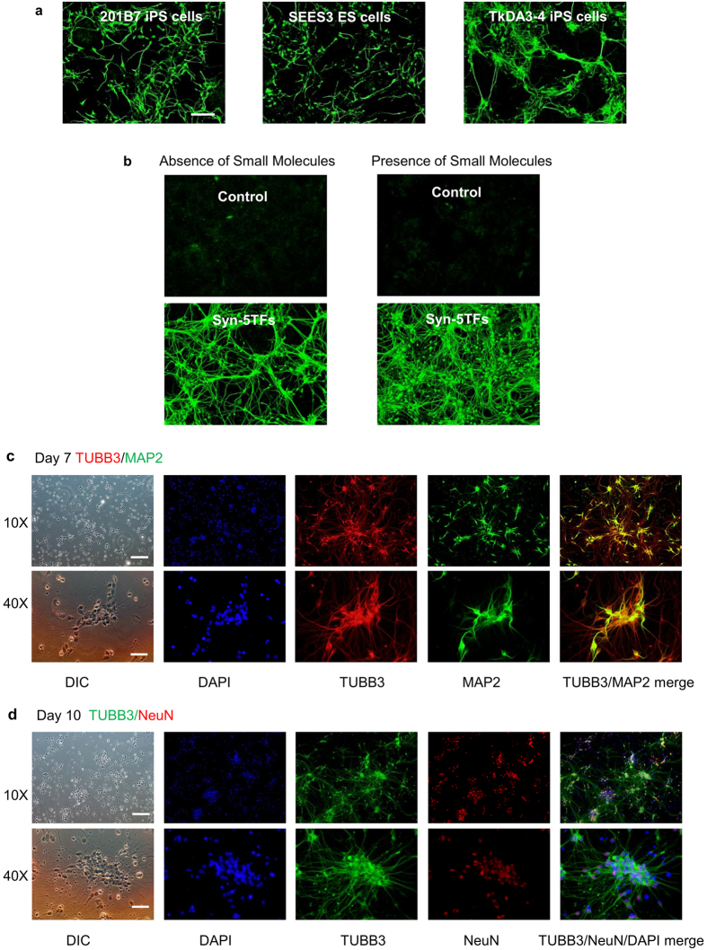
Figure 3
Induction of neurogenesis in human pluripotent stem cells by syn-5TFs mRNA cocktail.
( a ) Syn-5TFs-induces the efficient differentiation of human ES (SEES3) and iPS (201B7 and TkDA3-4) lines into neuronal cells. Representative images of TUBB3 staining at Day 5 are shown. The efficien...
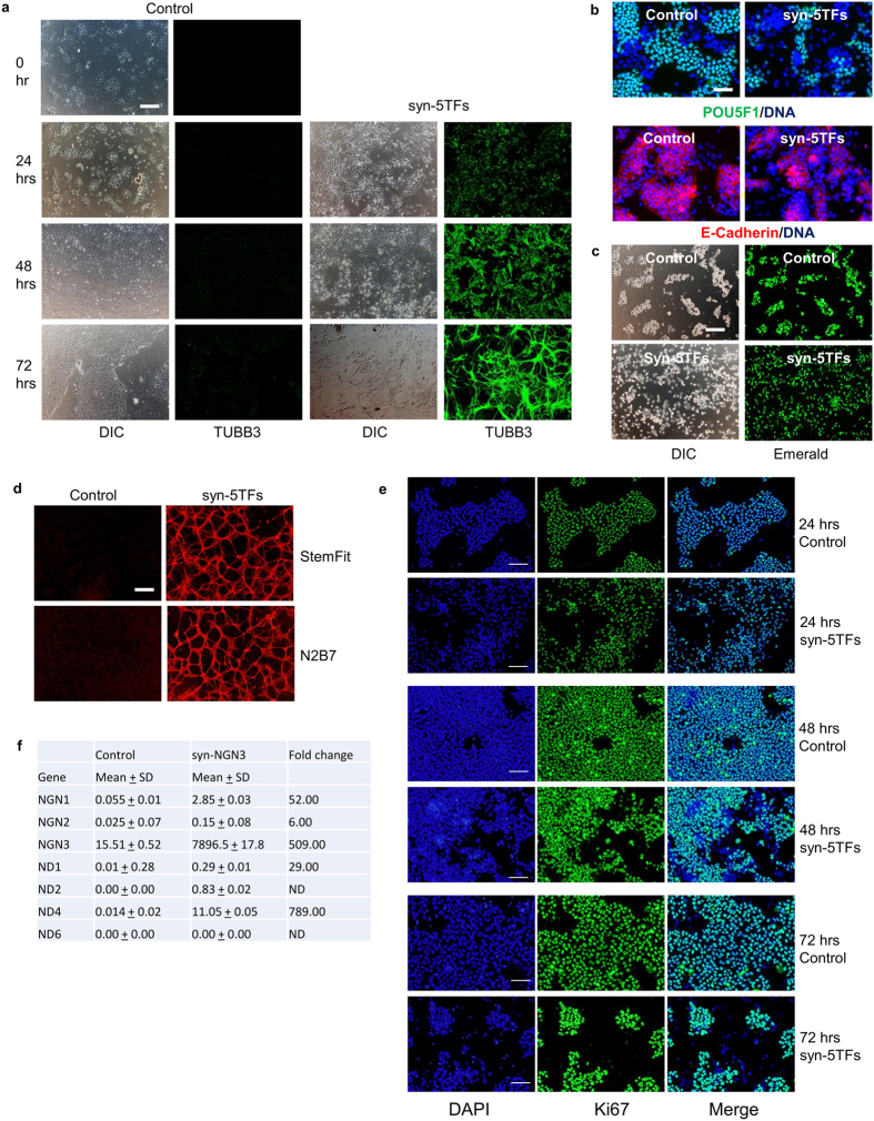
Figure 4
Kinetics and morphological changes during neuronal differentiation induced by the syn-5TFs cocktail.
( a ) Kinetics of neuronal TUBB3 expression during syn-5TFs mRNA cocktail-induced TkDA3-4 iPS cell differentiation. Scale bar indicates 200u2009u03bcm. ( b ) Expression of pluripotent markers 24u2009h...
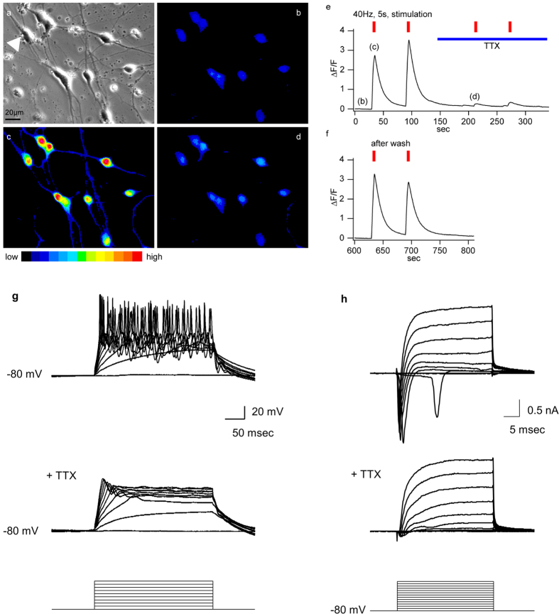
Figure 5
Syn-5TFs-induced neurons are functional.
( au2013f ) Electric stimulation-evoked calcium transients in neurons induced by syn-5TFs on Day 7. Fluo-4 loaded cells before electric stimulation in DIC microscopy ( a ) and fluorescence microscopy ...
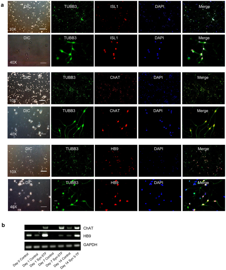
Figure 6
Syn-5TFs-induced motor neurons and their functional characterization.
( a ) Syn-5TFs differentiate human iPS cells into motor neurons. Day 7 neurons stained with ISL1 (top two panels), ChAT (middle two panels), and HB9 (bottom two panels). Upper row of each panel shows ...
Figure images are served from the NIH/NLM PubMed Central Open Access Subset or Europe PMC; copyright remains with the publishers and authors.
💬 Discussion
0 commentsNo comments yet. Be the first to start a discussion!
Leave a Comment