Abstract
Three PEG molecules (PEG-methacrylate, -diacrylate and -dimethacrylate) were incorporated into galactose-based polyacrylate hydrogels and their relative abilities to reduce non-specific protein adsorption in immunoassays were determined. Highly crosslinked hydrogels containing amine-terminated functionalities were formed and used to covalently attach antibodies specific for staphylococcal enterotoxin B (SEB). Patterned arrays of immobilized antibodies in the PEG-modified hydrogels were created with a PDMS template containing micro-channels for use in sandwich immunoassays to detect SEB. Different concentrations of the toxin were applied to the hydrogel arrays, followed with a Cy3-labeled tracer antibody specific for the two toxins. Fluorescence laser scanning confocal microscopy of the tracer molecules provided both qualitative and quantitative measurements on the detection sensitivity and the reduction in non-specific binding as a result of PEG incorporation. Results showed the PEG-modified hydrogel significantly reduced non-specific protein binding with a detection limit for SEB of 1 ng/mL. Fluorescence signals showed a 10-fold decrease in the non-specific binding and a 6-fold increase in specific binding of SEB.
🔬 Techniques
✨ Fluorophores
🧪 Sample Preparation
🏭 Microscope Brands
🧪 Reagent Suppliers
🏛️ Research Organizations (ROR)
Affiliated research institutions:
📋 Methods
Antibodies, antigens and reagents Poly(ethylene glycol) (n) methacrylate n = 526; Poly(ethylene glycol) (n) diacrylate n = 400; Poly(ethylene glycol) (n) dimethacrylate n = 400 and N -(3-aminopropyl) methacrylamide were purchased from Polysciences, Inc. (Warrington, PA, USA) and are shown in Figure 1 . Antibodies and antigens were obtained from the following sources: Staphylococcal enterotoxin B (SEB), rabbit and sheep anti-SEB polyclonal IgGs from Toxin Technology, Inc. (Sarasota, FL, USA); antibodies specific for ricin (two clones; RIC-07-AG1 and RIC-03-AG1) and ricin antigen from Naval Medical Research Center (NMRC) (Silver Spring, MD, USA); bis (sulfosuccinimidyl) suberate (BS 3 ) from Pierce Chemical Corp. (Rockford, IL, USA); N, N′-methylene bis-acrylamide, sodium persulfate and TEMED from BioRad Laboratories (Hercules, CA, USA); and 3-(trimethoxysilyl) propylmethacrylate and dichlorodimethylsilane from Sigma-Aldrich-Fluka (Milwaukee, WI, USA). Fluorescence labeling of antibodies specific for SEB and ricin with Cy3 dye (Amersham Biosciences, Piscataway, NJ, USA) was performed according to the protocol supplied by the manufacturer, except that 3 mg of protein was labeled for each packet of dye rather than 1 mg. Appropriate precautions were taken when using toxins and other hazardous reagents. Analyte solutions were treated with a 20% bleach solution before disposal. Contaminated disposables were placed in biohazard containers and later incinerated.
Preparation of PEG –modified polyacrylate hydrogel films
Hydrogel thin-films were prepared as previously reported [ 27 ]. Briefly, hydrogels were covalently attached to glass microscope slides through an acrylic group (3-(trimethoxysilyl) propyl methacrylate (MTPTS) that was previously applied to the glass surface. A solution of the galactose monomer, 6-acryloyl-β- O -methyl galactopyranoside [ 28 ], was prepared in 18MΩ Milli-Q water to a final concentration of 10% (w/v). The monomer solution was then added to N -(3-aminopropyl) methacrylamide at 25% (w/w) of the galactose monomer concentration. N, N -methylene bis-acrylamide (Bis) cross-linker at 3% (w/w) of the monomer concentration was dissolved in 100 μL of 18MΩ Milli-Q H 2 O and subsequently added to the galactose monomer-amine mixture. Formation of the hydrogel was accomplished through a free radical polymerization process using the initiator sodium persulfate (0.55 mg, 1.8 μmol). Poly(ethylene glycol) methacrylate, poly(ethylene glycol) diacrylate or poly(ethylene glycol) dimethacrylate was added to the monomer solution at a final concentration of 10% (v/v) and vortexed briefly. TEMED (1.5 μL, 8.2 nmol), which served as the catalyst, was added to the mixture followed by a brief nitrogen purge. A droplet of the hydrogel solution (110 μL) was placed on the methacrylate-treated slide and covered with the dichlorodimethyl (DCDM) silane-treated slide, clamped on both ends and allowed to polymerize overnight in an inert atmosphere (nitrogen). After gel polymerization, the DCDM-treated slide was removed, revealing a highly crosslinked PEG-modified polyacrylate hydrogel containing an amine-terminated moiety for antibody attachment. The resulting polymer has wt ratio of galactose-6-acrylate: 3-APM: Bis: PEG of about 10:2.5:0.3:10. Slides were briefly immersed in Milli-Q water (1.0 min), air-dried at room temperature, then stored semi-hydrated at 4 °C until further use.
Show full methods section
Antibodies, antigens and reagents Poly(ethylene glycol) (n) methacrylate n = 526; Poly(ethylene glycol) (n) diacrylate n = 400; Poly(ethylene glycol) (n) dimethacrylate n = 400 and N -(3-aminopropyl) methacrylamide were purchased from Polysciences, Inc. (Warrington, PA, USA) and are shown in Figure 1 . Antibodies and antigens were obtained from the following sources: Staphylococcal enterotoxin B (SEB), rabbit and sheep anti-SEB polyclonal IgGs from Toxin Technology, Inc. (Sarasota, FL, USA); antibodies specific for ricin (two clones; RIC-07-AG1 and RIC-03-AG1) and ricin antigen from Naval Medical Research Center (NMRC) (Silver Spring, MD, USA); bis (sulfosuccinimidyl) suberate (BS 3 ) from Pierce Chemical Corp. (Rockford, IL, USA); N, N′-methylene bis-acrylamide, sodium persulfate and TEMED from BioRad Laboratories (Hercules, CA, USA); and 3-(trimethoxysilyl) propylmethacrylate and dichlorodimethylsilane from Sigma-Aldrich-Fluka (Milwaukee, WI, USA). Fluorescence labeling of antibodies specific for SEB and ricin with Cy3 dye (Amersham Biosciences, Piscataway, NJ, USA) was performed according to the protocol supplied by the manufacturer, except that 3 mg of protein was labeled for each packet of dye rather than 1 mg. Appropriate precautions were taken when using toxins and other hazardous reagents. Analyte solutions were treated with a 20% bleach solution before disposal. Contaminated disposables were placed in biohazard containers and later incinerated.
Preparation of PEG –modified polyacrylate hydrogel films
Hydrogel thin-films were prepared as previously reported [ 27 ]. Briefly, hydrogels were covalently attached to glass microscope slides through an acrylic group (3-(trimethoxysilyl) propyl methacrylate (MTPTS) that was previously applied to the glass surface. A solution of the galactose monomer, 6-acryloyl-β- O -methyl galactopyranoside [ 28 ], was prepared in 18MΩ Milli-Q water to a final concentration of 10% (w/v). The monomer solution was then added to N -(3-aminopropyl) methacrylamide at 25% (w/w) of the galactose monomer concentration. N, N -methylene bis-acrylamide (Bis) cross-linker at 3% (w/w) of the monomer concentration was dissolved in 100 μL of 18MΩ Milli-Q H 2 O and subsequently added to the galactose monomer-amine mixture. Formation of the hydrogel was accomplished through a free radical polymerization process using the initiator sodium persulfate (0.55 mg, 1.8 μmol). Poly(ethylene glycol) methacrylate, poly(ethylene glycol) diacrylate or poly(ethylene glycol) dimethacrylate was added to the monomer solution at a final concentration of 10% (v/v) and vortexed briefly. TEMED (1.5 μL, 8.2 nmol), which served as the catalyst, was added to the mixture followed by a brief nitrogen purge. A droplet of the hydrogel solution (110 μL) was placed on the methacrylate-treated slide and covered with the dichlorodimethyl (DCDM) silane-treated slide, clamped on both ends and allowed to polymerize overnight in an inert atmosphere (nitrogen). After gel polymerization, the DCDM-treated slide was removed, revealing a highly crosslinked PEG-modified polyacrylate hydrogel containing an amine-terminated moiety for antibody attachment. The resulting polymer has wt ratio of galactose-6-acrylate: 3-APM: Bis: PEG of about 10:2.5:0.3:10. Slides were briefly immersed in Milli-Q water (1.0 min), air-dried at room temperature, then stored semi-hydrated at 4 °C until further use.
Immobilization of antibodies
Antibodies specific for SEB and ricin were immobilized and patterned in stripes onto PEG-modified hydrogel films using a PDMS patterning template containing six channels, each measuring 22 mm (l)×1.5 mm (w)×2.5 mm (h) [ 25 , 26 ]. The crosslinker, bis (sulfosuccinimidyl) suberate (BS 3 ) (2.5 mM) in 10 mM Na-phosphate buffer, pH 6.0, was injected into each channel and allowed to incubate for 30 min at RT. Covalent attachment of the crosslinker to the polymerized 3D hydrogel was achieved through the NHS-ester moiety of the crosslinker to the amine-terminated methacrylate group contained within the internal network of hydrogel. Each channel was rinsed with phosphate buffered saline, pH 7.4 (PBS, 0.6 mL), followed by the injection of rabbit anti-SEB in PBS (or mouse anti-ricin for ricin immunoassays) into the respective lanes. An incubation period of 1h at RT was performed followed with subsequent PBS rinse cycles. At each cycle the antibody was withdrawn carefully and used again for repeated treatments. This 30-min crosslinking and 60-min antibody cycle was performed six times in each micro-channel with the final cycle of antibody treatment allowed to incubate overnight at 4°C. At the conclusion of the final cycle, the antibody solution was withdrawn from the channels, the channels rinsed with PBS (0.6 mL), treated with PBS (0.6 mL) and subsequently blocked with 2% bovine serum albumin (BSA) in PBS (0.6 mL) for 1h at RT.
Sandwich immunoassay for SEB
SEB (in PBS containing 0.5% Tween ® 20 and 1.0 mg/mL BSA, PBSTB) was applied to the hydrogel slabs at concentrations 0 μg/mL to 1.0 μg/mL using a 6-channel PDMS template with its channels oriented perpendicular to the stripes of immobilized antibodies. The toxin solution was incubated (static) for 1h at RT to allow binding to the immobilized antibody. Each channel was then evacuated of toxin, rinsed with PBSTB (1.0 mL) and incubated for 30 min with Cy3-sheep anti-SEB (10 μg/mL in PBSTB). Following the tracer incubation, the channel was evacuated and received a final rinse with PBSTB (1.0 mL).
Fluorescence signals of bound
Cy3-labeled antibody were then measured using laser confocal microscopy and correlated to toxin concentration. Detection limits were defined as the lowest tested toxin concentration whose net signal was at least 3 standard deviations above both the negative control (no toxin) and the background (no capture antibody).
📊 Figures
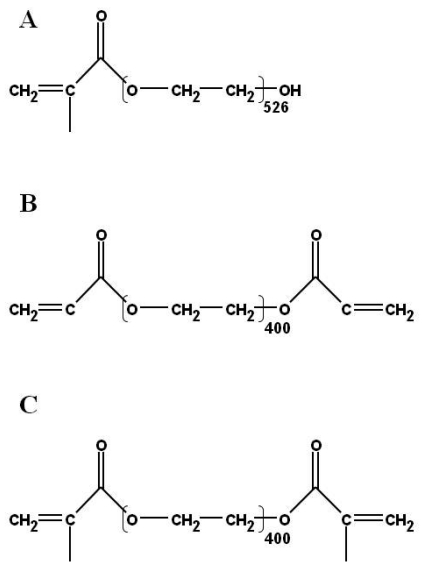
Figure 1.
Chemical structures of (a) Poly(ethylene glycol) methacrylate, (b) Poly(ethylene glycol) diacrylate, and (c) Poly(ethylene glycol) dimethacrylate.
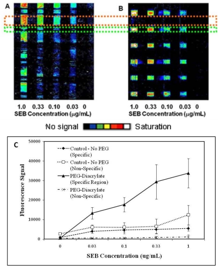
Figure 2.
Patterned fluorescence array images of sandwich immunoassay for SEB using galactose-based hydrogels. (a) Representative image of SEB immunoassay using hydrogel containing no PEG. (b) Representative im...
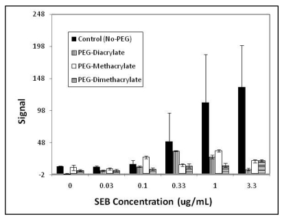
Figure 3.
Fluorescence measured from non-specific binding in sandwich immunoassays for SEB. Unmodified hydrogels, PEG-diacrylate, PEG-methacrylate and PEG-dimethacrylate modified hydrogels were compared (i.e., ...
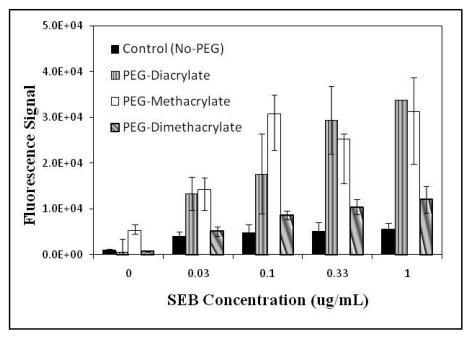
Figure 4.
Comparison of net fluorescence signal responses obtained in sandwich immunoassays using control (no PEG) and three different PEG-incorporated hydrogels. The three PEG candidates ([img]), PEG-diacrylat...
Figure images are served from the NIH/NLM PubMed Central Open Access Subset or Europe PMC; copyright remains with the publishers and authors.
💬 Discussion
0 commentsNo comments yet. Be the first to start a discussion!
Leave a Comment