Abstract
Centriolar satellites comprise cytoplasmic granules that are located around the centrosome. Their molecular identification was first reported more than a quarter of a century ago. These particles are not static in the cell but instead constantly move around the centrosome. Over the last decade, significant advances in their molecular compositions and biological functions have been achieved due to comprehensive proteomics and genomics, super-resolution microscopy analyses and elegant genetic manipulations. Centriolar satellites play pivotal roles in centrosome assembly and primary cilium formation through the delivery of centriolar/centrosomal components from the cytoplasm to the centrosome. Their importance is further underscored by the fact that mutations in genes encoding satellite components and regulators lead to various human disorders such as ciliopathies. Moreover, the most recent findings highlight dynamic structural remodelling in response to internal and external cues and unexpected positive feedback control that is exerted from the centrosome for centriolar satellite integrity.
🔬 Techniques
🧪 Sample Preparation
🔬 Cell Lines
🏛️ Research Organizations (ROR)
Affiliated research institutions:
📊 Figures
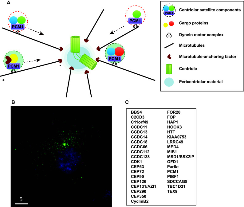
Fig.u00a01
Centriolar satellites and their components. a Schematic presentation of the centrosome and centriolar satellites. PCM1 is a structural platform of centriolar satellites, which are localised along the ...
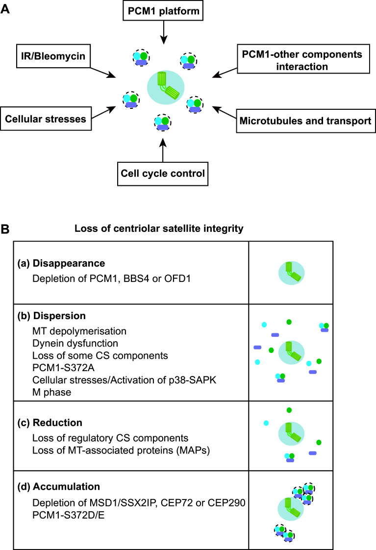
Fig.u00a02
Factors and requirements that ensure centriolar satellite integrity. a Centriolar satellite organisation is regulated by a number of both intrinsic and external cues. b Outcomes of centriolar satellit...
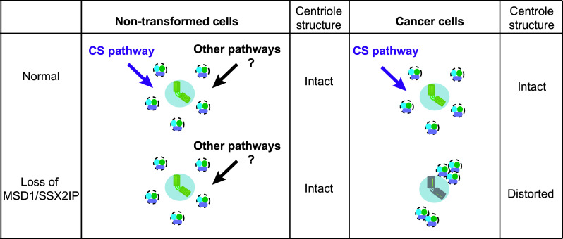
Fig.u00a03
Multiple pathways ensure centrosome assembly, which is compromised in cancer cells. In non-transformed cells ( left ), centriolar assembly is apparently normal upon depletion of MSD1/SSX2IP, although ...
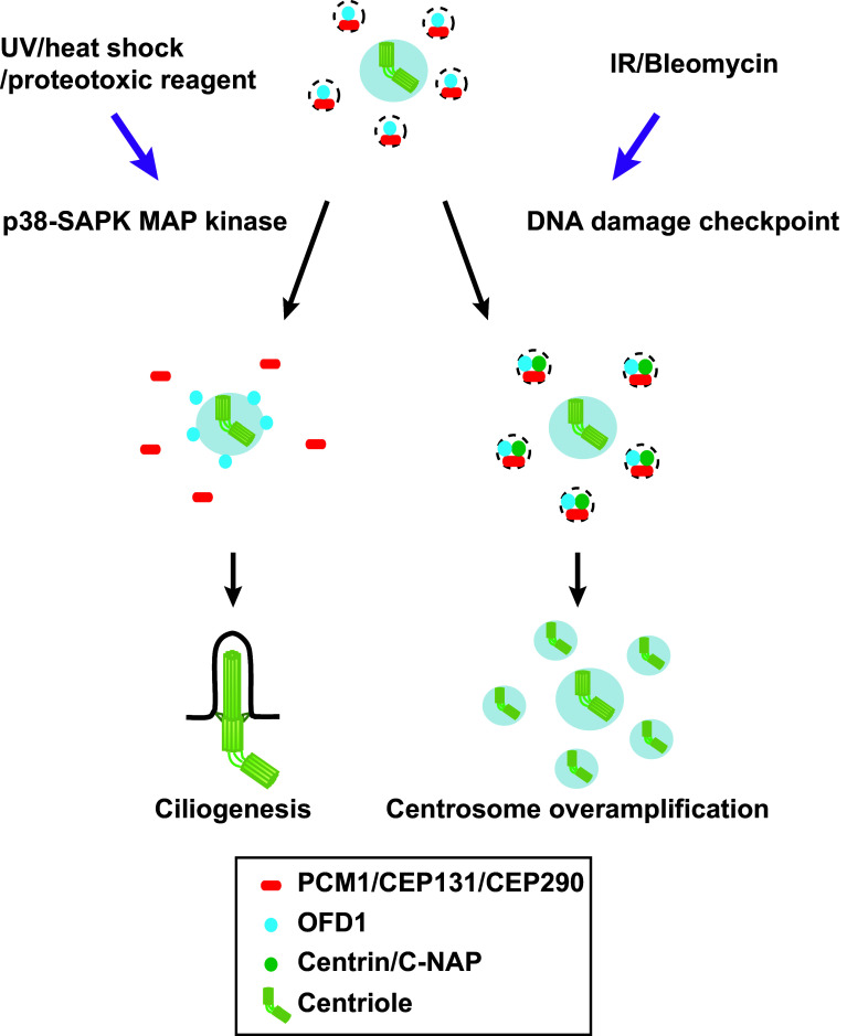
Fig.u00a04
Centriolar satellite remodelling upon exposure to various cellular stresses. Several cellular stresses including exposure to UV light, heat shock and proteotoxic reagents activate the p38-SAPK MAP pat...
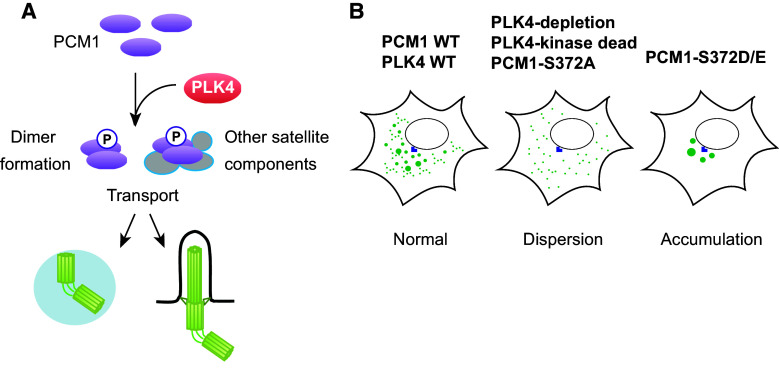
Fig.u00a05
Phosphoregulation of PCM1 through PLK4. a PCM1 is phosphorylated through PLK4. PLK4 phosphorylates the conserved S372 residue within PCM1. This phosphorylation plays a critical role in PCM1u2013PCM1 d...
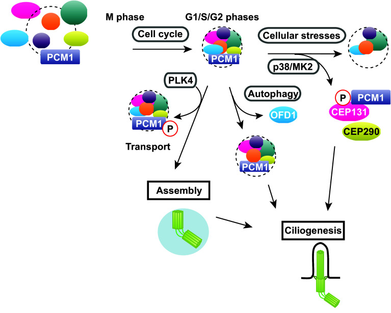
Fig.u00a06
Diverse forms of centriolar satellites in the context of various cues and cell cycle stages. Centriolar satellite integrity is regulated by many factors. Centriolar satellite organisation is changed d...
Figure images are served from the NIH/NLM PubMed Central Open Access Subset or Europe PMC; copyright remains with the publishers and authors.
💬 Discussion
0 commentsNo comments yet. Be the first to start a discussion!
Leave a Comment