Abstract
Remdesivir is a nucleoside analog approved by the US FDA for treatment of COVID-19. Here, we present a 3.9-Å-resolution cryo-EM reconstruction of a remdesivir-stalled RNA-dependent RNA polymerase complex, revealing full incorporation of 3 copies of remdesivir monophosphate (RMP) and a partially incorporated fourth RMP in the active site. The structure reveals that RMP blocks RNA translocation after incorporation of 3 bases following RMP, resulting in delayed chain termination, which can guide the rational design of improved antiviral drugs.
🔬 Techniques
🔭 Microscopes
🧬 Organisms
💻 Software
✨ Fluorophores
🏭 Microscope Brands
🧪 Reagent Suppliers
💻 Software Details
💻 Code & Software
💾 Data Repositories
🏛️ Research Organizations (ROR)
Affiliated research institutions:
📋 Methods
Key resources table REAGENT or RESOURCE SOURCE IDENTIFIER
Bacterial and virus strains BL21
E. coli New England Biolabs Cat. # C2530H Chemicals, peptides, and recombinant proteins SARS-CoV-2 NSP8 Dangerfield et al., 2020 N/A SARS-CoV-2 NSP12/7/8 Complex Dangerfield et al., 2020 N/A Remdesivir triphosphate Gilead Sciences Inc. GS-443902; CAS: 1355149-45-9 Deposited data Coordinates of SARS-CoV-2 nsp7-8-12:template:primer:(RMP 4 ) This paper PDB: 7L1F Cryo-EM map of SARS-CoV-2 nsp7-8-12:template:primer:(RMP 4 ) This paper EMD-23109 Supplemental information Mendeley Data https://doi.org/10.17632/ry9rk6tmcn.1 Oligonucleotides RNA Primer: 5′-[FAM]-GUC AUUCUCCUAAGAAGCUA-3′ Integrated DNA Technologies N/A RNA Template: 5′-CUAUCCCC AUGUGAUUUUAAUAGCUU CUUAGGAGAAUGAC-3′ Integrated DNA Technologies N/A Cy3 Internal Standard DNA: 5′-[Cy3]-CCGTGAGTTGGTTG GACGGCTGCGAGGC-3′ Integrated DNA Technologies N/A Recombinant DNA pcI ts,ind+ -(NSP8) Dangerfield et al., 2020 Deposited to Addgene – 160656 pQE-(NSP12)-pcI ts,ind+ -(NSP7-NSP8) Dangerfield et al., 2020 Deposited to Addgene–160540 pG-Tf2 Takara Biosciences Cat. # 3340 Software and algorithms KinTek Explorer v10 KinTek Corp. https://kintekexplorer.com Gene Mapper v5 ThermoFisher Scientific https://www.thermofisher.com/order/catalog/product/4370784#/4370784 SerialEM Mastronarde, 2005 https://bio3d.colorado.edu/SerialEM/ Warp Tegunov and Cramer, 2019 http://www.warpem.com/warp/# CryoSparc Punjani et al., 2017 https://cryosparc.com/ Coot Emsley and Cowtan, 2004 https://www2.mrc-lmb.cam.ac.uk/personal/pemsley/coot ChimeraX Goddard et al., 2018 https://www.cgl.ucsf.edu/chimerax/ DeepEMhancer Sanchez-Garcia et al., 2020 https://github.com/rsanchezgarc/deepEMhancer PHENIX Adams et al., 2010 https://www.phenix-online.org/documentation/index.html COSMIC 2 Cianfrocco et al., 2017 https://cosmic-cryoem.org/ eLBOW Moriarty et al., 2009 https://www.phenix-online.org/documentation/reference/elbow.html Other C-flat CF-2/2 200 mesh grids Protochips, Inc. CF-2/2-2Cu-50 Resource availability Lead contact Further information and requests for resources and reagents should be directed to and will be fulfilled by the lead contact, Kenneth A. Johnson ( kajohnson@utexas.edu ).
Show full methods section
Key resources table REAGENT or RESOURCE SOURCE IDENTIFIER
Bacterial and virus strains BL21
E. coli New England Biolabs Cat. # C2530H Chemicals, peptides, and recombinant proteins SARS-CoV-2 NSP8 Dangerfield et al., 2020 N/A SARS-CoV-2 NSP12/7/8 Complex Dangerfield et al., 2020 N/A Remdesivir triphosphate Gilead Sciences Inc. GS-443902; CAS: 1355149-45-9 Deposited data Coordinates of SARS-CoV-2 nsp7-8-12:template:primer:(RMP 4 ) This paper PDB: 7L1F Cryo-EM map of SARS-CoV-2 nsp7-8-12:template:primer:(RMP 4 ) This paper EMD-23109 Supplemental information Mendeley Data https://doi.org/10.17632/ry9rk6tmcn.1 Oligonucleotides RNA Primer: 5′-[FAM]-GUC AUUCUCCUAAGAAGCUA-3′ Integrated DNA Technologies N/A RNA Template: 5′-CUAUCCCC AUGUGAUUUUAAUAGCUU CUUAGGAGAAUGAC-3′ Integrated DNA Technologies N/A Cy3 Internal Standard DNA: 5′-[Cy3]-CCGTGAGTTGGTTG GACGGCTGCGAGGC-3′ Integrated DNA Technologies N/A Recombinant DNA pcI ts,ind+ -(NSP8) Dangerfield et al., 2020 Deposited to Addgene – 160656 pQE-(NSP12)-pcI ts,ind+ -(NSP7-NSP8) Dangerfield et al., 2020 Deposited to Addgene–160540 pG-Tf2 Takara Biosciences Cat. # 3340 Software and algorithms KinTek Explorer v10 KinTek Corp. https://kintekexplorer.com Gene Mapper v5 ThermoFisher Scientific https://www.thermofisher.com/order/catalog/product/4370784#/4370784 SerialEM Mastronarde, 2005 https://bio3d.colorado.edu/SerialEM/ Warp Tegunov and Cramer, 2019 http://www.warpem.com/warp/# CryoSparc Punjani et al., 2017 https://cryosparc.com/ Coot Emsley and Cowtan, 2004 https://www2.mrc-lmb.cam.ac.uk/personal/pemsley/coot ChimeraX Goddard et al., 2018 https://www.cgl.ucsf.edu/chimerax/ DeepEMhancer Sanchez-Garcia et al., 2020 https://github.com/rsanchezgarc/deepEMhancer PHENIX Adams et al., 2010 https://www.phenix-online.org/documentation/index.html COSMIC 2 Cianfrocco et al., 2017 https://cosmic-cryoem.org/ eLBOW Moriarty et al., 2009 https://www.phenix-online.org/documentation/reference/elbow.html Other C-flat CF-2/2 200 mesh grids Protochips, Inc. CF-2/2-2Cu-50 Resource availability Lead contact Further information and requests for resources and reagents should be directed to and will be fulfilled by the lead contact, Kenneth A. Johnson ( kajohnson@utexas.edu ).
Materials availability
This study did not generate new unique reagents.
Data and code availability
The cryo-EM structure and associated atomic coordinates are available at the Electron Microscopy Data Bank and Protein Data Bank with accession codes EMD:23109 and PDB: 7L1F , respectively. Method details Preparation of protein The SARS CoV-2 RdRp (nsp12/7/8) was expressed and purified without tags to provide a highly active enzyme for transient kinetic and structural studies using cryo-electron microscopy (cryo-EM). Tag-free nsp12/7/8 complex and nsp8 were expressed in E. coli and purified as described in detail previously ( Dangerfield et al., 2020 , 2021 ). A detailed summary of each expression and purification is given below. Nsp8 was expressed in BL21 E. coli harboring the plasmid pcI ts,ind+ -(NSP8). Cells were grown in Terrific Broth (2.4% [w/v] yeast extract, 2% [w/v] tryptone, 0.4% [v/v] glycerol, 17 mM KH 2 PO 4 , 72 mM K 2 HPO 4 ) with 100 μg/ml ampicillin for selection. Protein expression was induced by shifting the culture temperature from 30°C to 42°C for 20 minutes, followed by 3 hours at 38°C before harvesting the cells. The cells were lysed by sonication, then the lysate was clarified by centrifugation and separated on the following columns: Q Sepharose-FF, SP Sepharose-FF, HiTrap Blue-FF, Superdex 200. The protein was then dialyzed into Storage Buffer-1 (50 mM Tris-HCl pH 7.5, 0.1 mM EDTA, 20 mM KCl, 1 mM DTT, 50% [v/v] glycerol), aliquoted, and stored at −80°C. The three peptides for the nsp12/7/8 complex were co-expressed in BL21 E. coli harboring the plasmids pG-Tf2 and pQE-(NSP12)-pcI ts,ind+ -(NSP7-NSP8). Cells were grown in Terrific Broth with 30 μg/ml kanamycin and 20 μg/ml chloramphenicol for selection at 30°C. Protein expression was induced by addition of tetracycline to 10 ng/ml for 20 minutes, followed by addition of IPTG to 0.5 mM and nalidixic acid to 50 μg/ml, then incubation overnight at 16°C. Harvested cells were lysed by sonication, then the lysate was clarified by centrifugation and separated on the following columns: Q Sepharose-FF, HiTrap Blue-FF, Heparin Sepharose 6-FF, SP Sepharose-FF, Superdex 200. The protein was then dialyzed into Storage Buffer-2 (50 mM Tris-HCl pH 7.5, 0.1 mM EDTA, 50 mM NaCl, 1 mM DTT, 50% [v/v] glycerol), aliquoted, and stored at −80°C.
RNA substrates and other materials
RNA samples were prepared in DEPC treated water (Ambion). RNA substrates were purchased from Integrated DNA technologies with RNase free HPLC purification and resuspended in Annealing Buffer (10 mM Tris-HCl pH 7, 50 mM NaCl, 0.1 mM EDTA). Concentration of each oligo was determined by absorbance at 260 nm using the extinction coefficients 222,360 M -1 cm -1 and 403,100 M -1 cm -1 for the FAM-20 nt primer and the 40 nt template, respectively. The double stranded RNA substrate was prepared by mixing each oligo at a 1:1 molar ratio, heating to 75°C for 3 minutes, then cooling slowly to room temperature over approximately 2 hours. Oligonucleotides were stored at −20°C. UTP was purchased from New England Biolabs. Remdesivir triphosphate (GS-443902) was a kindly provided by Gilead Sciences and concentration was determined by absorbance at 245 nm using the extinction coefficient 24,100 M -1 cm -1 ( Dangerfield et al., 2020 ). Remdesivir triphosphate was stored at −20°C.
Extension reactions and analysis by capillary electrophoresis
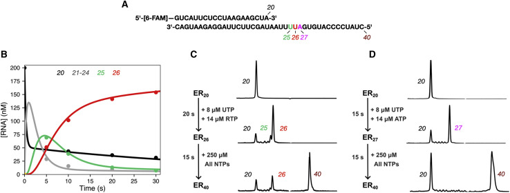
All reactions were conducted in Reaction Buffer (40 mM Tris-HCl pH 7, 50 mM NaCl, 5 mM MgCl 2 , 1 mM DTT) ( Dangerfield et al., 2020 ). Concentrations of enzyme, RNA and nucleotides are given in the legend for Figure 1 . (concentrations given are the final concentrations after mixing). Reactions were performed at room temperature (∼25°C) in Reaction Buffer after allowing the RdRp complex and RNA to equilibrate for approximately 30 minutes. Samples were quenched by adding 0.6 M EDTA to a final concentration of 0.4 M. One μL of each sample was diluted into 10 μL of HiDi formamide (ThermoFisher) containing 1 nM Cy3 internal standard DNA oligo (5′-[Cy3]-CCGTGAGTTGGTTGGACGGCTGCGAGGC-3′, purchased from Integrated DNA Technologies) in a 96 well plate. Samples were analyzed on an Applied Biosystems 3130xl Genetic Analyzer instrument equipped with a 36 cm capillary array (ThermoFisher) and nanoPOP-6 polymer (Molecular Cloning Laboratories). The oven temperature was set to 65°C. Before injecting samples, a pre-run electrophoresis step was performed at 15 kV for 3 minutes. Samples were injected at 3.6 kV for 12 s, then the voltage was ramped up to 15 kV over 40, 15 s steps. Fluorescence was monitored for 800 s using the G5 dye set and all peak intensities were within the linear range of the instrument. Peaks were integrated with GeneMapper 5 software (ThermoFisher). Cryo-EM sample preparation, data collection and processing Samples were prepared in Reaction Buffer. A solution of 3.33 μM nsp12/7/8, 4.67 μM nsp8, 3.33 μM FAM-20/40 RNA was mixed with 14 μM remdesivir triphosphate (RTP) and 7.5 μM UTP to start the reaction at room temperature (∼25°C) (concentrations of reaction components are given after mixing). The reaction was allowed to proceed for 10-20 s before application to glow discharged holey carbon grids (C-flat 4/2, Protochips Inc.), blotted for 0.5 s with a blot force of 4 and rapidly plunged into liquid ethane using an FEI Vitrobot MarkIV. Data was collected on an FEI Titan Krios cryo-electron microscope equipped with a K3 Summit direct electron detector (Gatan, Pleasanton, CA). Images were recorded with SerialEM ( Mastronarde, 2005 ), with a pixel size of 1.1Å over a defocus range of −1.5 to −2.5 μm. 2446 movies were recorded at 13.3 electrons/pixel/second for 6 s (80 frames) to give a total dose of 80 electrons/pixel. CTF correction, motion correction and particle picking were performed in real-time using WARP ( Tegunov and Cramer, 2019 ), resulting in 2,340,544 particles, which were uploaded to cryoSPARC v2 ( Punjani et al., 2017 ). Particles were subjected to multiple rounds of 3D classification in cryoSPARC, and a final set of 116,748 particles was refined to a global resolution of 3.89 Å based on the 0.143 FSC criterion. Map sharpening was performed using DeepEMhancer ( Sanchez-Garcia et al., 2020 ) as implemented in COSMIC 2 ( Cianfrocco et al., 2017 ). For modeling, the published structure of nsp7-8-12 (PDB: 7bv2 ( Yin et al., 2020 )) was used as a starting model. Non-proteinaceous molecules (i.e., RNA, ligands) were removed, and the template:primer duplex was built de novo in Coot ( Emsley and Cowtan, 2004 ). Restraints for RMP were generated using eLBOW ( Moriarty et al., 2009 )), and structures were subjected to real-space refinement using Phenix (which resulted in a map-to-model FSC of 4.0Å at the 0.5 threshold) ( Afonine et al., 2018 ). Figures were prepared using ChimeraX ( Goddard et al., 2018 ).
Quantification and statistical analysis
Structural statistics are provided in Table 1 . Table 1 Cryo-EM data collection and processing statistics Data collection and processing Magnification 22,500× Voltage (kV) 300 Electron exposure (e − /Å) 80 Defocus range (μM) −1.5 to −2.5 Symmetry imposed C1 Initial particle images 2,340,544 Final particle images 116,748 Map resolution (Å) 3.89 FSC threshold 0.143 Map resolution range (Å) 3.3 to >8 Refinement Initial model used (PDB code) PDB: 7BV2 Model resolution (Å) 3.9 FSC threshold 0.5 Map sharpening B factor (Å 2 ) 114.5 Model composition Nonhydrogen atoms 8,820 Residues (protein/RNA) 1,009/35 B factors (Å 2 ), min/max/mean Protein 60.2/114.2/92.6 RNA 81.2/219.3/145.3 RMSDs Bond lengths (Å) 0.008 Bond angles (°) 1.388 Validation MolProbity score 1.66 clashscore 4.15 Poor rotamers (%) 0 Ramachandran plot (%) Favored 92.58 Allowed 7.42 Disallowed 0 FSC, Fourier shell correlation.
Materials availability
This study did not generate new unique reagents.
Method details Preparation of protein The SARS CoV-2 RdRp (nsp12/7/8) was expressed and purified without tags to provide a highly active enzyme for transient kinetic and structural studies using cryo-electron microscopy (cryo-EM). Tag-free nsp12/7/8 complex and nsp8 were expressed in E. coli and purified as described in detail previously ( Dangerfield et al., 2020 , 2021 ). A detailed summary of each expression and purification is given below. Nsp8 was expressed in BL21 E. coli harboring the plasmid pcI ts,ind+ -(NSP8). Cells were grown in Terrific Broth (2.4% [w/v] yeast extract, 2% [w/v] tryptone, 0.4% [v/v] glycerol, 17 mM KH 2 PO 4 , 72 mM K 2 HPO 4 ) with 100 μg/ml ampicillin for selection. Protein expression was induced by shifting the culture temperature from 30°C to 42°C for 20 minutes, followed by 3 hours at 38°C before harvesting the cells. The cells were lysed by sonication, then the lysate was clarified by centrifugation and separated on the following columns: Q Sepharose-FF, SP Sepharose-FF, HiTrap Blue-FF, Superdex 200. The protein was then dialyzed into Storage Buffer-1 (50 mM Tris-HCl pH 7.5, 0.1 mM EDTA, 20 mM KCl, 1 mM DTT, 50% [v/v] glycerol), aliquoted, and stored at −80°C. The three peptides for the nsp12/7/8 complex were co-expressed in BL21 E. coli harboring the plasmids pG-Tf2 and pQE-(NSP12)-pcI ts,ind+ -(NSP7-NSP8). Cells were grown in Terrific Broth with 30 μg/ml kanamycin and 20 μg/ml chloramphenicol for selection at 30°C. Protein expression was induced by addition of tetracycline to 10 ng/ml for 20 minutes, followed by addition of IPTG to 0.5 mM and nalidixic acid to 50 μg/ml, then incubation overnight at 16°C. Harvested cells were lysed by sonication, then the lysate was clarified by centrifugation and separated on the following columns: Q Sepharose-FF, HiTrap Blue-FF, Heparin Sepharose 6-FF, SP Sepharose-FF, Superdex 200. The protein was then dialyzed into Storage Buffer-2 (50 mM Tris-HCl pH 7.5, 0.1 mM EDTA, 50 mM NaCl, 1 mM DTT, 50% [v/v] glycerol), aliquoted, and stored at −80°C.
RNA substrates and other materials
RNA samples were prepared in DEPC treated water (Ambion). RNA substrates were purchased from Integrated DNA technologies with RNase free HPLC purification and resuspended in Annealing Buffer (10 mM Tris-HCl pH 7, 50 mM NaCl, 0.1 mM EDTA). Concentration of each oligo was determined by absorbance at 260 nm using the extinction coefficients 222,360 M -1 cm -1 and 403,100 M -1 cm -1 for the FAM-20 nt primer and the 40 nt template, respectively. The double stranded RNA substrate was prepared by mixing each oligo at a 1:1 molar ratio, heating to 75°C for 3 minutes, then cooling slowly to room temperature over approximately 2 hours. Oligonucleotides were stored at −20°C. UTP was purchased from New England Biolabs. Remdesivir triphosphate (GS-443902) was a kindly provided by Gilead Sciences and concentration was determined by absorbance at 245 nm using the extinction coefficient 24,100 M -1 cm -1 ( Dangerfield et al., 2020 ). Remdesivir triphosphate was stored at −20°C.
Extension reactions and analysis by capillary electrophoresis
All reactions were conducted in Reaction Buffer (40 mM Tris-HCl pH 7, 50 mM NaCl, 5 mM MgCl 2 , 1 mM DTT) ( Dangerfield et al., 2020 ). Concentrations of enzyme, RNA and nucleotides are given in the legend for Figure 1 . (concentrations given are the final concentrations after mixing). Reactions were performed at room temperature (∼25°C) in Reaction Buffer after allowing the RdRp complex and RNA to equilibrate for approximately 30 minutes. Samples were quenched by adding 0.6 M EDTA to a final concentration of 0.4 M. One μL of each sample was diluted into 10 μL of HiDi formamide (ThermoFisher) containing 1 nM Cy3 internal standard DNA oligo (5′-[Cy3]-CCGTGAGTTGGTTGGACGGCTGCGAGGC-3′, purchased from Integrated DNA Technologies) in a 96 well plate. Samples were analyzed on an Applied Biosystems 3130xl Genetic Analyzer instrument equipped with a 36 cm capillary array (ThermoFisher) and nanoPOP-6 polymer (Molecular Cloning Laboratories). The oven temperature was set to 65°C. Before injecting samples, a pre-run electrophoresis step was performed at 15 kV for 3 minutes. Samples were injected at 3.6 kV for 12 s, then the voltage was ramped up to 15 kV over 40, 15 s steps. Fluorescence was monitored for 800 s using the G5 dye set and all peak intensities were within the linear range of the instrument. Peaks were integrated with GeneMapper 5 software (ThermoFisher). Cryo-EM sample preparation, data collection and processing Samples were prepared in Reaction Buffer. A solution of 3.33 μM nsp12/7/8, 4.67 μM nsp8, 3.33 μM FAM-20/40 RNA was mixed with 14 μM remdesivir triphosphate (RTP) and 7.5 μM UTP to start the reaction at room temperature (∼25°C) (concentrations of reaction components are given after mixing). The reaction was allowed to proceed for 10-20 s before application to glow discharged holey carbon grids (C-flat 4/2, Protochips Inc.), blotted for 0.5 s with a blot force of 4 and rapidly plunged into liquid ethane using an FEI Vitrobot MarkIV. Data was collected on an FEI Titan Krios cryo-electron microscope equipped with a K3 Summit direct electron detector (Gatan, Pleasanton, CA). Images were recorded with SerialEM ( Mastronarde, 2005 ), with a pixel size of 1.1Å over a defocus range of −1.5 to −2.5 μm. 2446 movies were recorded at 13.3 electrons/pixel/second for 6 s (80 frames) to give a total dose of 80 electrons/pixel. CTF correction, motion correction and particle picking were performed in real-time using WARP ( Tegunov and Cramer, 2019 ), resulting in 2,340,544 particles, which were uploaded to cryoSPARC v2 ( Punjani et al., 2017 ). Particles were subjected to multiple rounds of 3D classification in cryoSPARC, and a final set of 116,748 particles was refined to a global resolution of 3.89 Å based on the 0.143 FSC criterion. Map sharpening was performed using DeepEMhancer ( Sanchez-Garcia et al., 2020 ) as implemented in COSMIC 2 ( Cianfrocco et al., 2017 ). For modeling, the published structure of nsp7-8-12 (PDB: 7bv2 ( Yin et al., 2020 )) was used as a starting model. Non-proteinaceous molecules (i.e., RNA, ligands) were removed, and the template:primer duplex was built de novo in Coot ( Emsley and Cowtan, 2004 ). Restraints for RMP were generated using eLBOW ( Moriarty et al., 2009 )), and structures were subjected to real-space refinement using Phenix (which resulted in a map-to-model FSC of 4.0Å at the 0.5 threshold) ( Afonine et al., 2018 ). Figures were prepared using ChimeraX ( Goddard et al., 2018 ).
RNA substrates and other materials
RNA samples were prepared in DEPC treated water (Ambion). RNA substrates were purchased from Integrated DNA technologies with RNase free HPLC purification and resuspended in Annealing Buffer (10 mM Tris-HCl pH 7, 50 mM NaCl, 0.1 mM EDTA). Concentration of each oligo was determined by absorbance at 260 nm using the extinction coefficients 222,360 M -1 cm -1 and 403,100 M -1 cm -1 for the FAM-20 nt primer and the 40 nt template, respectively. The double stranded RNA substrate was prepared by mixing each oligo at a 1:1 molar ratio, heating to 75°C for 3 minutes, then cooling slowly to room temperature over approximately 2 hours. Oligonucleotides were stored at −20°C. UTP was purchased from New England Biolabs. Remdesivir triphosphate (GS-443902) was a kindly provided by Gilead Sciences and concentration was determined by absorbance at 245 nm using the extinction coefficient 24,100 M -1 cm -1 ( Dangerfield et al., 2020 ). Remdesivir triphosphate was stored at −20°C.
Supplemental information Document S1. Figure S1 Document S2. Article plus supplemental information
📊 Figures
Figureu00a01
RdRp stalls after incorporation of 4 remdesivirs (A) The RNA substrate used in experiments consists of a 20-nt, 5u2032-[6-FAM]-labeled primer annealed to a 40-nt template. The sequence is from the 3u2...
Figureu00a02
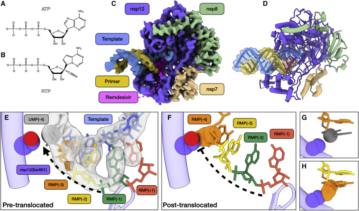
Structural basis of SARS-CoV-2 RdRp complex stalling by remdesivir (A) Chemical structures of adenosine triphosphate. (B) Chemical structure of remdesivir triphosphate. (C) Cryo-EM reconstruction of r...
Figure images are served from the NIH/NLM PubMed Central Open Access Subset or Europe PMC; copyright remains with the publishers and authors.
💬 Discussion
0 commentsNo comments yet. Be the first to start a discussion!
Leave a Comment