Abstract
Abstract p97, also known as valosin-containing protein (VCP) or Cdc48, plays a central role in cellular protein homeostasis. Human p97 mutations are associated with several neurodegenerative diseases. Targeting p97 and its cofactors is a strategy for cancer drug development. Despite significant structural insights into the fungal homolog Cdc48, little is known about how human p97 interacts with its cofactors. Recently, the anti-alcohol abuse drug disulfiram was found to target cancer through Npl4, a cofactor of p97, but the molecular mechanism remains elusive. Here, using single-particle cryo-electron microscopy (cryo-EM), we uncovered three Npl4 conformational states in complex with human p97 before ATP hydrolysis. The motion of Npl4 results from its zinc finger motifs interacting with the N domain of p97, which is essential for the unfolding activity of p97. In vitro and cell-based assays showed that the disulfiram derivative bis-(diethyldithiocarbamate)-copper (CuET) can bypass the copper transporter system and inhibit the function of p97 in the cytoplasm by releasing cupric ions under oxidative conditions, which disrupt the zinc finger motifs of Npl4, locking the essential conformational switch of the complex.
🔬 Techniques
🧬 Organisms
✨ Fluorophores
🔬 Cell Lines
🏭 Microscope Brands
🧪 Reagent Suppliers
📷 Detectors
💻 Software Details
💾 Data Repositories
🏛️ Research Organizations (ROR)
Affiliated research institutions:
📋 Methods
Protein expression and purification
Full-length human p97 including mutant A232E/E578Q, Ufd1, Npl4, as well as wild-type and mutant Npl4 fragments (residues 129–580) were overexpressed in E. coli BL21-DE3-RIPL cells. p97 was cloned into pET-47b vector with an N-terminal cleavable His-tag. All mutants of p97 were generated by site-directed mutagenesis using wild-type p97 as a template (Supplementary Table 3 ). Two plasmids containing full-length untagged Npl4 and C-terminal His-tagged Ufd1 were purchased from Addgene. Npl4 fragments were expressed in pET-28a vector with an N-terminal His-SUMO tag. Purification protocols were similar to those published before 47 . Briefly, E. coli cells were grown in LB with auto-induction supplement for 16 h at 30 °C. Cells were pelleted at 5000 × g and resuspended in Lysis Buffer (50 mM Tris, pH 8.0, 300 mM KCl, 20 mM imidazole, 1 mM DTT, and 1 mM MgCl 2 ). To purify the Npl4/Ufd1 complex, the resuspended cells of respective construct were mixed at this step. 1 mM PMSF was added to the suspensions, and cells were lysed by sonication. Lysates were cleared by centrifugation for 30 min at 30,000 × g . then the supernatants were flowed through a Ni-NTA gravity column twice at 4 °C. Beads were washed with Wash Buffer (50 mM Tris, pH 8.0, 150 mM NaCl, 20 mM imidazole and 1 mM MgCl 2 ). The His-tagged proteins were eluted in Elution Buffer (50 mM Tris, pH 8.0, 300 mM NaCl, and 400 mM imidazole). For p97 and Npl4 fragments, HRV3C protease and SUMO protease (Ulp1p) were added to the eluate, respectively, and dialyzed in SEC Buffer (25 mM Tris, pH 8.0, 150 mM NaCl, 1 mM MgCl 2 , and 0.5 mM Tris(2-carboxyethyl)phosphine (TCEP)). Npl4/Ufd1 complex were directly dialyzed in the SEC Buffer. Npl4/Ufd1 complex and Npl4 fragments were further purified by a Superdex 200 size-exclusion column (GE Healthcare) equilibrated in the SEC Buffer. p97 was purified by a Superose 6 column (GE Healthcare) equilibrated in the SEC Buffer.
Show full methods section
Protein expression and purification
Full-length human p97 including mutant A232E/E578Q, Ufd1, Npl4, as well as wild-type and mutant Npl4 fragments (residues 129–580) were overexpressed in E. coli BL21-DE3-RIPL cells. p97 was cloned into pET-47b vector with an N-terminal cleavable His-tag. All mutants of p97 were generated by site-directed mutagenesis using wild-type p97 as a template (Supplementary Table 3 ). Two plasmids containing full-length untagged Npl4 and C-terminal His-tagged Ufd1 were purchased from Addgene. Npl4 fragments were expressed in pET-28a vector with an N-terminal His-SUMO tag. Purification protocols were similar to those published before 47 . Briefly, E. coli cells were grown in LB with auto-induction supplement for 16 h at 30 °C. Cells were pelleted at 5000 × g and resuspended in Lysis Buffer (50 mM Tris, pH 8.0, 300 mM KCl, 20 mM imidazole, 1 mM DTT, and 1 mM MgCl 2 ). To purify the Npl4/Ufd1 complex, the resuspended cells of respective construct were mixed at this step. 1 mM PMSF was added to the suspensions, and cells were lysed by sonication. Lysates were cleared by centrifugation for 30 min at 30,000 × g . then the supernatants were flowed through a Ni-NTA gravity column twice at 4 °C. Beads were washed with Wash Buffer (50 mM Tris, pH 8.0, 150 mM NaCl, 20 mM imidazole and 1 mM MgCl 2 ). The His-tagged proteins were eluted in Elution Buffer (50 mM Tris, pH 8.0, 300 mM NaCl, and 400 mM imidazole). For p97 and Npl4 fragments, HRV3C protease and SUMO protease (Ulp1p) were added to the eluate, respectively, and dialyzed in SEC Buffer (25 mM Tris, pH 8.0, 150 mM NaCl, 1 mM MgCl 2 , and 0.5 mM Tris(2-carboxyethyl)phosphine (TCEP)). Npl4/Ufd1 complex were directly dialyzed in the SEC Buffer. Npl4/Ufd1 complex and Npl4 fragments were further purified by a Superdex 200 size-exclusion column (GE Healthcare) equilibrated in the SEC Buffer. p97 was purified by a Superose 6 column (GE Healthcare) equilibrated in the SEC Buffer.
Preparation of polyubiquitinated Ub-Eos
Ub-Eos was constructed by fusing two tandem ubiquitin to the N-terminus of fluorescent protein mEos3.2. The polyubiquitinated Ub-Eos was prepared similar to previously described 16 . Briefly, ubiquitination reaction was carried out with final concentrations of 20 μM Ub-Eos, 1 μM E1, 20 μM gp78RING-Ube2g2, and 500 μM ubiquitin in 20 mM Hepes, pH 7.4, 150 mM KCl, 10 mM ATP, and 10 mM MgCl 2 at 37 °C overnight. To purify ubiquitinated Eos3.2 from free ubiquitin chains, the reaction mixture was incubated with Ni-NTA resin, eluted with 300 mM imidazole, and run over a Superdex 200 size-exclusion column equilibrated in the SEC Buffer. Fractions with long ubiquitin chain (over 10 Ubs) were pooled and concentrated with centrifugal filter units, followed by flash frozen. p97 complex formation To form p97-Npl4/Ufd1 complex, Npl4/Ufd1 was added at a three folds molar excess to p97 hexamer, and proteins were incubated with 1 mM ATPγS for 60 min before gel filtration. After gel filtration, all proteins were flash frozen in liquid nitrogen. For p97-Npl4/Ufd1-Ub-Eos complex, a mutant p97 bearing A232E and E578Q mutations was used instead of wild-type p97. To form the ternary complex, p97 (A232E/ E578Q)-Npl4/Ufd1 complex was first prepared as described above, then polyubiquitinated Ub-Eos was added at a two-fold molar excess to p97 (A232E/E578Q)-Npl4/Ufd1 complex. After gel filtration, all proteins were flash frozen in liquid nitrogen. Specimen preparation for single-particle cryo-EM For p97-Npl4/Ufd1 complex, before preparing grids for cryo-EM, the complex was concentrated to ~20 mg/mL, and incubated with 5 mM ATPγS (90% pure, Sigma) for 30 min at room temperature. For p97 (A232E/E578Q)-Npl4/Ufd1-Ub-Eos complex, the sample was concentrated to 20 mg/mL without adding additional nucleotide. For p97-Npl4/Ufd1 complex in the presence of cupric ion, the complex was concentrated to ~20 mg/mL, and incubated with 5 mM ATPγS (90% pure, Sigma) and 100 μM cupric ion for 30 min at room temperature. To relief the preferred orientations, IGEPAL CA-630 (Sigma) was added to the samples to a final concentration of 0.05% immediately before grid freezing. The freezing was performed using a Vitrobot mark IV (Thermo Fisher) operating at 8 °C and 100% humidity. Samples (3.5 μL) were applied to a non-glow-discharged Quantifoil Au 1.2/1.3 grid. The grid was blotted for 1 s using standard Vitrobot filter paper (Ted Pella, 47000-100) and then plunge frozen in liquid ethane.
Data collection for single-particle cryo-EM
Optimized frozen grids were shipped to National Cryo-Electron Microscopy Facility for data collection. All datasets were acquired as movie stacks with a Titan Krios electron microscope operating at 300 kV, equipped with either a Gatan K2 Summit or K3 direct detection camera. A single stack typically consists of 40 frames with a total exposure around 50 electrons/Å 2 . The defocus range was set at −1.0 to −2.5 μm. See Supplementary Table 1 for the details.
Image processing
Movie stacks were subjected to beam-induced motion correction using MotionCor2 48 . CTF parameters for each micrograph were determined by CTFFIND4 49 . The following particle picking, two- and three-dimensional classifications, and three-dimensional refinement were performed in RELION-3 50 . Briefly, particle picking was done by manually choosing ~2000 particles and generating templates through 2D classification for the following automatic picking. False-positive particles or particles classified in poorly defined classes were discarded after 2D classification. The initial 3D classification was performed on a binned dataset with the previously reported p97 structures as the reference model 15 . The detailed data processing flows are shown in Supplementary Figs. 1 , 3, and 9 . To make sure that the 3D classification did not miss any major conformations, additional runs were performed asking for different number of classes and using different regularization parameters (T ranges from 2 to 6), which all ended up with the same number of major classes. Since we did not observe a dependent on the regularization parameter, the results from the default value (T = 4) were shown in the figures. To improve the resolution of Npl4/Ufd1 part of the map, masked refinement, focused classification, and multibody refinement in RELION were performed, but the resulting maps were not improved. Data processing statistics are summarized in Supplementary Table 1 . Reported resolutions are based on Fourier shell correlation (FSC) using the FSC = 0.143 criterion. Local resolution was determined using ResMap 51 with half-reconstructions as input maps. Histograms and directional FSC plots for the cryo-EM maps generated using the online 3DFSC server ( https://3dfsc.salk.edu/upload/ ) 52 were summarized in Supplementary Fig. 10 . Model building, refinement, and validation Model building was based on the existing crystal structures of human p97 5 (PDB code: 3CF3). A homology model of human Npl4 was built using SWISS-MODEL 53 based on the crystal structure of thermophilic fungi Npl4 13 (PDB code: 6CDD). The existing p97 model and the homology model were first docked into the cryo-EM maps as rigid bodies using UCSF Chimera 54 . The p97 part was then manually adjusted residue-by-residue to fit the density using COOT 55 , and was subjected to global refinement and minimization in real space using the real space refine module in Phenix 56 . The Npl4 part was kept as a rigid body during the process. The statistics of model refinement and geometry (p97 part) is shown in Supplementary Table 2 . Cys reactivity validation through iodoacetyl-modified probe-based LC-MS/MS The quantitative chemical proteomics was performed as recently described 57 . 200 μL Npl4 (0.11 mg/mL in 25 mM Hepes, pH 7.6, 150 mM NaCl,) was directly incubated with 1 μL CuCl 2 (2 mM) or 1 μL ddH 2 O for 30 min at 25 °C, respectively. After the treatment with or without copper, iodoacetamide (IA probe) was added to a final concentration of 100 μM, and incubated for 60 min at 25 °C. Then, 10 μL DTT (200 mM) was added to each sample at 37 °C for 30 min to quench the reaction. Each solution was supplemented with 8 M Urea and then diluted with 1 mL 100 mM triethylammonium bicarbonate buffer (TEAB) to a final urea concentration of 2 M. 10 μL mass spectrometry grade trypsin (0.5 μg/μL, ratio of samples: trypsin = 1: 40 (w/w), Promega, V5280) was used to digest protein samples overnight at 37 °C. 8 µL of 4% (v/v) ‘light’ (Sigma, F1635) or ‘heavy’ formaldehyde-13C, d2 (Sigma, 596388) was added to the reaction with or without the copper treatment, respectively. 8 µL of sodium cyanoborohydride (0.6 M) were added and the reaction was incubated at 25 °C for 1 h before quenching with 32 µL of 1% ammonia followed by 16 µL of 5% formic acid. The corresponding ‘light’ and ‘heavy’ sample were combined and centrifuged (1400 × g , 2 min). Mixed peptides were dried and stored at −20 °C until LC-MS/MS analysis. LC-MS/MS data were analyzed by ProLuCID 58 with static modification of cysteine (+57.0215 Da) and variable oxidation of methionine (+15.9949 Da). The isotopic modifications (+28.0313 and +34.0631 Da for light and heavy labeling respectively) were set as static modifications on the N-terminal of a peptide and lysine residues. Additional 357.17223 Da of IA probe was set as variable modifications on cysteines. The searching results were filtered by DTASelect 59 and peptides were also restricted to fully tryptic with a defined peptide false-positive rate of 1%. The ratios (L/H) of reductive dimethylation were quantified by the CIMAGE software as described before 60 .
Substrate unfolding assay
The polyubiquitinated, photo-converted Ub-Eos was prepared as described 17 . Experiments were carried out in Reaction Buffer (20 mM Hepes, pH 7.4, 150 mM KCl, 20 mM MgCl2, 1 mg/mL BSA) supplemented with an ATP regeneration mixture (5 mM ATP, 30 mM creatine phosphate, and 50 μg/mL creatine phosphokinase). Proteins were pre-incubated in a 96-well plate (Fisherbrand FB012931 ) for 10 min at 37 °C before adding the ATP regeneration mixture to initiate the reaction. Final concentrations of the reactants were 20 nM substrate, 400 nM p97 in hexamer, and 300 nM Npl4/Ufd1. Fluorescence signal was monitored using a TECAN safire2 plate reader at 540 nm excitation and 580 nm emission wavelength and 30 s intervals for 60 min. Each reaction was repeated three times. Background fluorescence was measured by mixing the same amount of substrate with 6 M guanidine-HCl and was subtracted from the average of the experimental groups. Normalized fluorescence and the initial velocity of the reaction (first 20 data points corresponding to 10 min of reaction) were plotted and fitted using OriginPro (OriginLab). Isothermal titration calorimetry Isothermal titration calorimetry measurements were carried out on an ITC200 Microcalorimeter (GE Healthcare). p97 and Npl4 fragments were dialyzed in a buffer containing 25 mM HEPES (pH 7.4) and 150 mM NaCl. Sufficient amount of 1 mM Cu(Gly) 2 in the injection syringe was titrated to the sample cell containing 0.02 mM of p97 or Npl4 fragments to achieve a complete binding isotherm. All binding experiments were performed at constant temperature of 25 °C. A total of 20 injections of 2.0 μL were dispensed with a 5-second addition time and a spacing of 120 s. All experiments were repeated at least three times. Data were analyzed and the titration curves were fitted using MicroCal Origin software assuming a single binding site mode.
Peptide synthesis
All peptides used in this work were synthesized using standard Fmoc SPPS protocols under microwave conditions (CEM Liberty Blue) 61 . Rink Amide AM resin was first swelled in DMF for 10 min. Every coupling cycle was executed programmatically. In general, the deprotected condition is 10% piperidine in DMF with 0.1 M Oxyma (1 min at 90 °C) and the amino acid coupled condition is 4 folds of 0.2 M Fmoc-protected amino acid, 1.0 M DIC, and 1.0 M Oxyma in DMF (10 min at 50 °C for His and Cys, 90 °C for other residues). After the completion of SPPS, the peptide-resin was transferred into customized sand core funnel and treated with the cleavage cocktail (TFA/H 2 O/thioanisole/EDT 87.5/5/5/2.5, v/v/v/v) for 2 h at room temperature. Crude peptides were precipitated with cold diethyl ether and dissolved in water (containing 0.1% TFA) mixed with acetonitrile (containing 0.1% TFA) and purified by semi-preparative RP-HPLC.
Measurement of tBHP induced oxidatively modified proteins
Two experimental groups were designed, namely the tBHP group and the Control group. A549 cells in the tBHP group were pretreated with 2.5 mM tBHP (Sigma, 458139) for 2 h, and the control group was treated with ddH 2 O instead. Cells were then washed twice with ice-cold PBS and harvested with the addition of 100 μL NP-40 lysis buffer (50 mM Tris-HCl, pH 8.0, 150 mM NaCl, 1.0% NP-40, 0.1% Triton X-100, and cocktail protease inhibitors). Cell lysates were analyzed for oxidatively modified proteins using the Oxyblot Protein Oxidation Detection Kit (Millipore, S7150). Protein concentrations were measured using BCA assay (BioRad) to ensure that equal amount of samples were subjected to the Oxyblot kit.
Measurement of intracellular copper level
Three experimental groups were designed, namely CuCl 2 group, CuET group and CuET (tBHP) group. Cells in the CuET (tBHP) group were pretreated with 2.5 mM tBHP (Sigma, 458139) for 2 h, which was reported to increase the intracellular ROS level by 40% 21 . The other two groups of cells were pretreated with ddH 2 O. After pretreatment, the three groups of cells were treated with 5 μM CuCl 2 , 5 μM CuET and 5 μM CuET for 3 h, respectively. Then, cells were harvested using trypsin-EDTA solution, and washed twice with metal-free Hanks’ balanced salt solution (HBSS). Next, 300 μL lysis buffer (Beyotime, P0013) was added to each group of cell pellets. Aliquots of the three cell suspensions were collected to measure the protein content. Finally, cell suspensions were digested with 5 mL concentrated nitric acid and then analyzed for copper content using ICP-MS.
Measurement of intracellular bioavailable copper
The intracellular bioavailable copper content was evaluated by the phosphorylated level of ERK1/2 according to the previous study 22 . Four experimental groups were designed, namely DMSO group, CuET group, tBHP group and CuET (tBHP) group. First, tBHP group and CuET (tBHP) group were pretreated with 2.5 mM tBHP (Sigma, 458139) for 2 h, and the other two groups were treated with ddH 2 O. After pretreatment, the four groups were treated with DMSO, 5 μM CuET, DMSO and 5 μM CuET for 3 h, respectively. Then the cells were washed twice with PBS and harvested with the addition of 100 μL phosphosafe extraction reagent buffer (Merck, lot: 3388). After centrifugation, the cell lysates were mixed with 4× loading buffer and heated at 95 °C for 10 min. The phosphorylation level of ERK1/2 were then detected using Western blot (pERK1/2 antibody, Cell Signaling, 9101 S, 1:1000 dilution) and evaluated using ImageJ 62 . MTT Assay for CuET and copper toxicity The MTT assay to evaluate CuET toxicity under oxidative conditions in cancer cells was modified from previous protocols 63 . Briefly, cells were plated on 96-well tissue culture plates (Fisher, FB012931 ) at 5000 cell per well and grown at 5% CO 2 at 37 °C. The next day, oxidative stress was achieved by treating the cells with growth medium containing 0, 25, or 50 μM TBH70X (Sigma, 458139). After 6 h, the TBH70X-containing medium was removed and replaced with medium containing 0, 0.1 or 0.25 μM CuET. The DMSO control contained 100 μL culture medium with 0.05 μL DMSO (Fisher, BP231100 ). After 18 h of CuET treatment, 20 μL of 5 mg/mL MTT (Sigma, M2128) was added to each well and the cells were further incubated for 3.5 h in 37 °C incubator. To dissolve the formazan crystals, the culture medium was carefully removed and 150 μL of MTT solvent (4 mM HCl, 0.1% NP-40 in isopropanol) was added to each well with 15 min shaking at room temperature. The absorbance was measured at 590 nm with a reference wavelength of 620 nm with a SAFIRE II plate reader (Tecan, Männedorf, Switzerland). Each experimental group contained four biological replicates with the error bar indicating standard deviation. The cell survival rate was calculated by the following equation: cell survival rate (%) = (absorbance of experimental group/absorbance of control group) × 100%. Reporting summary Further information on research design is available in the Nature Research Reporting Summary linked to this article.
Supplementary information Supplementary Information Peer Review File Reporting Summary
📊 Figures
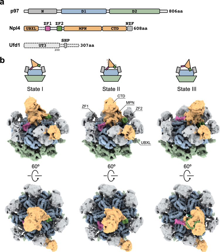
Fig. 1
Three conformational states of human p97 in complex with Npl4/Ufd1.
a Domain architecture of human p97, Npl4, and Ufd1. Unresolved parts are shown with dotted lines. b Single-particle cryo-EM maps (unsharpened, 0.014 threshold) of human p97 in complex with Npl4/Ufd1. ...
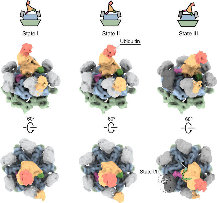
Fig. 2
Three conformational states of human p97 in complex with Npl4/Ufd1 and polyubiquitinated Ub-Eos.
Single-particle cryo-EM maps (unsharpened, 0.009 threshold) of human p97 in complex with Npl4/Ufd1 and polyubiquitinated Ub-Eos. Three conformational states were resolved using 3D classification. Each...
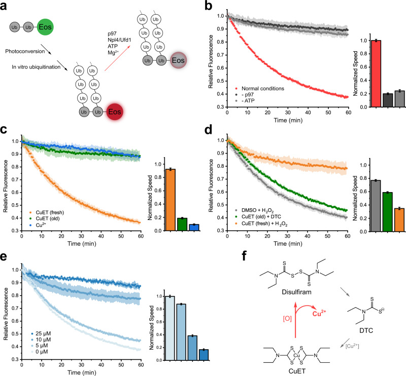
Fig. 3
Copper released from the disulfiram derivative CuET under oxidative conditions inhibits the unfolding activity of p97.
a A diagram showing the established substrate unfolding assay of p97. The unfolding of ubiquitinated Eos corresponds to a decrease in the fluorescence signal (red arrow). b The unfolding activity of w...
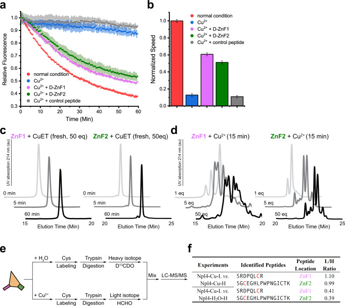
Fig. 4
Copper interacts with the zinc finger motifs of Npl4.
a Zinc finger motifs of Npl4 rescue the unfolding activity of p97 in the presence of cupric ions. b The initial velocity corresponds to panel a . The curves are presented as mean valuesu2009u00b1u2009...
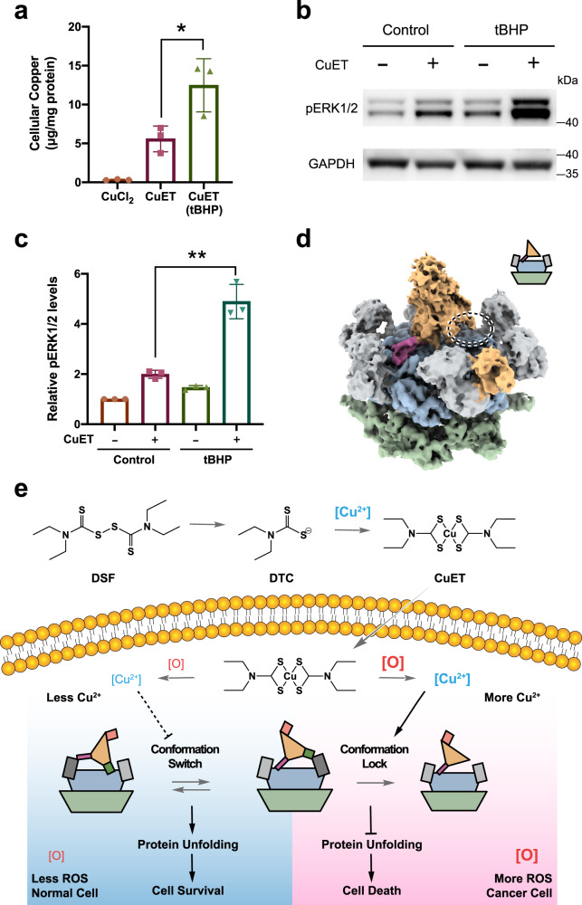
Fig. 5
Conformational lock induced by copper released from CuET.
a Quantification of cellular copper level using ICP-MS. A549 cells were treated with 5u2009u03bcM CuCl 2 , 5u2009u03bcM CuET, or 5u2009u03bcM CuET (pretreated with 2.5u2009mM tBHP), respectively ( n =...
Figure images are served from the NIH/NLM PubMed Central Open Access Subset or Europe PMC; copyright remains with the publishers and authors.
💬 Discussion
0 commentsNo comments yet. Be the first to start a discussion!
Leave a Comment